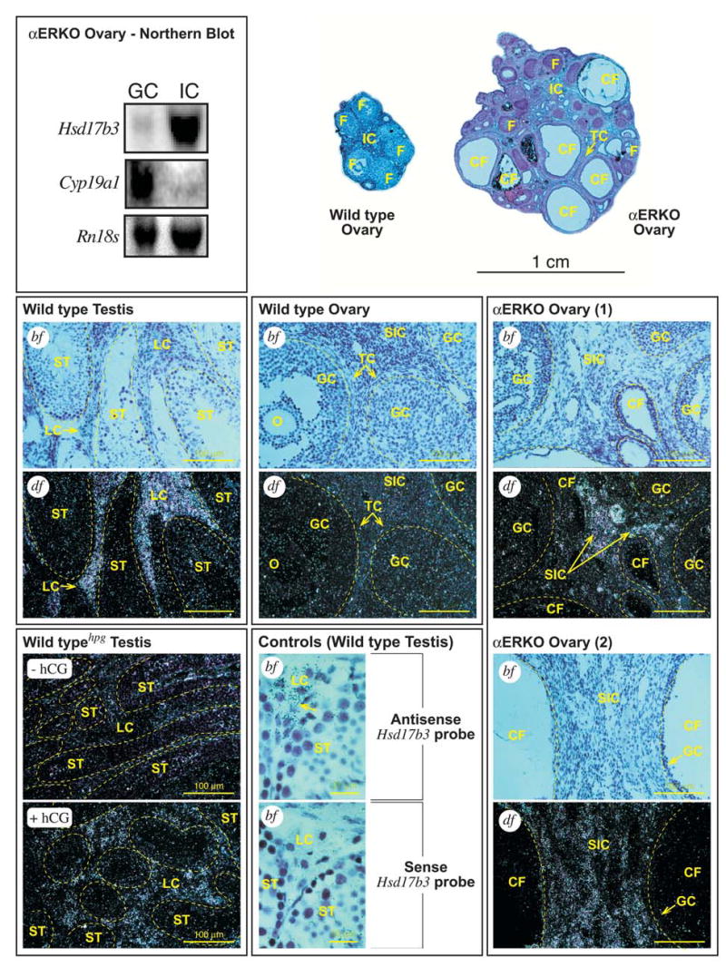
FIG. 4.

Northern blot and in situ hybridization (ISH) for Hsd17b3 mRNA in ovary and testes. (top, left) Northern blot analysis on total RNA from pooled αERKO ovaries that were fractionated into granulosa (GC) and stromal-interstitial (IC) cell compartments, demonstrating that Hsd17b3 expression in αERKO ovaries is localized to the stromal-interstitial compartment. The blot was stripped and reprobed for transcripts of a granulosa cell-specific gene (Cyp19a1) to indicate the cellular purity of each ovarian fraction. A photograph of ethidium-stained Rn18s (18S rRNA) bands demonstrates equal loading levels between lanes. (top, right) Cross-sections from representative adult wild type (WT) and αERKO ovaries photographed at low magnification. The WT ovary exhibits several healthy, large follicles (F) and a normal stromal-interstitial region (IC). In contrast, the αERKO ovary exhibits some smaller, relatively healthy follicles (F) but several enlarged, cystic follicles (CF). Also indicated in the αERKO ovary is a region of hypertrophied thecal cells (TC). (middle, bottom) Shown are representative photomicrographs (bf, bright field photomicrograph; df, dark-field photomicrograph) of tissue sections from wild type testes, wild type ovaries and αERKO ovaries (as labeled) that were subjected to ISH for Hsd17b3 mRNAs. [Wild type Testis] Photomicrographs of serial sections from a representative adult wild type testis show specific hybridization of the Hsd17b3 probe to the Leydig cells (LC) only, while hybridization levels among cells within the seminiferous tubules (ST), as indicated by dashed border, are at or below background. [Wild type Ovary] Photomicrographs of serial sections from a representative wild type ovary indicate no specific hybridization of the Hsd17b3 probe in the oocyte (O), granulosa (GC), secondary interstitial (SIC), thecal (TC) cells or any other cell type. Follicles are indicated by dashed border. [αERKO Ovary] Photomicrographs of a pair of serial sections from two representative αERKO ovaries indicates specific hybridization of the Hsd17b3 probe to the secondary interstitial cells (SIC) of the ovary, while hybridization levels among the granulosa cells of normal and cystic follicles (CF) are at or below background. Follicles are indicated by dashed border. [Wild typehpg Testis] Dark-field photomicrographs of testes sections from WThpg males following treatment with either vehicle (-hCG) or human choriogonadotropin (+hCG) to induce Hsd17b3 expression in the Leydig cells (see Materials and Methods). The untreated WThpg testis exhibits little detectable Hsd17b3 mRNA whereas the testis of the hCG-treated WThpg male exhibits significant induction of Hsd17b3 expression that is localized to the interstitial areas where Leydig cells (LC) are found. Seminiferous tubules are indicated by dashed border. [Controls - Wild type Testis] Photomicrographs of serial sections from a WT testis probed with either the Hsd17b3 sense (negative control) or anti-sense (positive control) probes to illustrate specific hybridization of the anti-sense probe only to the Leydig cells (LC), as indicated by the numerous silver grains (arrow).
