Abstract
Two cases of transient acute cardiomyopathy occurring in the immediate aftermath of intense emotional stress and without any identified aetiology are described. These two case reports, mimicking cases of acute cardiomyopathy described in patients with pheochromocytoma, suggest the possibility in man of acute catecholamine induced cardiomyopathy related to major emotional stress alone, a phenomenon so far reported only in animal experimental models. Keywords: acute cardiomyopathy; catecholamines; stress
Full Text
The Full Text of this article is available as a PDF (118.7 KB).
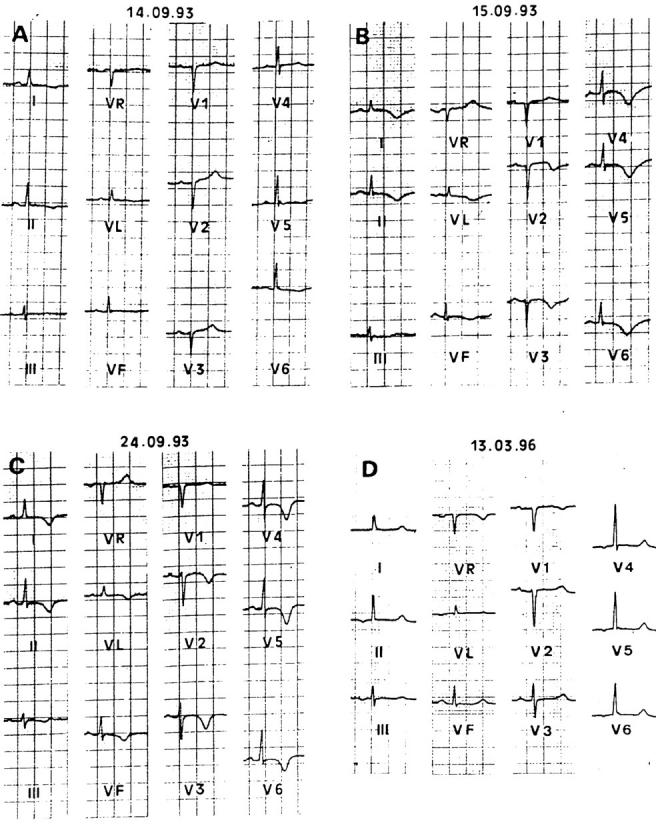
Figure 1 .

Electrocardiograms recorded in patient 1 at admission (A), one day (B), and eight days (C) after admission, and after three years of follow up (D). (A) ECG shows R wave low amplitude in right precordial leads and T wave flattening or inversion in lateral and inferior leads. (B) and (C) Diffuse repolarisation abnormalities with QT interval prolongation and marked negative T waves (pseudoischaemic) are evident. Note that in (C) R wave amplitude in right precordial leads has returned to normal. (D) ECG is normal.
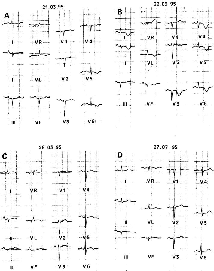
Figure 2 .
Electrocardiograms recorded in patient 2 at admission (A), one day (B) and seven days (C) after admission, and after four months of follow up (D). Similar abnormalities and evolution as in figure 1 are seen with as well as left axis deviation. Pseudoischaemic repolarisation abnormalities are near normal at the end of the first week (C). (D) ECG is normal apart from a less important left axis deviation.


