Full Text
The Full Text of this article is available as a PDF (124.1 KB).
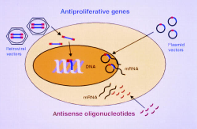
Figure 1 .
Representation of a cell showing three possible approaches to influence protein formation and thus reduce cell proliferation using gene therapy. Augmentation of antiproliferative genes can be by retroviral integration of the therapeutic gene into native DNA or by transfection with expression plasmid vectors. Inhibition of naturally occurring growth stimulators can be through antisense oligonucleotides.
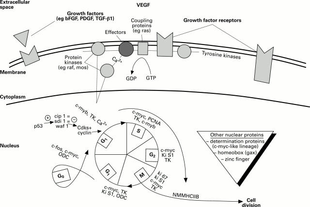
Figure 2 .
Extracellular, membrane and intracellular control points for cell division, showing some of the protein interactions where gene therapy might act. bFGF, basic fibroblast growth factor; Cdks, cyclin dependent kinases; gax, growth arrest homeobox; M, mitosis; PCNA, peripheral cell nuclear antigen; RB, retinoblastoma protein; S, DNA synthesis; sdi 1, senescent cell derived inhibitor 1; TK, thymidine kinase; TGF-β1, transforming growth factor β1; VEGF, vascular endothelial derived growth factor; PDGF, platelet derived growth factor; cip 1, Cdk interacting protein; ODC, ornithin olecarboxylase; NMMHC II B, non-muscle myosin heavy chain II B; waf 1, wild-type p53 activated fragment.


