Abstract
Macrophage migration inhibitory factor (MIF) is a ubiquitous cytokine that functions in reproduction and plays an important role in sperm maturation and motility. Here we reveal a correlation between MIF levels in human seminal fluid and fertility status. We identify an abnormal biphasic profile of MIF in the seminal fluid of patients with impaired sperm parameters. Our findings may be of interest for the development of a diagnostic method for fertility status.
INTRODUCTION
Macrophage migration inhibitory factor (MIF) is a proinflammatory cytokine with an important pathogenic role in several diseases, including sepsis, septic shock, and diabetes (1–4). The MIF molecule is also a d-dopachrome tautomerase, which catalyzes the conversion of the red color solution of l-dopachrome methyl ester into a colorless solution of indole derivatives (5). Identification of this activity has facilitated the development of novel MIF antagonists, because MIF tautomerase activity correlates well with its biological activity (4,6–8).
Recent studies have indicated a broader scope for MIF activities, including a role in reproduction (9,10), because it affects sperm maturation and spermatozoa motility (11,12). MIF, secreted by the Leydig cells of the testis, is a constituent of seminal fluid and a cytoskeleton element of the mid- and principal piece of the sperm tail (12,13–15). MIF is highly expressed in the epididymis and has been shown to be an important factor in sperm maturation (11). Increased sperm-associated MIF has been associated with poor sperm motility (12).
Based on these observations, we hypothesized that MIF levels in seminal fluid may be indicative of spermatozoa quality, and therefore male fertilizing ability. Here we reveal a correlation between MIF levels in human seminal fluid and fertility status. We identify an abnormal biphasic profile of MIF in the seminal fluid of patients with impaired sperm parameters. Our findings may be of interest for the development of a diagnostic method for fertility status.
MATERIALS AND METHODS
Seminal Fluid Collection
Human semen samples were collected after three to five days of abstinence of sexual activity from healthy and infertile individuals. Sperm parameters were evaluated according to the World Health Organization criteria (1999). All individuals signed a consent form to allow the use of 200 mL of their seminal fluid in this study. All samples were collected and shipped to the US for analysis. All data was coded to protect subject identity. This research is therefore not considered research with human subjects as defined by 45CFR46.102 law (US Department of Health and Human Services).
Determination of the l-Dopachrome Tautomerase Activity of the Seminal Fluids
The supernatants of the seminal fluids were analyzed for MIF tautomerase activity using l-dopachrome methyl ester (1,2). l-Dopachrome methyl ester was prepared at 2.4 mM concentration through oxidation of l-3,4-dihydroxyphenylalanine methyl ester with sodium periodate. Activity was determined at room temperature by adding dopachrome methyl ester (0.3 mL) to a cuvette containing 50 mL of the above supernatants and measuring the time required to decolorize l-dopachrome methyl ester.
Sperm Preparation for Testing Exogenous MIF Effects on Sperm Motility and Apoptosis
Only samples with sperm concentration at least 20 × 106/mL, total motility ≥50%, progressive motility at least 40%, viability at least 80% and, leukocyte concentration <1 × 10⁶/mL were used. Samples from each subject were tested separately. After 30-min liquefaction at 37°C, motile spermatozoa were isolated using a swim-up procedure, as reported (9). The final pellet was resuspended in Biggers, Whitten, and Wittingham (BWW) medium at a concentration of 20–30 × 106 spermatozoa/mL. An aliquot containing 5 × 10⁶ spermatozoa was then incubated with increasing concentrations of MIF (0, 5, 10, 20 μg/mL) for 3 h. At the end of the incubation, total and progressive sperm motility was evaluated and, by flow cytometry analysis, the following parameters were assessed: sperm chromatin packaging quality following staining with propidium iodide (PI); DNA fragmentation, using the terminal deoxynucleotidyl transferase-mediated fluorescein-dUTP nick end labeling (TUNEL) assay; externalization of phosphatidylserine (PS) on the outer leaflet of the cell membrane using the annexin V/PI assay; and mitochondrial membrane potential assessed by the cationic dye JC-1.
Flow Cytometry
Flow cytometry analysis was performed using the flow cytometer EPICS XL (Coulter Electronics, IL, Italy) equipped with a 488-nm argon laser as light source. According to the assay, three fluorescent detectors were used to measure the fluorescence corresponding to the green color (FL-1 detector 525-nm wavelength band), orange color (FL-2 detector 620-nm wavelength band), and red color (FL-3 detector 620-nm wavelength band). Twenty thousand events were measured for each sample at a flow rate of 50 to 100 events/s and analyzed using SISTEM II Software, version 3.0. The debris was gated out by drawing a region on the forward vs. side scatter dot plot enclosing the population of cells of interest. The compensations and the settings were adapted according to the assay utilized.
PI Staining
Spermatozoa were centrifuged at 500g for 10 min at room temperature, the supernatant was removed, and the cells stained with PI as previously reported (10). Briefly, 1 × 10⁶ spermatozoa were gently incubated in 1 mL PBS containing 50 μg/mL PI (Sigma Chemical, Milan, Italy), 0.1% sodium citrate, 0.1% Nonidet P40 (Sigma Chemical), and 100 kU/mL RNAse type A (Sigma Chemical) in the dark at room temperature. After 30 min, flow cytometry analysis was performed. For this assay, only one of the three fluorescent detectors available was used to measure the fluorescence corresponding to the red color of PI (FL-3 detector). To gate out, and thus exclude from the analysis, doublets and cell aggregates, a doublet discrimination module was used. The peak width was estimated using the coefficient of variation (CV) of the signals within each peak.
TUNEL Assay
DNA fragmentation, the final result of the apoptotic process, was evaluated by attaching to the dUTP breaks, conjugated to a fluorochrome, in a reaction catalyzed by exogenous terminal deoxynucleotidyl transferase (TdT), as reported (10). Briefly, spermatozoa were labeled with the Apoptosis Mebstain kit (Beckman Coulter, IL, Milan, Italy). To obtain a negative control, TdT was omitted from the reaction mixture. The positive control was obtained by pretreating the spermatozoa with 1 μg/mL deoxyribonuclease I (RNAse-free; Sigma Chemical) at 37°C for 60 min before labeling. Ten thousand events were measured for each sample at a flow rate of 500 events/s. The debris was eliminated by the same procedure described above. Light-scattering and fluorescence data were obtained at a fixed-gain setting in logarithmic mode. The percentage of FITC-labeled spermatozoa was determined in the FL-1 detector of the flow cytometer.
Annexin V/PI Assay
Staining with annexin V/PI was performed using a commercial kit (Annexin V-FITC Apoptosis detection kit; Sigma Chemical). An aliquot of cells containing about 0.5 × 10⁶/mL was resuspended in 500 μL binding buffer, labeled with 10 μL Annexin V-FITC plus 20 μL PI, incubated for 10 min in the dark according to the manufacturer’s instructions, and immediately analyzed by flow cytometry. Samples were evaluated by FL-1 (FITC) and FL-3 (PI) detectors. The cell population of interest was gated on the basis of the forward and side scatter properties. The different labeling patterns in the bivariate PI/annexin V analysis identified the different cell populations, where FITC-negative and PI-negative were designated as viable cells, FITC-positive and PI-negative as apoptotic cells, and FITC-positive and PI-positive as late apoptotic or necrotic cells.
JC-1 Staining
The cell suspension was adjusted to a density of 1 × 10⁶cells/mL and incubated for 10 min at 37°C in the dark with JC-1 probe (Molecular Probes, Milan, Italy). This molecule, which is able to selectively enter into the mitochondria, exists in a monomeric form emitting at 527 nm after excitation at 490 nm. Depending on the membrane potential, however, JC1 is able to form aggregates that are associated with a large shift in emission (590 nm). Thus, the color of the dye changes reversibly from green to greenish orange as the mitochondrial membrane becomes more polarized. At the end of the incubation period, cells were washed in PBS and analyzed by flow cytometry. The photomultiplier (PMT) value of the detector in FL-1 was set at 529 V and in FL-2 at 543 V, FL1-FL2 compensation was 12%, and FL2-FL1 compensation was 28%. A minimum of 10,000 events/sample were analyzed.
Statistical Analysis
Results are reported as mean ± SEM. The data were analyzed by one-way analysis of variance (ANOVA) followed by the Duncan’s multiple range test. The software SPSS 9.0 for Windows was used for statistical evaluation (SPSS, Chicago IL, USA). A statistically significant difference was accepted when the P value was lower than 0.05.
RESULTS AND DISCUSSION
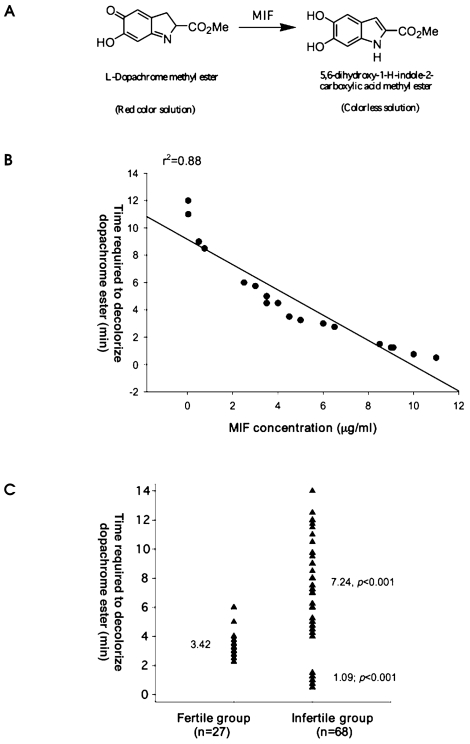
We first studied MIF levels in human seminal fluid and the corresponding MIF tautomerase activity as a measure of MIF bioactivity. The d-dopachrome tautomerase activity of MIF catalyzes the conversion of the red color solution of l-dopachrome methyl ester into a colorless solution of indole derivatives (Figure 1A) (5). A broad range of MIF protein levels in human semen (n = 18) were measured, which correlated with MIF tautomerase activity (Figure 1B) (r2 = 0.88). These data indicate that MIF is in an active form in the human seminal fluid and MIF tautomerase activity can be used as a measure of MIF bioavailability.
Figure 1.
(A) Structure of dopachrome methyl ester, a chromogenic substrate used in enzymatic assays of MIF, and its tautomerized colorless product. (B) Relationship between seminal fluid MIF concentrations and the dopachrome decolorizing rate (tautomerase activity) (n = 18, r2 = 0.88); Seminal fluid exhibited dopachrome tautomerase activity. (C) MIF tautomerase activity in seminal fluids obtained from normozoospermic subjects (controls, n = 27) and patients with abnormal sperm parameters (n = 68, P < 0.001).
We then examined the biological significance of the observed differences in the MIF tautomerase activity in seminal fluid by measuring this enzyme activity in normal control subjects and patients with abnormal sperm parameters, according to the World Health Organization guidelines. We analyzed seminal fluids from healthy individuals (n = 27) and patients (n = 68) who gave informed consent. We found that 50 uL control seminal fluid can tautomerize 1 mM l-dopachrome methyl ester between 2.25 and 6 min, with an average of 3.42 min (Figure 1C). We then examined individuals with azoospermia or patients with severe oligozoospermia (<1,000,000 spermatozoa/mL); the time required to convert the l-dopachrome ranged between 30 s and 1.5 min, with an average value of 1.09 min, indicating a high level of MIF (Figure 1C). Seminal fluid of other oligozoospermic patients as well as of patients with normal sperm counts, but with asthenozoospermia, required an average time of 7.24 min for the tautomerase reaction, within the range 4 to 14 min (Figure 1C). These data identify a biphasic profile of MIF tautomerase activity in patients with abnormal sperm parameters, which is either below or above the range of MIF tautomerase activity, typical of normozoospermic individuals.
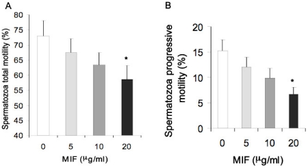
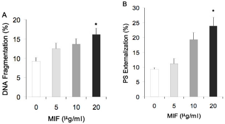
Higher MIF levels determined in men with azoospermia or severe oligozoospermia might be associated with adverse effects of this molecule related to reduced sperm motility as indicated by Frenette et al. (12). We consistently found that MIF (1 to 20 μg/mL) treatment of healthy spermatozoa dose-dependently decreased sperm total and progressive motility (Figure 2). Additionally, it increased the percentage of apoptotic cells determined by chromatin damage, DNA fragmentation, and phosphatidyl-serine translocation (Figure 3). Low MIF concentrations are indicative of impaired fertilizing ability, which corresponds with the finding that MIF is required for sperm maturation (11). It has recently been shown that MIF binds zinc through its CXXC area. Increased MIF level may cause depletion of free zinc, thereby affecting sperm maturation (11). To determine whether zinc binding to the MIF had an effect on MIF tautomerase activity, we measured enzyme activity of MIF in the presence of increasing zinc concentrations. We found no significant effect of zinc ions (0.1 to 100 μM) on MIF tautomerase activity (data not shown).
Figure 2.
Effects of increasing concentrations of MIF on total (A) and progressive (B) motility of spermatozoa obtained from normozoospermic subjects (n = 8). *P < 0.05.
Figure 3.
Effects of increasing concentrations of MIF on chromatin integrity (PI), externalization of phosphatidyl-serine (PS), and DNA fragmentation (TUNEL) of spermatozoa obtained from normozoospermic subjects (n = 8). *P < 0.05.
This study reveals a correlation between MIF bioavailability in the semen and the quality of spermatozoa, which is associated with male fertility status. The intriguing question of whether the abnormal MIF tautomerase activity could be further explored to differentiate the symptoms among infertile individuals remains to be addressed in future studies. Our data are of interest for the development of diagnostic tools for fertility status.
Footnotes
Online address: http://www.molmed.org
REFERENCES
- 1.Calandra T, Roger T. Macrophage migration inhibitory factor: a regulator of innate immunity. Nat Rev Immunol. 2003;3:791–800. doi: 10.1038/nri1200. [DOI] [PMC free article] [PubMed] [Google Scholar]
- 2.Riedemann NC, Guo RF, Ward PA. Novel strategies for the treatment of sepsis. Nat Med. 2003;9:517–24. doi: 10.1038/nm0503-517. [DOI] [PubMed] [Google Scholar]
- 3.Cvetkovic I, et al. Critical role of macrophage migration inhibitory factor activity in experimental autoimmune diabetes. Endocrinology. 2005;146:2942–51. doi: 10.1210/en.2004-1393. [DOI] [PubMed] [Google Scholar]
- 4.Al-Abed Y, et al. ISO-1 binding to the tautomerase active site of MIF inhibits its pro-inflammatory activity and increases survival in severe sepsis. J Biol Chem. 2005;280:36541–4. doi: 10.1074/jbc.C500243200. [DOI] [PubMed] [Google Scholar]
- 5.Rosengren E, et al. The immunoregulatory mediator macrophage migration inhibitory factor (MIF) catalyzes a tautomerization reaction. Mol Med. 1996;2:143–9. [PMC free article] [PubMed] [Google Scholar]
- 6.Lubetsky JB, et al. The tautomerase active site of macrophage migration inhibitory factor is a potential target for discovery of novel anti-inflammatory agents. J Biol Chem. 2002;277:24976–82. doi: 10.1074/jbc.M203220200. [DOI] [PubMed] [Google Scholar]
- 7.Dios A, et al. Inhibition of MIF bioactivity by rational design of pharmacological inhibitors of MIF tautomerase activity. J Med Chem. 2002;45:2410–6. doi: 10.1021/jm010534q. [DOI] [PubMed] [Google Scholar]
- 8.Senter PD, et al. Inhibition of macrophage migration inhibitory factor (MIF) tautomerase and biological activities by acetaminophen metabolites. Proc Natl Acad Sci U S A. 2002;99:144–9. doi: 10.1073/pnas.011569399. [DOI] [PMC free article] [PubMed] [Google Scholar]
- 9.Arcuri F, Cintorino M, Vatti R, Carducci A, Liberatori S, Paulesu L. Expression of macrophage migration inhibitory factor transcript and protein by 1st-trimester human trophoblasts. Biol Reprod. 1999;60:1299–303. doi: 10.1095/biolreprod60.6.1299. [DOI] [PubMed] [Google Scholar]
- 10.Arcuri F, et al. Macrophage migration inhibitory factor in the human endometrium: expression and localization during the menstrual cycle and early pregnancy. Biol Reprod. 2001;64:1200–5. doi: 10.1095/biolreprod64.4.1200. [DOI] [PubMed] [Google Scholar]
- 11.Eickhoff R, et al. Influence of macrophage migration inhibitory factor (MIF) on the zinc content and redox state of protein-bound sulphydryl groups in rat sperm: indications for a new role of MIF in sperm maturation. Mol Hum Reprod. 2004;10:605–11. doi: 10.1093/molehr/gah075. [DOI] [PubMed] [Google Scholar]
- 12.Frenette G, Legare C, Saez F, Sullivan R. Macrophage migration inhibitory factor in the human epididymis and semen. Mol Hum Reprod. 2005;11:575–82. doi: 10.1093/molehr/gah197. [DOI] [PubMed] [Google Scholar]
- 13.Utleg AG, et al. Proteomic analysis of human prostasomes. Prostate. 2003;56:150–61. doi: 10.1002/pros.10255. [DOI] [PubMed] [Google Scholar]
- 14.Meinhardt A, et al. Macrophage migration inhibitory factor production by Leydig cells: evidence for a role in the regulation of testicular function. Endocrinology. 1996;137:5090–5. doi: 10.1210/endo.137.11.8895383. [DOI] [PubMed] [Google Scholar]
- 15.Frenette G, Tremblay RR, Dube JY, Lazure C, Lemay M. High concentrations of the macrophage migration inhibitory factor in human seminal plasma and prostatic tissues. Arch Androl. 1998;41:185–93. doi: 10.3109/01485019808994890. [DOI] [PubMed] [Google Scholar]


