

A 59-year-old woman presented with severe exertional dyspnea 1 year after having experienced a myocardial infarction and subsequent Dressler's syndrome. Physical examination revealed a displaced apical impulse that mimicked a double beat and an unusual systolic murmur that overlay the cardiac apex. Cardiac catheterization revealed a large pseudoaneurysm of the posterolateral left ventricular wall. The pseudoaneurysm was substantially larger than the left ventricular cavity (Fig. 1). Coronary angiography showed an occluded circumflex marginal branch, which was probably the origin of the pseudoaneurysm's formation. Computed tomography showed the pseudoaneurysm's size and anatomy (Fig. 2). The patient underwent surgical repair1 and primary closure of the pseudoaneurysm via median sternotomy with cardiopulmonary bypass (Fig. 3). Histologic examination of the pseudoaneurysmal wall revealed prominent fibrovascular tissue and the conspicuous absence of normal anatomic layers of myocardium (Fig. 4). It is likely that the pathophysiologic characteristics of Dressler's syndrome played a protective role, helping to contain the rupture or leak that had caused the formation of the pseudoaneurysm.

Fig. 1 Cardiac catheterization reveals a large pseudoaneurysm of the posterolateral left ventricular wall. The cavity of the pseudoaneurysm (arrowheads) is substantially larger than that of the left ventricle, as is seen in the anteroposterior view (A) and the lateral view (B).Real-time motion images are available at texasheart.org/journal.
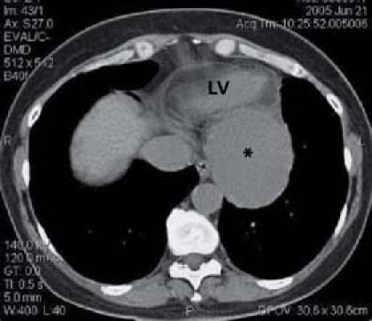
Fig. 2 A computed tomographic scan of the chest shows a large pseudoaneurysm (*) of the posterolateral left ventricular (LV) wall.
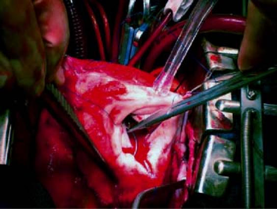
Fig. 3 Intraoperative photograph shows the surgical repair of the pseudoaneurysm.
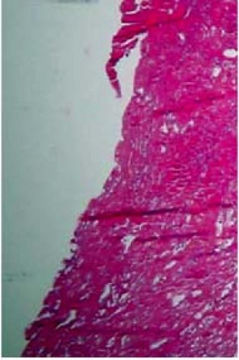
Fig. 4 Photomicrograph of the pseudoaneurysmal wall (H & E, orig. ×40), with prominent fibrovascular tissue. Normal anatomic layers of myocardium are absent.
Supplementary Material
Footnotes
Address for reprints: Nikola Dobrilovic, MD, Section of Cardiothoracic Surgery, Yale University School of Medicine, 333 Cedar Street, FMB 121, New Haven, CT 06510. E-mail: ndobrilovic@hotmail.com
Reference
- 1.Treasure T. False aneurysm of the left ventricle. Heart 1998; 80:7–8. [PMC free article] [PubMed]
Associated Data
This section collects any data citations, data availability statements, or supplementary materials included in this article.