

Abstract
Acidification, which occurs in some pathological conditions, such as ischemia and hypoxia often induces neurotoxicity. The activation of acid sensing ion channels (ASICs), which are highly permeable to calcium, has been considered the main target responsible for calcium overload in ischemic/hypoxia brain. However, the influence of extracellular proton on GABAergic synaptic transmission is not well understood. In the rat (aged 6–12 postnatal days) hippocampal CA3 neurons dissociated with an enzyme free, mechanical method, we show that raising the extracellular pH (to 8.5) or lowering it (to 6.0) significantly increased or decreased, respectively, the frequency and the amplitude of spontaneous inhibitory postsynaptic currents mediated by γ-aminobutyric acid A (GABAA) receptors. Interestingly, these modifications were not altered by amiloride (100 μM, an antagonist for ASICs), tetrodotoxin (0.5 μM, a sodium channel blocker), cadmium (100 μM, a non-selective blocker for voltage gated calcium channels), or a medium containing low calcium (0.5 mM). Significantly, changes in extracellular pH biphasically altered the peak amplitude of the currents elicited by extracellular GABA in CA3 neurons dissociated with enzyme. Raising the extracellular pH (to 8.5) or lowering it (to 6.5) shifted the concentration-response curves of GABA to the left or right, respectively, without altering the maximal responses. These data suggest that proton alters the apparent affinity of GABA receptors for agonist. Thus, extracellular proton modifies GABAergic synaptic transmission both presynaptically and postsynaptically, and this could be independent of ASICs and voltage gated calcium channels. Our finding may constitute a new mechanism underlying acidification-induced neurotoxicity.
Keywords: acidification, patch clamp, neurotoxicity, rat
1. Introduction
Complete oxidation of glucose is critical for the normal brain function. However, under certain pathological conditions, such as ischemia and hypoxia, oxygen depletion forces the brain to switch to anaerobic glycolysis. Accumulation of lactic acid and protons reduce extracellular pH (Calrk et al., 1993; Siesjo et al., 1996), and interrupt the normal neuronal activity (Xiong et al., 2004). A rapid drop of extracellular pH has been shown to excite a variety of neurons, and the activation of acid sensing ion channels (ASICs) is considered the major cause (Xiong et al., 2004). On the other hand, changes in extracellular pH have been shown to modulate the NMDA receptor (Tang et al., 1990), γ-aminobutyric acid A (GABAA) receptor (Pasternack et al., 1996), glycine receptor (Li et al., 2003), and voltage gated calcium channels (VGCC) (Delisle and Satin, 2000).
GABA is a major inhibitory neurotransmitter in the adult mammalian CNS (Cherubini and Conti, 2001). Changes in extracellular pH were previously reported to modulate postsynaptic GABAA receptors in rat hippocampal neurons (Mozrzymas et al., 2003; Pasternack et al., 1996), granule cells (Krichek and Smart, 2001), and in expression systems such as Xenopus oocytes (Robello et al., 2000). Extracellular proton is found to induce desensitization of GABAA receptors and to modify the affinity of these receptors to the agonist. Furthermore, these effects of change in extracellular pH depended on the subunit composition of the GABAA receptor (Krishek et al., 1996, 1998).
A variety of results have been reported on the effects of changes in extracellular pH on synaptic transmissions. Changes in extracellular pH modify the frequency of glycinergic inhibitory postsynaptic currents (IPSCs) in the spinal cord (Li et al., 2003), but not the miniature GABAergic IPSCs in cultured hippocampal neurons (Mozrzymas et al., 2003).
The objective of this study is to test the effect of changes in extracellular pH on GABAergic transmission. We show here that changes in extracellular pH biphasically modulate the frequency and the amplitude of the spontaneous GABAergic IPSCs in CA3 neurons dissociated with an enzyme free, mechanical method. Furthermore, we show that the effects of changes in extracellular pH on GABAergic sIPSCs are independent of amiloride sensitive ASICs, extracellular calcium, and VGCCs. In addition, we show that changes in extracellular pH alter the affinity of GABA receptors for the agonist. These findings provide a mechanism for acidification-induced neurotoxicity other than through ASICs activation.
2. Results
2.1. Changes in extracellular pH modulate spontaneous GABAergic IPSCs (sIPSCs) on hippocampal CA3 neurons
Whole-cell currents were recorded from mechanically dissociated hippocampal CA3 neurons (Fig. 1). Spontaneous IPSCs (sIPSCs) were recorded at a holding potential of −50 mV in the presence of APV (50 μM) and DNQX (10 μM), which eliminate glutamate receptor-mediated synaptic transmission. Under these conditions, bicuculline (10 μM) reversibly abolished all the spontaneous postsynaptic events, indicating that they were GABAA receptor-mediated IPSCs (Fig. 2A).
Fig. 1.
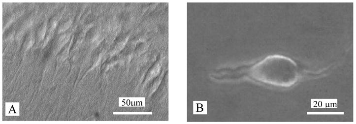
Photomicrograph of rat hippocampal CA3 neurons. This photo was obtained by a Nikon E600FN upright microscope with the aid of a near-infrared CCD camera. A) hippocampal CA3 neurons from a P12 rat were observed under differential interference contrast illumination (40 X, water immersion objective). B) a photo of a hippocampal CA3 neuron mechanically dissociated from a P12 rat was obtained by a Leica DMIRB invert microscope (40X objective). The much-reduced dendritic arbors of such neurons facilitate the space clamp. These nerve-bouton preparations often preserve functional synaptic boutons.
Fig. 2.

Effect of changes in pH on GABAergic sIPSCs. A) Spontaneous IPSCs recorded from a hippocampal neuron were completely blocked by 10 μM BIC. For this and the following figures, all IPSCs were recorded in the presence of DNQX (10 μM) and APV (50 μM) using whole cell recording method at a holding potential of −50 mV. B, Extracellular pH 8.5 potentiates frequency and amplitude of sIPSCs. B2 and C2, the averaged trace (of 15 to 55 events in one-minute period) of single IPSCs (in expanded time scale) before and after the changes of extracellular pH. C) An extracellular pH of 6.5 inhibits the frequency and the amplitude of sIPSCs. D) Cumulative curves show the effect of extracellular pH on the frequency (D1) and the amplitude (D2) of sIPSCs. E) The effect of pH ranging from 6.0 to 8.5 on the frequency and the amplitude of sIPSCs. Each column was plotted as mean ± S.E.M from 6 neurons. * p < 0.05, ** p < 0.001, paired t-test for pH 6.5 or 8.5 vs. pH 7.4.
As shown in Figure 2, an increase in the extracellular pH from 7.4 to 8.5 increased the frequency and the amplitude of sIPSCs by 173 ± 34% (p < 0.001, n = 6) and by 33 ± 5% (p < 0.05, n = 6), respectively, whereas a drop in extracellular pH from 7.4 to 6.5 inhibited the frequency and the amplitude of sIPSCs by 40 ± 8% (p < 0.001, n = 6) and by 27 ± 8% (p < 0.05, n = 6), respectively. A further drop in extracellular pH to 6.0 further inhibited the frequency and the amplitude of sEPSCs, by 69 ± 7% (p < 0.001, n = 6) and by 37 ± 6% (p < 0.05, n = 6), respectively. These data suggest that extracellular proton alters sIPSCs may involve both presynaptic and postsynaptic mechanisms.
2.2. ASICs do not play a major role in proton modulation of GABAergic sIPSCs
The expression of functional ASICs has been found in the hippocampal CA1 and CA3 subfields (Baron et al., 2001; Waldmann et al., 1997). To determine the contribution of ASICs in extracellular proton modulation of GABAergic sIPSCs in CA3 neurons, we tested the effect of pH changes on sIPSCs in the presence of 100 μM amiloride, a selective antagonist of ASICs. In the presence of 100 μM amiloride, an increase in extracellular pH from 7.4 to 8.5 increased the frequency and the amplitude of sIPSCs, by 149 ± 17% (p < 0.001, n = 5) and by 40 ± 3% (p < 0.05, n = 5), respectively, which were at the same level as that in the absence of amiloride (p > 0.05, n = 5) (Fig. 3). Similarly, a pH drop to 6.5 induced inhibition of the frequency (by 31 ± 3%, p < 0.001, n = 5) and the amplitude (by 21 ± 5%, p < 0.05, n = 5) of sIPSCs in the presence of 100 μM amiloride, which was not significantly different from that in the absence of amiloride (p > 0.05, n = 5). These data suggest that ASICs do not play a major role in the effects of changes in extracellular pH on GABAergic sIPSCs under our experimental conditions.
Fig. 3.

A) Amiloride, a blocker of ASICs did not block modulation of sIPSCs induced by changes in pH. pH 8.5 (left) potentiates whereas pH 6.5 (right) inhibits the frequency and the amplitude of sIPSCs in the presence of 100 μM amiloride. B) Effect of amiloride on the frequency and the amplitude of sIPSCs induced by changes in pH. Each column was plotted as mean ± S.E.M from 5 neurons. * p < 0.05, ** p < 0.001, paired t-test for pH 6.5 or 8.5 vs. pH 7.4.
2.3. Effects of proton on miniature IPSCs (mIPSCs)
Next, we examined the effect of pH changes on mIPSCs in the presence of tetrodotoxin (TTX, 1 μM), which eliminates action potential-induced spontaneous events. As shown in Figure 3A, a drop of extracellular pH from 7.4 to 6.5 significantly inhibited the frequency and the amplitude of mIPSCs. This is further illustrated in Figure 4B with the significant shift of the cumulative probability plot of the intervals between successive mIPSCs (B1, K - S test, rightward shift, p < 0.05) and of the amplitude of mIPSCs (B2, K - S test, left ward shift, p < 0.05), as well as by the accompanying histogram (insets). In the 5 neurons tested, a drop of extracellular pH from 7.4 to 6.5 inhibited the frequency and amplitude of mIPSCs by 33 ± 7% (p < 0.05) and 29 ±2% (p < 0.05), respectively. The effects are not significantly different from those in the absence of TTX (p > 0.05, n = 5). These data suggest that TTX-sensitive sodium channels are not involved in the modification of mIPSCs by extracellular proton.
Fig. 4.

Changes in extracellular pH modify mIPSCs. A) Typical traces showing that the effect of pH 6.5 on mIPSCs. B) Cumulative plots for inter-event interval cumulative probability plots for inter- event interval (B1, * p < 0.05, K - S test) and amplitude (B2, p > 0.05, K - S test) of GABAergic mIPSCs. Insets: pooled data from 5 neurons show that pH 6.5 inhibits mIPSC frequency and amplitude. * p < 0.05, paired t-test for pH 6.5 vs. pH 7.4)
2.4. Effects of extracellular calcium on proton-induced inhibition of sIPSCs
We first assessed the contribution of voltage-gated calcium channels (VGCCs) to the effect of extracellular proton on the frequency of sIPSCs. We treated neurons with an extracellular solution of pH 6.5 in the absence and presence of cadmium (100 μM), a nonselective VGCC blocker. A drop of pH from 7.4 to 6.5 inhibited the frequency and the amplitude of sIPSCs by 29 ± 7% (p < 0.001, n = 4) and by 24 ± 9% (p < 0.05, n = 4), respectively, in the presence of cadmium. The effects are similar to those in the absence of cadmium (p > 0.05, n = 4, Fig. 5A, B). Thus, proton-induced inhibition of sIPSCs was not dependent on VGCCs.
Fig. 5.

VGCCs and extracellular Ca2+ are not involved in the effect of changes in pH on sIPSCs. Sample traces showing that pH 6.5 inhibits the frequency and the amplitude of sIPSCs in the presence of 100 μM Cd2+ (A) or 0.5 μM Ca2+ (C). Cumulative plots for inter-event interval are shown in B1 (* p < 0.05, K - S test) and D1 (* p < 0.05, K - S test). Cumulative plots for amplitude are shown in B2 (* p < 0.05, K - S test) and D2 (* p < 0.05, K - S test). Inset: pooled data from 5 cells shows that pH 6.5 inhibits frequency and amplitude of sIPSCs in the presence of 0.5 μM Ca2+ or Cd2+.* p < 0.05, ** p < 0.001, paired t-test for pH 6.5 vs. pH 7.4.
We then determined whether calcium influx was required for the action of proton on sIPSCs. We tested the effect of pH drop to 6.5 on sIPSCs in extracellular solution containing 2 mM (normal) and 0.5 mM calcium (low calcium). The perfusion of low calcium extracellular solution reduced sIPSC frequency by 57 ± 12% (n = 5), compared with sIPSC frequency in the normal extracellular solution. In the low calcium extracellular solution, a pH drop from 7.4 to 6.5 further inhibited the frequency and the amplitude of sIPSCs by 35 ± 7% (p < 0.001, n = 5) and 27 ± 6% (p < 0.05, n = 5), respectively. The decrease in extracellular calcium concentration did not significantly change the effects of extracellular proton on sIPSCs (p > 0.05, n = 5) (Fig. 5C, D). These data indicate that proton-induced inhibition of GABAergic sIPSCs was independent of extracellular calcium.
2.5. Changes in extracellular pH biphasically modify currents elicited by exogenous GABA
To further characterize the effects of extracellular proton on GABAA receptors, we tested the effect of changes in extracellular pH on currents induced by GABA (IGABA) recorded from CA3 neurons dissociated with enzyme, at a holding potential of −50 mV. The neurons were pretreated by extracellular solutions with different pH for 20 seconds to desensitize ASICs. As shown in Figure 6, currents elicited by 3 μM GABA were inhibited by 51 ± 5% (p < 0.001, n = 4, Fig. 6A1) in response to a drop of extracellular pH to 6.5, and potentiated by 110 ± 3% (p < 0.001, n = 4, Fig. 6A2) when extracellular pH was raised to 8.5. However, when elicited by the nearly saturating concentration of GABA (300 μM), the GABAA current was inhibited by only 24 ± 7% (p < 0.05, n = 4, Fig. 6A3) in the extracellular solution with the pH of 6.5 and was not altered (5 ± 4%, p > 0.05, n = 4, Fig. 6A4) in the extracellular solution with pH of 8.5.
Fig. 6.

Changes in pH biphasically modify GABA induced currents. Typical traces showing the effect of pH 6.5 and pH 8.5 on currents evoked by 3 μM (A1, A2) and 300 μM GABA(A3, A4). B) Concentration-response curves of GABA in pH 7.4 (open circles) and pH 6.5 (close circles). Each point is the mean ± SEM from 3 to 7 cells. The data were normalized to the peak value of IGABA elicited by 3 μM GABA. Solid lines are least square fit of the Michaelis-Menten equation to the experimental data: I = (IMAX * Cn)/(Cn + EC50 n), where I, IMAX, C) EC50 and n are IGABA, maximal IGABA, GABA concentration, the concentration at which IGABA is 50% of maximum and the Hill coefficient, respectively. The EC50 was 25 μM and 56 μM for GABA in pH 7.4 and in pH 6.5, respectively. The EC50 was 24 μM and 10 μM for GABA in pH 7.4 and in pH 8.5, respectively.
Evaluation of the GABA concentration-response relation in the pH 6.5 suggests that extracellular proton reduces the affinity of the GABAAR for its agonist. That is, a drop of pH from 7.4 to 6.5 shifted the curve expressing IGABA as a function of GABA concentration (Fig. 6B) to the right. The apparent EC50 for GABA in pH 7.4 and pH 6.5 was 25 and 56 μM, respectively.
On the other hand, as shown in Fig. 6C, an increase of pH from 7.4 to 8.5 shifted the GABA concentration-response curve to the left. The apparent EC50 for GABA in pH 7.4 and pH 6.5 was 24 and 10 μM, respectively. These suggest that a raise of pH from 7.4 to 8.5 increases the affinity of the GABAA receptor for its agonist.
3. Discussion
We reported here that the acidification and alkalization of extracellular solutions modulated GABAergic synaptic transmission in hippocampal CA3 area, both presynaptically and postsynaptically. The modification was independent of ASICs. Some non-specific modulators, such as a pH jump may be involved in the presynaptic effect of proton on GABA release. Moreover, the postsynaptic effects of protons were mediated by the changes in the affinity of GABAA receptor for agonist.
The hippocampus is involved in memory processes and in the physiopathology of ischemia/hypoxia (Schmidt-Kastner and Freund, 1991). Hippocampal CA3 neurons receive GABAergic inputs which control neuronal excitability. These characteristics make the hippocampus an ideal place to study the modulation on GABAergic transmission by protons. Moreover, the fact that hippocampal CA3 neurons can be isolated along with attached GABAergic terminal boutons (Akaike and Moorhouse, 2003) provides an opportunity to evaluate the effect of changes of extracellular pH on GABA release under a well-controlled experimental condition.
3.1. Changes of extracellular pH biphasically modulate the frequency and the amplitude of GABAergic sIPSCs
One of our major findings is that GABAergic transmission is very sensitive to the changes in extracellular pH, independent of amiloride sensitive ASICs. Changes of pH biphasically modulated the frequency and the amplitude of GABAergic sIPSCs and mIPSCs. Alkalization to pH 8.5 significantly enhanced the frequency and the amplitude of the GABAergic sIPSCs, while acidification significantly decreased these parameters of sIPSCs. Spontaneous postsynaptic currents are events that represent the release of presynaptic vesicles. Changes in frequency come from the presynaptic action, while changes in amplitude result from the postsynaptic action (Li et al., 1998). Our results suggest that the modulation of sIPSCs induced by pH changes involves both presynaptic and postsynaptic mechanisms. This means that changes of extracellular pH modulate both the release of GABA and the function of the postsynaptic GABAA receptors.
Interestingly, a biphasic effect induced by changes in pH was observed on glycinergic mIPSCs in the spinal cord (Li et al., 2003). Nevertheless, a previous study on culture hippocampal neurons failed to observe an effect of a drop of pH (to 6) in extracellular solution on the frequency of mIPSCs (Mozrzymas et al., 2003). While the mechanism underlying the difference warrants further study, an apparent difference between that study and our study is that the preparations used are different. It will be interested to determine whether the mechanical dissociation procedure (or culture treatment) makes the neurons more (or less) sensitive to the change of the extracellular pH.
3.2. Changes in pH modulate GABA release through non-specific mechanisms
The observation that 100 μM amiloride, which blocks ASICs, did not alter pH change-induced modulation of GABAergic sIPSCs indicates that ASICs are not involved. It was reported that acidification inhibits voltage-dependent calcium channels (Krafte and Kass, 1988; Kwan and Kass, 1993), which control neurotransmitter release in the CNS (Poncer et al., 1997; Takahashi and Momiyama, 1993). However, in mechanically dissociated CA3 neurons, cadmium did not attenuate the modulation of sIPSCs induced by pH changes. This indicates that voltage-dependent calcium channels are not involved in the effects associated with pH change-induced modulation of sIPSCs under our experimental conditions.
Changes in the pH of extracellular solutions can induce a very small shift in the intracellular pH (Kaila, 1994), which may contribute to the results we observed. However, under our experimental conditions, the significant effects on sIPSCs were observed immediately after the change of perfusates of different pH. Therefore, it seems unlikely that the small variations in internal pH could account for the effects on sIPSCs induced by the changes in extracellular pH. Furthermore, GABA itself causes acidification when CO2/HCO3- are present (Kalia and Voipio, 1987). The effect of GABA on the intracellular pH depends on the concentrations of HCO3 (Kaila et al., 1990). However, in our recording solution, CO2/HCO3 was not present (See ‘Experimental procedures’). Therefore, if GABA induces acidification, it will be minimal under our experimental conditions.
Synaptic vesicles in nerve terminal have an acidic pH (5.2–5.5), which is much lower than the surrounding extracellular medium (Fuldner and Stadler, 1982). The difference is called a pH jump, which can be one of the factors controlling transmitter release from synaptic vesicles (Ahdut-Hacohen et al., 2004). A large pH jump causes an increase in the open probability of the non-specific ion channels in synaptic vesicles, allows ions to enter the vesicle thus facilitating the ion exchange process, and finally enhances transmitter release. Whereas a narrow pH jump decreases transmitter release. In the present study, the biphasic modulation of sIPSC frequency by extracellular changes in pH provides strong electrophysiological evidence for this pH jump mechanism.
3.3. Changes in pH alter the affinity of GABAA receptors to GABA
In the present study, we found that changes in pH biphasically modified the amplitude of sIPSCs and mIPSCs. The modifications were independent of ASICs because these modifications were not blocked by amiloride, and sustained in the process of perfusion without desensitization. It also indicates that changes in extracellular pH may directly act upon GABAA receptors.
Controversial results were reported on pH change-induced modulation of mammalian GABAA receptors, depending on the experimental conditions, animal species, brain regions, subunit compositions, and concentrations of GABA (Feng and Macdonald, 2004; Huang et al., 1999, 2004; Krishek et al., 1996 Krishek et al., 1998, 2001; Li et al., 2003; Mozrzymas et al., 2003; Pasternack et al., 1996; Robello e tal., 2000; Smart, 1992; Vyklicky et al., 1993; Wilkins et al., 2002; 2005; Zha et al., 1998). Li and colleagues reported that changes in pH exert different effects on glycine current in spinal cord neurons depending on the timing of proton application (Li et al., 2003). To eliminate a possible effect of ASICs on IGABA, we applied GABA after more than 20 second incubation with proton solution. This 20 second period allowed ASICs to be desensitized, mostly, if not completely (Xiong et al., 2004). Under this experimental condition, we observed that changes in pH had similar effects on IGABA in the absence and presence of amiloride. This observation suggests a possible direct effect of pH changes on GABAA receptors. This possibility is supported by the following observations. First, IGABA was inhibited by a decrease in extracellular pH, but enhanced by an increase in extracellular pH. Second, pH changes induced stronger effects on IGABA elicited by a sub-saturating concentration of GABA (3 μM) compared to changes of IGABA elicited by a saturating concentration of GABA (300 μM). Third, a pH drop and a raise respectively cause a rightward and a leftward shift of GABA concentration-response curve. These evidence points to the possibility that acidification and alkalization respectively decreases and increases the affinity of the GABAA receptors to GABA.
In conclusion, acidification and alkalization modulates GABAergic synaptic transmission both presynaptically and postsynaptically, and independent of ASICs. In addition, some non-specific modulators such as a pH jump are probably involved in the effect of pH change on GABA release. Moreover, the postsynaptic effects are mediated by altering the affinity of GABAA receptor to GABA. This may be an additional mechanism underlying acidification and alkalization induced neurotoxicity.
4. Experimental procedures
4.1. Preparation
The care and use of animals, and the experimental protocol were approved by the Institutional Animal Care and Use Committee of the University of Medicine and Dentistry of New Jersey. The brain slices were prepared as described previously (Ye et al., 2004, 2006; Zhou et al., 2006). In brief, rats, aged 6–12 postnatal days (P), were anesthetized and then killed by decapitation, and the semisphere of the brain was quickly excised and coronally sliced (300 μm) with a VF-100 Slicer (Precisionary Instruments, Greenville, NC). This was done in ice-cold modified glycerol-based artificial cerebrospinal fluid (GACSF) saturated with 95%O2/5% CO2 (carbogen) containing (in mM): 250 glycerol, 2.5 KCl, 1.2 NaH2PO4, 1.2 MgCl2, 2.4 CaCl2, 26 NaHCO3, and 11 glucose. Hippocampus slices were then kept in carbogen-saturated ACSF containing (in mM): 126 NaCl, 1.6 KCl, 1.25 NaH2PO4, 1.5 MgCl2, 2 CaCl2, 25 NaHCO3, and 10 glucose at room temperature (22–24°C) for at least one hour before use.
Neurons, with functional terminals, were obtained by mechanical dissociation as described previously (Akaike and Moorhouse, 2003; Ye et al., 2004; Zhou et al., 2006). Briefly, slices were transferred to a 35 mm culture dish (Falcon, Rutherford, NJ) filled with a standard extracellular solution containing (mM): 140 NaCl, 5 KCl, 2 CaCl2, 1 MgCl2, 10 HEPES, and 10 glucose (320 mOsm, pH set to 7.3 with Tris base). The region of hippocampal CA3 was identified with an inverted microscope (Nikon, Tokyo, Japan). A heavily fire-polished glass pipette with a ~50 μm tip in diameter was fixed on a homemade device. Then, the pipette was positioned by a manipulator to slightly touch the surface of the hippocampal CA3 region. The individual neurons were dissociated by horizontal vibration at a frequency of 15–20 Hz, with a range from 0.1 to 0.3 mm, for 2–5 minutes. The slice was then removed. Within 20 minutes, the isolated neurons (3–10 per dish) adhered to the bottom of the dish and were ready for electrophysiological recording. These mechanically dissociated neurons often persevered some functional nerve terminals (Akaike and Moorhouse, 2003; Ye et al., 2004; Zhou et al., 2006). The postsynaptic elements of neurons isolated in this manner consist of a soma and processes of either short or medium length (Fig. 1B).
Some experiments were conducted on enzymatically dissociated hippocampal CA3 neurons (Ye et al., 1999). Hippocampal slices were first incubated in oxygenated standard solution containing 0.3 mg/ml papain (from papaya latex; Sigma, St. Louis, MO) at room temperature for 15 minutes. The slices were then incubated in enzyme-free standard extracellular solution. The hippocampal CA3 region was cut out under an inverted microscope and single cells were dissociated by trituration using two fire-polished glass pipettes with gradually narrower diameters. The cells settled to the bottom of the culture dish within 20 min and were ready for electrophysiological recordings.
4.2. Electrophysiological measurements
Whole-cell configurations were used to record electrical activity with an Axopatch 200B amplifier (Axon Instruments, Foster city, CA), via a Digidata 1322A analog-to-digital converter (Axon Instruments), and pClamp 9.2 software (Axon Instruments). Data were filtered at 1 kHz and sampled at 5 kHz.
The patch electrodes had a resistance of 3–5 MΩ when filled with pipette solution containing (in mM): 140 CsCl, 2 MgCl2, 4 EGTA, 0.4 CaCl2, 10 HEPES, 2 Mg-ATP, and 0.1 GTP. The pH was adjusted to 7.2 with Tris - base, and the osmolarity was adjusted to 280–300 mOsm with sucrose. Electrophysiological recordings were performed at room temperature (22–24 °C).
4.3. Drugs and solutions
Most of the chemicals including bicuculline (BIC), DL-2-amino-5-phosphono-valeric acid (APV), 6,7-dinitroquinoxaline-2, 3-dione (DNQX), tetrodotoxin (TTX), and papain were purchased from Sigma-Aldrich Inc (St. Louis, MO). All solutions were prepared on the day of the experiment. Chemicals were applied to dissociated neurons with a Y-tube. This exchanged the extracellular solution surrounding the neurons within 40 ms (Zhou et al., 2006).
4.4. Data analysis
Spontaneous inhibitory postsynaptic currents (sIPSCs) were analyzed with Clampfit 9.2 software (Molecular Devices Corporation, Sunnyvale, U.S.A.) as described previously (Zhou et al., 2006). Briefly, the sIPSCs were screened automatically using a template with an amplitude threshold of 5.5 pA. These were visually accepted or rejected based upon the rise and decay times. More than 95% of the sIPSCs, which were visually accepted, were screened using a suitable template. The amplitudes and intervals of sIPSCs in different conditions were also obtained. Their cumulative probability distributions were constructed using Clampfit 9.2. Following this, a Kolmogorov-Smirnov (K-S) test was used for evaluating the significance of drug effects. Differences in amplitude and frequency were tested by Student’s paired two-tailed t-test, unless indicated otherwise. Numerical values are presented as the mean ± standard error of the mean (SEM). Values of p < 0.05 were considered significant.
Acknowledgments
This work is made possible by the NIH grants AA11989, AA01595 and AT 001182 to JHY.
Footnotes
Publisher's Disclaimer: This is a PDF file of an unedited manuscript that has been accepted for publication. As a service to our customers we are providing this early version of the manuscript. The manuscript will undergo copyediting, typesetting, and review of the resulting proof before it is published in its final citable form. Please note that during the production process errors may be discovered which could affect the content, and all legal disclaimers that apply to the journal pertain.
References
- Ahdut-Hacohen R, Duridanova D, Meiri H, Rahamimoff R. Hydrogen ions control synaptic vesicle ion channel activity in Torpedo electromotor neurons. J Physiol. 2004;556:347–352. doi: 10.1113/jphysiol.2003.058818. [DOI] [PMC free article] [PubMed] [Google Scholar]
- Akaike N, Moorhouse AJ. Techniques: applications of the nerve-bouton preparation in neuropharmacology. Trends Pharmacol Sci. 2003;24:44–47. doi: 10.1016/s0165-6147(02)00010-x. [DOI] [PubMed] [Google Scholar]
- Baron A, Schaefer L, Lingueglia E, Champigny G, Lazdunski M. Zn2+ and H+ are coactivators of acid-sensing ion channels. J Biol Chem. 2001;276:35361–35367. doi: 10.1074/jbc.M105208200. [DOI] [PubMed] [Google Scholar]
- Cherubini E, Conti F. Generating diversity at GABAergic synapses. Trends Neurosci. 2001;24:155–162. doi: 10.1016/s0166-2236(00)01724-0. [DOI] [PubMed] [Google Scholar]
- Clark K, Stewart LC, Neubauer S, Balschi JA, Smith TW, Ingwall JS, Nedelac JF, Humphrey SM, Kleber AG, Springer CS. Extracellular volume and transsarcolemmal proton movement during ischemia and reperfusion: a 31P NMR Spectroscopic study of the isovolumic rat heart. Nmr Biomed. 1993;6:278–286. doi: 10.1002/nbm.1940060407. [DOI] [PubMed] [Google Scholar]
- Delisle BP, Satin J. pH modification of human T-type calcium channel gating. Biophys J. 2002;78:1895–905. doi: 10.1016/S0006-3495(00)76738-5. [DOI] [PMC free article] [PubMed] [Google Scholar]
- Feng HJ, Macdonald RL. Proton modulation of alpha-1-beta-3-delta GABAA receptor channel gating and desensitization. J Neurophysiol. 2004;92:1577–1585. doi: 10.1152/jn.00285.2004. [DOI] [PubMed] [Google Scholar]
- Fuldner HH, Stadler H. 31P-NMR analysis of synaptic vesicles, Status of ATP and internal pH. Eur J Biochem. 1982;121:519–524. doi: 10.1111/j.1432-1033.1982.tb05817.x. [DOI] [PubMed] [Google Scholar]
- Huang RQ, Dillon GH. Effect of extracellular pH on GABA-activated current in rat recombinant receptors and thin hypothalamic slices. J Neurophysiol. 1999;82:1233–1243. doi: 10.1152/jn.1999.82.3.1233. [DOI] [PubMed] [Google Scholar]
- Huang RQ, Chen Z, Dillon GH. Molecular basis for modulation of recombinant alpha-1-beta-2-gamma-2 GABAA receptors by protons. J Neurophysiol. 2004;92:883–894. doi: 10.1152/jn.01040.2003. [DOI] [PubMed] [Google Scholar]
- Kaila K. Ionic basis of GABAA receptor channel function in the nervous system. Prog Neurobiol. 1994;42:489–537. doi: 10.1016/0301-0082(94)90049-3. [DOI] [PubMed] [Google Scholar]
- Kaila K, Saarikoski J, Voipio J. Mechanism of action of GABA on intracellular pH and on surface pH in crayfish muscle fibres. J Physiol. 1990;427:241–260. doi: 10.1113/jphysiol.1990.sp018170. [DOI] [PMC free article] [PubMed] [Google Scholar]
- Kaila K, Voipio J. Postsynaptic fall in intracellular pH induced by GABA-activated bicarbonate conductance. Nature. 1987;330:163–165. doi: 10.1038/330163a0. [DOI] [PubMed] [Google Scholar]
- Krafte DS, Kass RS. Hydrogen ion modulation of Ca channel current in cardiac ventricular cells. Evidence for multiple mechanisms. J Gen Physiol. 1988;91:641–657. doi: 10.1085/jgp.91.5.641. [DOI] [PMC free article] [PubMed] [Google Scholar]
- Krishek BJ, Amato A, Connolly CN, Moss SJ, Smart TG. Proton sensitivity of the GABAA receptor is associated with the receptor subunit composition. J Physiol (Lond) 1996;492:431–443. doi: 10.1113/jphysiol.1996.sp021319. [DOI] [PMC free article] [PubMed] [Google Scholar]
- Krishek BJ, Moss SJ, Smart TG. Interaction of H+ and Zn2+ on recombinant and native rat neuronal GABAA receptors. J Physiol (Lond) 1998;507:639–652. doi: 10.1111/j.1469-7793.1998.639bs.x. [DOI] [PMC free article] [PubMed] [Google Scholar]
- Krishek BJ, Smart TG. Proton sensitivity of rat cerebellar granule cell GABAA receptors: dependence on neuronal development. J Physiol (Lond) 2001;530:219–233. doi: 10.1111/j.1469-7793.2001.0219l.x. [DOI] [PMC free article] [PubMed] [Google Scholar]
- Kwan YW, Kass RS. Interactions between protons and calcium near L-type calcium channels: evidence for independent channel-associated binding sites. Biophys J. 1993;65:1188–1195. doi: 10.1016/S0006-3495(93)81152-4. [DOI] [PMC free article] [PubMed] [Google Scholar]
- Li YF, Wu LJ, Li Y, Xu L, Xu TL. Mechanisms of H+ modulation of glycinergic response in rat sacral dorsal commissural neurons. J Physiol. 2003;552:73–87. doi: 10.1113/jphysiol.2003.047324. [DOI] [PMC free article] [PubMed] [Google Scholar]
- Li YX, Zhang Y, Lester HA, Schuman EM, Davidson N. Enhancement of neurotransmitter release induced by brain-derived neurotrophic factor in cultured hippocampal neurons. J Neurosci. 1998;18:10231–10240. doi: 10.1523/JNEUROSCI.18-24-10231.1998. [DOI] [PMC free article] [PubMed] [Google Scholar]
- Michaelson DM, Angel I. Determination of pH change in cholinergic synaptic vesicles: its effect on storage and release of acetylcholine. Life Sci. 1980;27:39–44. doi: 10.1016/0024-3205(80)90017-x. [DOI] [PubMed] [Google Scholar]
- Mozrzymas JW, Zarnowska ED, Pytel M, Mercik K. Modulation of GABA(A) receptors by hydrogen ions reveals synaptic GABA transient and a crucial role of the desensitization process. J Neurosci. 2003;23:7981–7992. doi: 10.1523/JNEUROSCI.23-22-07981.2003. [DOI] [PMC free article] [PubMed] [Google Scholar]
- Pasternack M, Smirnov S, Kaila K. Proton modulation of functionally distinct GABAA receptors in acutely isolated pyramidal neurons of rat hippocampus. Neuropharmacology. 1996;35:1279–1288. doi: 10.1016/s0028-3908(96)00075-5. [DOI] [PubMed] [Google Scholar]
- Poncer JC, McKinney RA, Gähwiler BH, Thompson SM. Either N- or P-type calcium channels mediate GABA release at distinct hippocampal inhibitory synapses. Neuron. 1997;18:463–472. doi: 10.1016/s0896-6273(00)81246-5. [DOI] [PubMed] [Google Scholar]
- Robello M, Barduzzi R, Cupello A. Modulation by extracellular pH of GABAA receptors expressed in Xenopus oocytes injected with rat brain mRNA. Int J Neurosci. 2000;103:41–51. doi: 10.3109/00207450009003251. [DOI] [PubMed] [Google Scholar]
- Schmidt-Kastner R, Freund TF. Selective vulnerability of the hippocampus in brain ischemia. Neuroscience. 1001;40:599–636. doi: 10.1016/0306-4522(91)90001-5. [DOI] [PubMed] [Google Scholar]
- Siesjo BK, Katsura K, Kristian T. Acidosis-related damage. Adv Neurol. 1996;71:209–233. [PubMed] [Google Scholar]
- Smart TG. A novel modulatory site for zinc on the GABA receptor complex in cultured rat neurons. J Physiol. 1992;447:587–625. doi: 10.1113/jphysiol.1992.sp019020. [DOI] [PMC free article] [PubMed] [Google Scholar]
- Takahashi T, Momiyama A. Different types of calcium channels mediate central synaptic transmission. Nature. 1993;366:156–158. doi: 10.1038/366156a0. [DOI] [PubMed] [Google Scholar]
- Tang CM, Presser F, Morad M. Amiloride selectively blocks the low threshold (T) calcium channel. Science. 1990;240:213–215. doi: 10.1126/science.2451291. [DOI] [PubMed] [Google Scholar]
- Vyklicky L, Philippi M, Kuffler DP, Orkand RK. GABAA membrane currents are insensitive to extracellular acidification in cultured sensory neurons of the frog. Physiol Res. 1993;42:313–317. [PubMed] [Google Scholar]
- Waldmann R, Bassilana F, de Weille J, Champigny G, Heurteaux C, Lazdunski M. Molecular cloning of a non-inactivating proton-gated Na+ channel specific for sensory neurons. J Biol Chem. 1997;272:20975–20978. doi: 10.1074/jbc.272.34.20975. [DOI] [PubMed] [Google Scholar]
- Wilkins ME, Hosie AM, Smart TG. Identification of a beta subunit TM2 residue mediating proton modulation of GABA type A receptors. J Neurosci. 2002;22:5328–5333. doi: 10.1523/JNEUROSCI.22-13-05328.2002. [DOI] [PMC free article] [PubMed] [Google Scholar]
- Wilkins ME, Hosie AM, Smart TG. Proton modulation of recombinant GABA(A) receptors: influence of GABA concentration and the beta subunit TM2-TM3 domain. J Physiol. 2005;567:365–377. doi: 10.1113/jphysiol.2005.088823. [DOI] [PMC free article] [PubMed] [Google Scholar]
- Xiong ZG, Zhu XM, Chu XP, Minami M, Hey J, Wei WL, MacDonald JF, Wemmie JA, Price MP, Welsh MJ, Simon RP. Neuroprotection in ischemia: blocking calcium-permeable acid-sensing ion channels. Cell. 2004;118:687–698. doi: 10.1016/j.cell.2004.08.026. [DOI] [PubMed] [Google Scholar]
- Ye JH, Schaefer R, Wu WH, Liu PL, Zbuzek VK, Mcardle JJ. Inhibitory effect of ondansetron on glycine response of dissociated rat hippocampal neurons. J Pharmacol Exp Ther. 1999;290:104–11. [PubMed] [Google Scholar]
- Ye JH, Wang F, Krnjevic K, Wang W, Xiong ZG, Zhang J. Presynaptic glycine receptors on GABAergic terminals facilitate discharge of dopaminergic neurons in ventral tegmental area. J Neurosci. 2004;24:8961–8974. doi: 10.1523/JNEUROSCI.2016-04.2004. [DOI] [PMC free article] [PubMed] [Google Scholar]
- Ye JH, Zhang J, Xiao C, Kong JQ. Patch-clamp studies in the CNS illustrate a simple new method for obtaining viable neurons in rat brain slices: Glycerol replacement of NaCl protects CNS neurons. J Neurosci Methods. 2006;158:251–259. doi: 10.1016/j.jneumeth.2006.06.006. [DOI] [PubMed] [Google Scholar]
- Zhai J, Peoples RW, Li C. Proton inhibition of GABA-activated current in rat primary sensory neurons. Pfluegers Arch. 1998;435:539–545. doi: 10.1007/s004240050550. [DOI] [PubMed] [Google Scholar]
- Zhou C, Xiao C, McArdle JJ, Ye JH. Mefloquine enhances nigral {gamma}-aminobutyric acid release via inhibition of cholinesterase. J Pharmacol Exp Ther. 2006;317:1155–1160. doi: 10.1124/jpet.106.101923. [DOI] [PubMed] [Google Scholar]