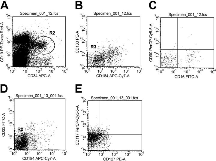
Figure 2.
Flow cytometric characterization of the expression of other primitive hematopoietic surface markers on CD34+/CD19+/CXCR4- cells. Low-density BM cells were stained for 6-color immunofluorescence as described in “Materials and methods.” Representative histograms of the flow cytometric phenotypic characterization of the CD34+/CD19+/CXCR4- cells are shown in dot plots A-E. Refer to Table 1 for the averaged fraction of the CD34+/CD19+/CXCR4- cells expressing combinations of primitive hematopoietic surface antigen. Dot plot A depicts CD34+CD19+ cells within R2. Cells contained in R2 were then analyzed for the expression of CXCR4 (CD184 in the figure) and CD133 (VEGFr2) in dot plot B. Essentially no CD34+/CD19+/CXCR4- cells were CD133+. CD34+/CD19+/CXCR4- cells were also analyzed for the expression of CD16 and CD90 (dot plot C). Whereas a small fraction of these cells expressed CD16, very few expressed CD90. Dot plot D depicts the expression of CD33 fraction of CD34+/CD19+/CXCR4- cells. Dot plot E shows the expression of CD127 and CD117 in the CD34+/CD19+/CXCR4- population. CD117 analysis found a significant increase in the mean fluorescence intensity, but the gated fraction of cells expressing CD177 is still small, as shown in Table 1.

