Abstract
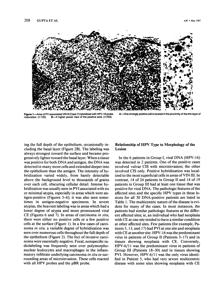
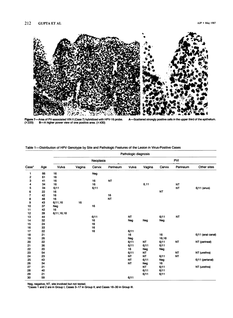
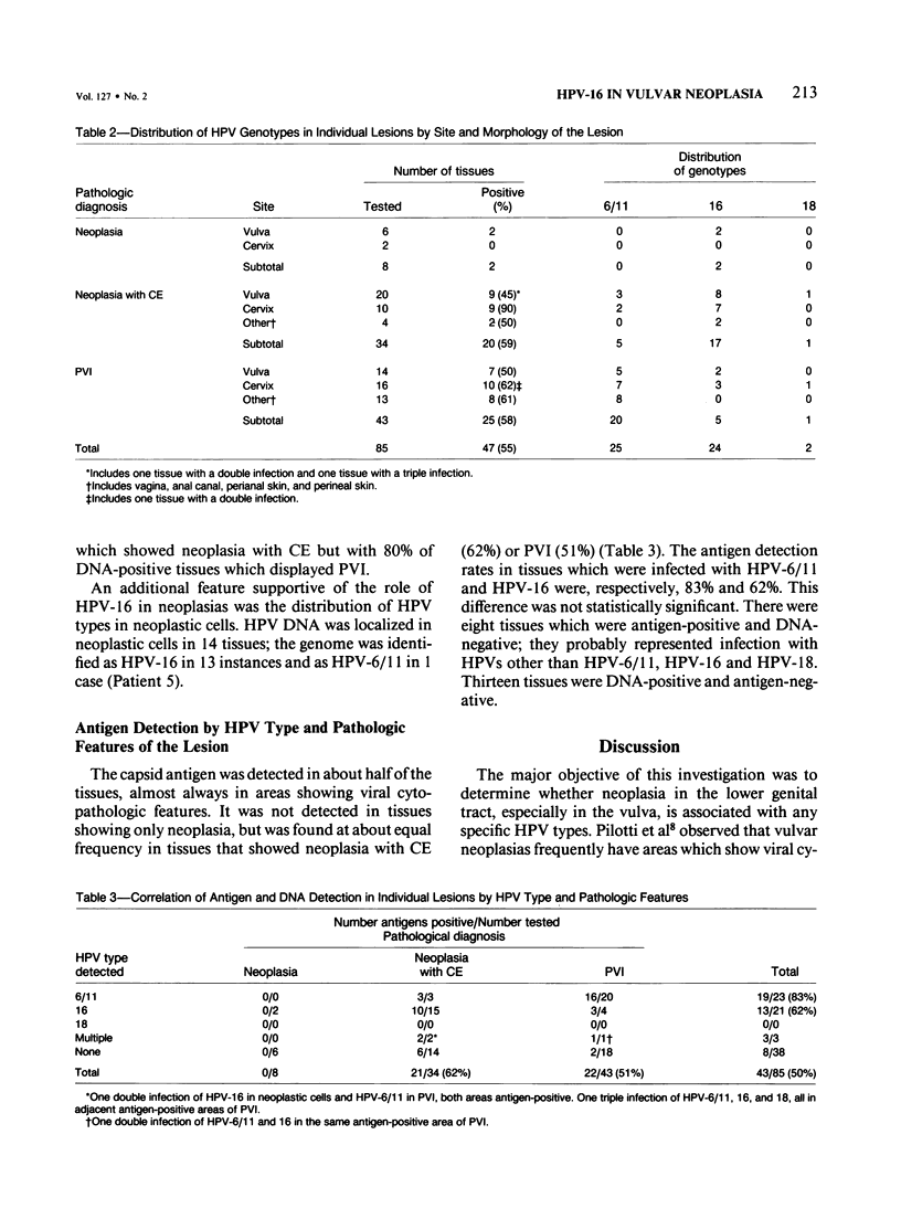
The authors examined paraffin sections from 85 genital tract tissues from 49 cases for the presence of human papillomavirus (HPV) Types 6/11, 16, and 18 by stringent in situ hybridization using 35S-labeled viral DNA probes, and for viral capsid antigen by the immunoperoxidase test. The cases, selected mostly on the basis of vulvar pathology, were distributed as follows: early neoplasia (Group I, 6 cases); early neoplasia with viral cytopathic effect (CE) (Group II, 24 cases); and papillomavirus infection (PVI) (Group III, 19 cases). Available tissues from all affected sites were examined when the disease was multicentric. One or more viral DNAs were identified in 58% of 77 tissues from Groups II and III and in 2 of 8 tissues from Group I. HPV-6/11, HPV-16 and HPV-18 DNAs were detected, respectively, in 25, 24, and 2 tissues; 3 tissues were infected simultaneously with either two or three viruses. Viral DNA was identified at more than one site in 14 of 30 DNA-positive patients; in 10 of these, a single type was detected at all sites in the same patient. The viral DNA was localized mostly in areas showing viral cytopathology. The presence of HPV-16 correlated with neoplasia. HPV-16 DNA was identified in the 2 virus-positive tissues showing neoplasia, in 17 of 20 (85%) of the DNA-positive tissues showing neoplasia with CE, and in 5 of 25 (20%) of the DNA-positive tissues showing PVI. Conversely, HPV-6/11 was found in 25% of the DNA-positive tissues showing neoplasia with CE and in 80% of the cases of PVI. An HPV genome was identified in neoplastic cells in 14 instances; in all but 1 case, the genome was HPV-16. The association of HPV-16 with neoplasia was seen for both vulvar and cervical lesions. Viral antigen was detected in 83% of lesions associated with HPV 6/11 and in 62% of lesions associated with HPV-16.
Full text
PDF
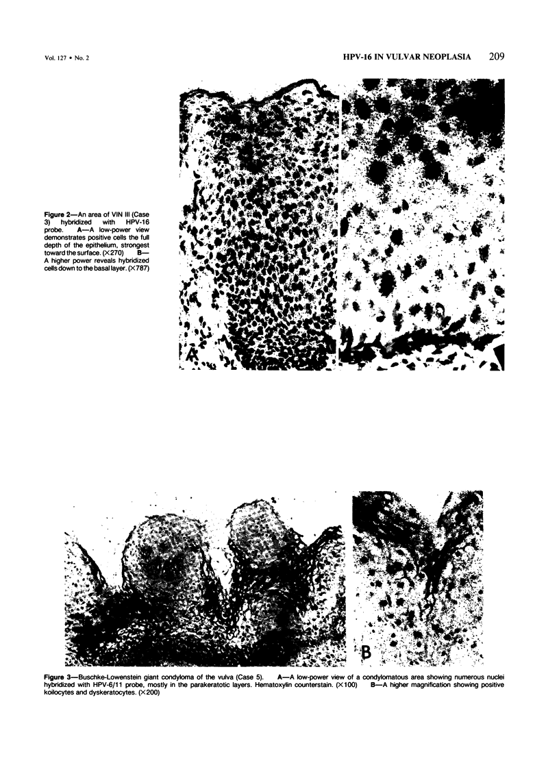
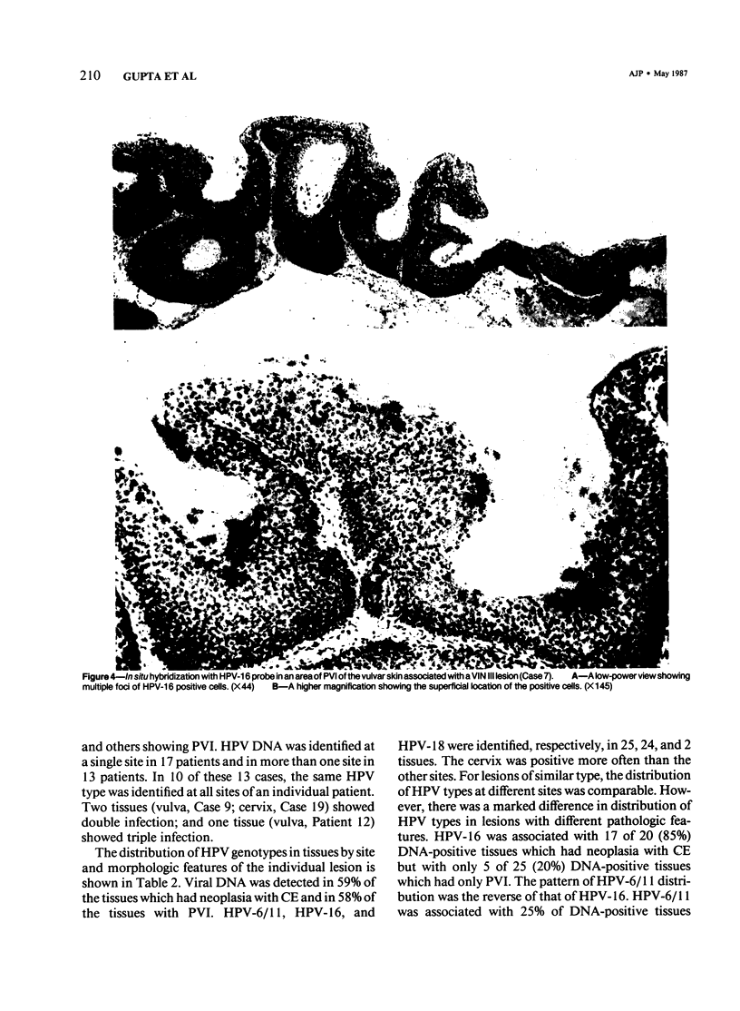
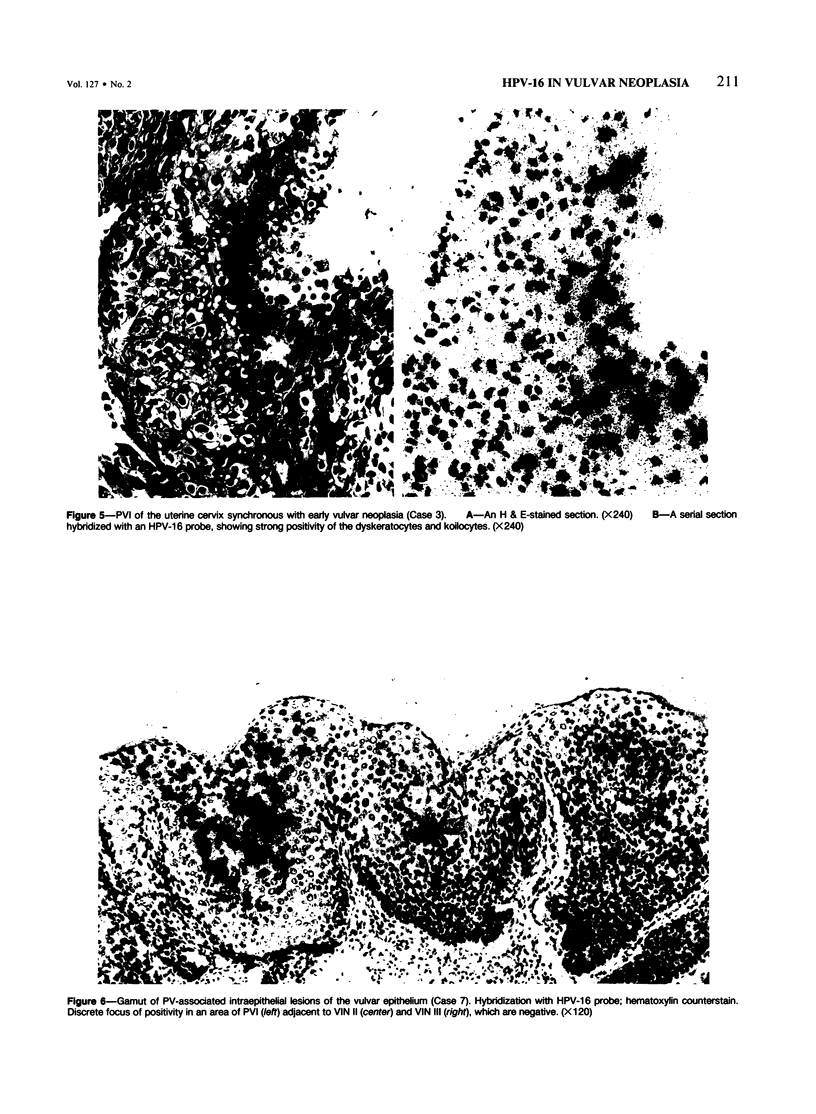
Images in this article
Selected References
These references are in PubMed. This may not be the complete list of references from this article.
- Beckmann A. M., Myerson D., Daling J. R., Kiviat N. B., Fenoglio C. M., McDougall J. K. Detection and localization of human papillomavirus DNA in human genital condylomas by in situ hybridization with biotinylated probes. J Med Virol. 1985 Jul;16(3):265–273. doi: 10.1002/jmv.1890160307. [DOI] [PubMed] [Google Scholar]
- Boshart M., Gissmann L., Ikenberg H., Kleinheinz A., Scheurlen W., zur Hausen H. A new type of papillomavirus DNA, its presence in genital cancer biopsies and in cell lines derived from cervical cancer. EMBO J. 1984 May;3(5):1151–1157. doi: 10.1002/j.1460-2075.1984.tb01944.x. [DOI] [PMC free article] [PubMed] [Google Scholar]
- Brahic M., Haase A. T. Detection of viral sequences of low reiteration frequency by in situ hybridization. Proc Natl Acad Sci U S A. 1978 Dec;75(12):6125–6129. doi: 10.1073/pnas.75.12.6125. [DOI] [PMC free article] [PubMed] [Google Scholar]
- Crum C. P., Braun L. A., Shah K. V., Fu Y. S., Levine R. U., Fenoglio C. M., Richart R. M., Townsend D. E. Vulvar intraepithelial neoplasia: correlation of nuclear DNA content and the presence of a human papilloma virus (HPV) structural antigen. Cancer. 1982 Feb 1;49(3):468–471. doi: 10.1002/1097-0142(19820201)49:3<468::aid-cncr2820490313>3.0.co;2-7. [DOI] [PubMed] [Google Scholar]
- Crum C. P., Fu Y. S., Levine R. U., Richart R. M., Townsend D. E., Fenoglio C. M. Intraepithelial squamous lesions of the vulva: biologic and histologic criteria for the distinction of condylomas from vulvar intraepithelial neoplasia. Am J Obstet Gynecol. 1982 Sep 1;144(1):77–83. doi: 10.1016/0002-9378(82)90398-2. [DOI] [PubMed] [Google Scholar]
- Crum C. P., Ikenberg H., Richart R. M., Gissman L. Human papillomavirus type 16 and early cervical neoplasia. N Engl J Med. 1984 Apr 5;310(14):880–883. doi: 10.1056/NEJM198404053101403. [DOI] [PubMed] [Google Scholar]
- Crum C. P., Mitao M., Levine R. U., Silverstein S. Cervical papillomaviruses segregate within morphologically distinct precancerous lesions. J Virol. 1985 Jun;54(3):675–681. doi: 10.1128/jvi.54.3.675-681.1985. [DOI] [PMC free article] [PubMed] [Google Scholar]
- Crum C. P., Nagai N., Levine R. U., Silverstein S. In situ hybridization analysis of HPV 16 DNA sequences in early cervical neoplasia. Am J Pathol. 1986 Apr;123(1):174–182. [PMC free article] [PubMed] [Google Scholar]
- Crum C. P. Vulvar intraepithelial neoplasia: the concept and its application. Hum Pathol. 1982 Mar;13(3):187–189. doi: 10.1016/s0046-8177(82)80176-7. [DOI] [PubMed] [Google Scholar]
- Gissmann L., Diehl V., Schultz-Coulon H. J., zur Hausen H. Molecular cloning and characterization of human papilloma virus DNA derived from a laryngeal papilloma. J Virol. 1982 Oct;44(1):393–400. doi: 10.1128/jvi.44.1.393-400.1982. [DOI] [PMC free article] [PubMed] [Google Scholar]
- Gissmann L., Wolnik L., Ikenberg H., Koldovsky U., Schnürch H. G., zur Hausen H. Human papillomavirus types 6 and 11 DNA sequences in genital and laryngeal papillomas and in some cervical cancers. Proc Natl Acad Sci U S A. 1983 Jan;80(2):560–563. doi: 10.1073/pnas.80.2.560. [DOI] [PMC free article] [PubMed] [Google Scholar]
- Gupta J. W., Gupta P. K., Shah K. V., Kelly D. P. Distribution of human papillomavirus antigen in cervicovaginal smears and cervical tissues. Int J Gynecol Pathol. 1983;2(2):160–170. doi: 10.1097/00004347-198302000-00007. [DOI] [PubMed] [Google Scholar]
- Gupta J., Gendelman H. E., Naghashfar Z., Gupta P., Rosenshein N., Sawada E., Woodruff J. D., Shah K. Specific identification of human papillomavirus type in cervical smears and paraffin sections by in situ hybridization with radioactive probes: a preliminary communication. Int J Gynecol Pathol. 1985;4(3):211–218. doi: 10.1097/00004347-198509000-00005. [DOI] [PubMed] [Google Scholar]
- Mathieu A., Avril M. F., Duvillard P., George M., Orth G., Riou G., Prade M., Gerbaulet A., Wolff J. P., Michel G. Tumeur de Buschke-Löwenstein. Trois localisations vulvaires. Association à l'HPV 6 dans un cas. Ann Dermatol Venereol. 1985;112(9):745–746. [PubMed] [Google Scholar]
- Okagaki T. Female genital tumors associated with human papillomavirus infection, and the concept of genital neoplasm-papilloma syndrome (GENPS). Pathol Annu. 1984;19(Pt 2):31–62. [PubMed] [Google Scholar]
- Pilotti S., Rilke F., Shah K. V., Delle Torre G., De Palo G. Immunohistochemical and ultrastructural evidence of papilloma virus infection associated with in situ and microinvasive squamous cell carcinoma of the vulva. Am J Surg Pathol. 1984 Oct;8(10):751–761. doi: 10.1097/00000478-198410000-00004. [DOI] [PubMed] [Google Scholar]
- Rastkar G., Okagaki T., Twiggs L. B., Clark B. A. Early invasive and in situ warty carcinoma of the vulva: clinical, histologic, and electron microscopic study with particular reference to viral association. Am J Obstet Gynecol. 1982 Aug 1;143(7):814–820. doi: 10.1016/0002-9378(82)90015-1. [DOI] [PubMed] [Google Scholar]
- Richart R. M. Cervical intraepithelial neoplasia. Pathol Annu. 1973;8:301–328. [PubMed] [Google Scholar]
- Stoler M. H., Broker T. R. In situ hybridization detection of human papillomavirus DNAs and messenger RNAs in genital condylomas and a cervical carcinoma. Hum Pathol. 1986 Dec;17(12):1250–1258. doi: 10.1016/s0046-8177(86)80569-x. [DOI] [PubMed] [Google Scholar]


