Abstract
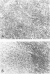
The authors studied 48 cases of well-differentiated lymphocytic neoplasms using a panel of monoclonal antibodies applied to frozen sections. Forty-seven tumors expressed monotypic immunoglobulin, one or more B-lineage antigens, and Ia (HLA-DR) antigen. Proliferation centers expressed the T9 antigen and increased numbers of Ki-67-positive cells. One tumor was of T-cell origin, had a cytotoxic/suppressor cell phenotype, and showed anomalous loss of Leu-1 antigen. Immunophenotypic findings were correlated to the clinical presentation and morphologic features of each neoplasm. Sixteen tumors were associated with peripheral lymphocytosis (greater than 4000/cu mm), 13 biopsies were obtained from extranodal sites, 16 tumors had proliferation centers, and 11 neoplasms had plasmacytoid features. The authors found no absolute and few statistically significant immunologic differences between the B-cell tumors according to their clinical presentation or morphologic features. Tumors associated with peripheral lymphocytosis more commonly expressed the Leu-1 antigen (P less than 0.01) and IgD (P less than 0.01) and less frequently were stained by BA-2 (P less than 0.05) and OKT9 (P less than 0.05). Plasmacytoid neoplasms more frequently expressed the Tac (P less than 0.01) and T9 antigens (P less than 0.05), and all expressed kappa light chain (P less than 0.05). Extranodal neoplasms more commonly expressed IgM (P less than 0.01). In contrast to the markedly different clinical presentation and morphologic appearance these tumors may have, the immunologic data suggest that B-cell small lymphocytic neoplasms are relatively homogeneous. For an individual case, immunophenotype does not predict clinical presentation or morphologic features.
Full text
PDF


Images in this article
Selected References
These references are in PubMed. This may not be the complete list of references from this article.
- Aisenberg A. C., Wilkes B. M., Harris N. L. Monoclonal antibody studies in non-Hodgkin's lymphoma. Blood. 1983 Mar;61(3):469–475. [PubMed] [Google Scholar]
- Dick F. R., Maca R. D. The lymph node in chronic lymphocytic leukemia. Cancer. 1978 Jan;41(1):283–292. doi: 10.1002/1097-0142(197801)41:1<283::aid-cncr2820410140>3.0.co;2-h. [DOI] [PubMed] [Google Scholar]
- Dick F., Bloomfield C. D., Brunning R. D. Incidence cytology, and histopathology of non-Hodgkin's lymphomas in the bone marrow. Cancer. 1974 May;33(5):1382–1398. doi: 10.1002/1097-0142(197405)33:5<1382::aid-cncr2820330525>3.0.co;2-2. [DOI] [PubMed] [Google Scholar]
- Dillman R. O., Beauregard J. C., Lea J. W., Green M. R., Sobol R. E., Royston I. Chronic lymphocytic leukemia and other chronic lymphoid proliferations: surface marker phenotypes and clinical correlations. J Clin Oncol. 1983 Mar;1(3):190–197. doi: 10.1200/JCO.1983.1.3.190. [DOI] [PubMed] [Google Scholar]
- Falini B., Schwarting R., Erber W., Posnett D. N., Martelli M. F., Grignani F., Zuccaccia M., Gatter K. C., Cernetti C., Stein H. The differential diagnosis of hairy cell leukemia with a panel of monoclonal antibodies. Am J Clin Pathol. 1985 Mar;83(3):289–300. doi: 10.1093/ajcp/83.3.289. [DOI] [PubMed] [Google Scholar]
- Garcia C. F., Warnke R. A., Weiss L. M. Follicular large cell lymphoma. An immunophenotype study. Am J Pathol. 1986 Jun;123(3):425–431. [PMC free article] [PubMed] [Google Scholar]
- Greil R., Gattringer C., Knapp W., Huber H. Growth fraction of tumour cells and infiltration density with natural killer-like (HNK1+) cells in non-Hodgkin lymphomas. Br J Haematol. 1986 Feb;62(2):293–300. doi: 10.1111/j.1365-2141.1986.tb02932.x. [DOI] [PubMed] [Google Scholar]
- Harris N. L., Bhan A. K. B-cell neoplasms of the lymphocytic, lymphoplasmacytoid, and plasma cell types: immunohistologic analysis and clinical correlation. Hum Pathol. 1985 Aug;16(8):829–837. doi: 10.1016/s0046-8177(85)80255-0. [DOI] [PubMed] [Google Scholar]
- Link M., Warnke R., Finlay J., Amylon M., Miller R., Dilley J., Levy R. A single monoclonal antibody identifies T-cell lineage of childhood lymphoid malignancies. Blood. 1983 Oct;62(4):722–728. [PubMed] [Google Scholar]
- Pangalis G. A., Nathwani B. N., Rappaport H. Malignant lymphoma, well differentiated lymphocytic: its relationship with chronic lymphocytic leukemia and macroglobulinemia of Waldenström. Cancer. 1977 Mar;39(3):999–1010. doi: 10.1002/1097-0142(197703)39:3<999::aid-cncr2820390302>3.0.co;2-r. [DOI] [PubMed] [Google Scholar]
- Papadimitriou C. S., Müller-Hermelink U., Lennert K. Histologic and immunohistochemical findings in the differential diagnosis of chronic lymphocytic leukemia of B-cell type and lymphoplasmacytic/lymphoplasmacytoid lymphoma. Virchows Arch A Pathol Anat Histol. 1979;384(2):149–158. doi: 10.1007/BF00427252. [DOI] [PubMed] [Google Scholar]
- Schwarting R., Stein H., Wang C. Y. The monoclonal antibodies alpha S-HCL 1 (alpha Leu-14) and alpha S-HCL 3 (alpha Leu-M5) allow the diagnosis of hairy cell leukemia. Blood. 1985 Apr;65(4):974–983. [PubMed] [Google Scholar]
- Spier C. M., Grogan T. M., Fielder K., Richter L., Rangel C. Immunophenotypes in "well-differentiated" lymphoproliferative disorders, with emphasis on small lymphocytic lymphoma. Hum Pathol. 1986 Nov;17(11):1126–1136. doi: 10.1016/s0046-8177(86)80418-x. [DOI] [PubMed] [Google Scholar]
- Stein H., Bonk A., Tolksdorf G., Lennert K., Rodt H., Gerdes J. Immunohistologic analysis of the organization of normal lymphoid tissue and non-Hodgkin's lymphomas. J Histochem Cytochem. 1980 Aug;28(8):746–760. doi: 10.1177/28.8.7003001. [DOI] [PubMed] [Google Scholar]
- Strickler J. G., Weiss L. M., Copenhaver C. M., Bindl J., McDaid R., Buck D., Warnke R. Monoclonal antibodies reactive in routinely processed tissue sections of malignant lymphoma, with emphasis on T-cell lymphomas. Hum Pathol. 1987 Aug;18(8):808–814. doi: 10.1016/s0046-8177(87)80055-2. [DOI] [PubMed] [Google Scholar]
- Swerdlow S. H., Murray L. J., Habeshaw J. A., Stansfeld A. G. Lymphocytic lymphoma/B-chronic lymphocytic leukaemia--an immunohistopathological study of peripheral B lymphocyte neoplasia. Br J Cancer. 1984 Nov;50(5):587–599. doi: 10.1038/bjc.1984.225. [DOI] [PMC free article] [PubMed] [Google Scholar]
- Uchiyama T., Broder S., Waldmann T. A. A monoclonal antibody (anti-Tac) reactive with activated and functionally mature human T cells. I. Production of anti-Tac monoclonal antibody and distribution of Tac (+) cells. J Immunol. 1981 Apr;126(4):1393–1397. [PubMed] [Google Scholar]
- Weiss L. M., Crabtree G. S., Rouse R. V., Warnke R. A. Morphologic and immunologic characterization of 50 peripheral T-cell lymphomas. Am J Pathol. 1985 Feb;118(2):316–324. [PMC free article] [PubMed] [Google Scholar]
- Wood G. S., Warnke R. A. The immunologic phenotyping of bone marrow biopsies and aspirates: frozen section techniques. Blood. 1982 May;59(5):913–922. [PubMed] [Google Scholar]
- van der Reijden H. J., van der Gaag R., Pinkster J., Rümke H. C., van't Veer M. B., Melief C. J., von dem Borne A. E. Chronic lymphocytic leukemia. Immunologic markers and functional properties of the leukemic cells. Cancer. 1982 Dec 15;50(12):2826–2833. doi: 10.1002/1097-0142(19821215)50:12<2826::aid-cncr2820501223>3.0.co;2-f. [DOI] [PubMed] [Google Scholar]


