Abstract
The Saccharomyces cerevisiae RAD18 gene is essential for postreplication repair but is not required for homologous recombination (HR), which is the major double-strand break (DSB) repair pathway in yeast. Accordingly, yeast rad18 mutants are tolerant of camptothecin (CPT), a topoisomerase I inhibitor, which induces DSBs by blocking replication. Surprisingly, mammalian cells and chicken DT40 cells deficient in Rad18 display reduced HR-dependent repair and are hypersensitive to CPT. Deletion of nonhomologous end joining (NHEJ), a major DSB repair pathway in vertebrates, in rad18-deficient DT40 cells completely restored HR-mediated DSB repair, suggesting that vertebrate Rad18 regulates the balance between NHEJ and HR. We previously reported that loss of NHEJ normalized the CPT sensitivity of cells deficient in poly(ADP-ribose) polymerase 1 (PARP1). Concomitant deletion of Rad18 and PARP1 synergistically increased CPT sensitivity, and additional inactivation of NHEJ normalized this hypersensitivity, indicating their parallel actions. In conclusion, higher-eukaryotic cells separately employ PARP1 and Rad18 to suppress the toxic effects of NHEJ during the HR reaction at stalled replication forks.
Genetic screening of Saccharomyces cerevisiae mutants that are sensitive to killing by UV light, ionizing radiation (IR), or both has defined the three major epistasis groups of repair genes, named after the mutant with the most prominent phenotype in each group, RAD3, RAD52, and RAD6 (reviewed in references 23 and 47). The RAD3 group corresponds to the nucleotide excision repair pathway, which eliminates UV-induced photoproducts. The RAD52 group genes, including RAD51 and RAD54, are involved in homologous recombination (HR), which plays a major role in double-strand break (DSB) repair following exposure to IR (reviewed in reference 43) and is essential for maintenance of chromosomal DNA by release from replication blockage in vertebrate cells (reviewed in reference 21). The RAD6/RAD18-dependent pathway is also required for completion of replication (reviewed in references 15, 16, and 31). Genes in this epistasis group are generally involved in filling postreplicative gaps in the DNA, and thus, this system is known as postreplication repair (PRR) (reviewed in references 15, 32, and 46). The PRR pathway consists of at least two subpathways: translesion DNA synthesis (TLS) and an error-free mechanism thought to be a form of template switch (19, 34). TLS employs specialized DNA polymerases, which are flexible enough to bypass damaged template DNA that stalls the replicative polymerases (reviewed in reference 39). Yeast rad6 and rad18 mutants are hypersensitive to a broad spectrum of DNA-damaging agents that interfere with DNA replication, such as UV, IR, cisplatin [cis-platinum(II) diaminodichloride], and methylmethane sulfonate (MMS), consistent with their central roles in the tolerance of DNA damage encountered during replication. Yeast genetic studies indicate that PRR and HR separately contribute to processing of DNA damage for release from replication blockage.
RAD6/RAD18 are central to PRR in budding yeast (reviewed in reference 6), and the amino acid sequences of both proteins are well conserved from yeast to human cells. Rad6 is a ubiquitin-conjugating enzyme (E2) (25), forming a tight complex with the Rad18 protein (4, 6). Rad18 has DNA-binding activity and may recruit the Rad6 protein to DNA lesions (3). Rad18 contains a RING finger motif, which is common to E3 ubiquitin ligases. The complex of Rad6 and Rad18 transfers ubiquitin onto PCNA, a clamp protein for DNA polymerases, in response to DNA damage (22, 55, 60). The ubiquitylation at Lys164 of PCNA appears to facilitate the recruitment of translesion DNA polymerases to DNA damage sites in mammalian cells, as well as in yeast (29, 60, 62). Although, like yeast, Rad18 deficiency in DT40 and murine embryonic stem cells results in hypersensitivity to a variety of DNA-damaging agents, it is unclear whether the function of Rad18 has been fully conserved during evolution (58, 63). In fact, PRR in vertebrates seems to be more complex than in yeast, not least because the number of TLS polymerases is larger (39) than in budding yeast (37). Accumulating evidence points to a degree of independence between Rad18 and at least three components of TLS, Pol kappa (κ), Rev3 (Polζ), and Rev1, in DT40 cells (40, 48, 53). This observation is in agreement with the finding that Rev3 deficiency compromises cellular proliferation more significantly than does Rad18 deficiency in both mouse and DT40 cells (42, 53, 61). Likewise, the mutation of Lys164 of PCNA to arginine (pcnaK164R) in DT40 cells results in a more pronounced phenotype than does the disruption of the RAD18 gene (2, 52). Therefore, Rad18 apparently has lost some regulatory roles in PRR during evolution from yeast to mammals.
DSBs occur as a consequence of collapsed replication forks at sites of replication blockage or due to the action of environmental factors, such as IR. There are two major DSB repair pathways, HR and nonhomologous end joining (NHEJ), the functions of which are conserved from yeast to mammals (reference 18; reviewed in references 24 and 49). HR relies on the presence of a stretch of undamaged homologous DNA to act as a template for repair and is therefore largely accurate. NHEJ is able to directly rejoin the ends of a break and often results in errors in the form of sequence deletions (35). The initial steps of NHEJ involve the association of the DNA end-binding proteins Ku70 and Ku80 with the break, followed by the recruitment of the catalytic subunit of the DNA-dependent protein kinase (DNA-PKcs). The Ku-DNA-PKcs complex ultimately recruits ligase IV, which completes the repair of the break (12).
In vertebrates, HR and NHEJ differentially contribute to DSB repair, depending on the nature of the DSB and the phase of the cell cycle (57). IR induces DSBs in the genomic DNA of packed chromatin structure. The resulting DSBs are repaired primarily by NHEJ in the G1 phase and partly by HR between a damaged and an intact sister chromatid in the late S and G2 phases (reference 35; reviewed in reference 64). Cells deficient in both HR and NHEJ are extremely sensitive to IR compared to the respective single mutants, indicating that the two DSB repair pathways are complementary to each other in the repair of IR-induced DSBs (11, 57). Furthermore, several studies suggest that the Ku heterodimer interferes with HR-dependent DSB repair in various organisms, indicating competition between NHEJ and HR (14, 17, 44). In addition to IR, DSBs are generated as a consequence of replication blockage. This type of DSB is induced by exposing cells to camptothecin (CPT), which causes the stabilization of a complex of topoisomerase I covalently linked to nicked DNA. Such complexes interrupt replication and cause DSBs in one of the sister chromatids (reviewed by 45, 50). The resulting DSBs are repaired primarily by HR with the other, intact sister chromatids rather than by NHEJ (50). Accordingly, HR-deficient cells, but not ku70 or ligaseIV cells, are hypersensitive to killing by CPT (reviewed in reference 10). Moreover, ku70 DT40 cells tend to be more resistant to CPT than wild-type cells, suggesting that NHEJ may even be toxic to the cells (1).
In this study, we evaluated DSB repair in rad18-deficient DT40 cells (63) (Table 1) , as well as Rad18-depleted SW480sn3 human cells (36). We present genetic evidence that, in addition to a role for Rad18 in DNA damage bypass and PRR, Rad18 has a direct role in DSB repair. The evidence includes hypersensitivity of rad18 DT40 cells, but not pcnaK164R mutant or rev3 DT40 cells, to CPT; reduced HR-dependent DSB repair in a recombination reporter substrate; and the complete suppression of these phenotypes by concomitant inactivation of Ku70. A strikingly similar phenotype is also observed in DT40 cells deficient in poly(ADP-ribose) polymerase 1 (PARP1) (20), which is activated at DSBs, as well as single-strand breaks, and mediates the covalent attachment of ADP-ribose moieties derived from NAD to target proteins (reviewed in reference 7). To explore the functional interactions between Rad18 and PARP1, we created rad18/parp1 double-mutant and rad18/parp1/ku70 triple-mutant clones and found that rad18/parp1 cells displayed a synergistic increase in CPT sensitivity and that this phenotype was also normalized by additional inactivation of the KU70 gene. In summary, like PARP1, Rad18 facilitates HR by inhibiting the toxic effect of NHEJ, particularly at the DSBs that occur as a consequence of replication blockage. Thus, Rad18 has acquired this new function, in parallel with its critical regulatory role in facilitating lesion bypass, during evolution from unicellular to multicellular organisms.
TABLE 1.
DT40 mutants used in this study
| Cell line | Selection marker for gene disruption
|
Reference or source | ||
|---|---|---|---|---|
| First gene | Second gene | Third gene | ||
| rad18 | puro/hyg | 63 | ||
| rev3 | his/bsr | 54 | ||
| rad30 | bsr/puro | 30 | ||
| ku70 | his/bsr | 57 | ||
| LigIV | hyg/puro | 1 | ||
| rad18/ku70 | puro/hyg | his/bsr | This study | |
| rad18/parp1 | his/hyg | puro/bsr | This study | |
| rad18/ligIV | hyg/puro | bsr/his | This study | |
| ku70/rev3 | his/bsr | puro/hyg | This study | |
| rad18/ku70/parp1 | puro/hyg | his/bsr | neo/ecogpt | This study |
| pcnaK164R | cre excised (bsr) | 2 | ||
MATERIALS AND METHODS
Cell culture, DNA transfection, and colony formation assay following genotoxic treatments.
Cells were cultured in RPMI 1640 supplemented with 10−5 M β-mercaptoethanol, 10% fetal calf serum, and 1% chicken serum (Sigma, St. Louis, MO) at 39.5°C. Methods for DNA transfection and genotoxic treatments have been described previously (57). The culture conditions for the mutant cells were essentially the same as those for parental DT40 cells. Genotoxic treatment and colony formation assays were performed as described previously (40). The constructs and selection cassettes used in the generation of the mutants in this study have also been described previously and are summarized in Table 1. SW480 is a human cell line derived from colon cancer; it is defective in p53 and is proficient in mismatch repair (36). This cell line was propagated in Dulbecco's modified Eagle's medium (Nissui Pharmaceutical, Tokyo, Japan) supplemented with 10% fetal calf serum at 37°C in a 5% CO2-95% air atmosphere. The cells were plated on glass bottom dishes at 50% confluence 24 h before transfection.
Chromosome analysis.
Analysis of chromosome abnormalities was performed as described previously (13, 54). Briefly, cells were treated for 3 h with medium containing 0.1 μg/ml Colcemid. Harvested cells were incubated in 1 ml of 75 mM KCl for 30 min at room temperature and fixed in 5 ml of a freshly prepared 3:1 mixture of methanol-acetic acid. The cell suspension was dropped onto an ice-cold wet glass slide and immediately flame dried. Then, the slides were stained with 3% Giemsa solution at pH 6.4 for 30 min. Chromosomal aberrations were assessed for at least 60 mitotic cells by microscope. Data for DT40 cells are presented as macrochromosomal (1 to 5 and Z) aberrations per 60 metaphase spreads (54).
siRNA transfection.
Short interfering RNA (siRNA), 19-mer RNA with a protruding 3′-TT sequence (total, 21-mer), was synthesized and purified by high-performance liquid chromatography (Hokkaido System Science). The sense sequence, 5′-ACTCAGTGTCCAACTTGCT-3′, which includes nucleotides 172 to 190 relative to the start codon, was used for designing siRNA for human Rad18 (hRad18). One hundred nanomolar siRNA was used for transfection. Transfection was performed using oligofectamine (Invitrogen), and as a control, cells were mock transfected with oligofectamine alone. Immunoblot assays with anti-hRad18 antibody and exposure of cells to cisplatin for colony formation assays were all performed 72 h after transfection. Determination of the cisplatin sensitivity of RNA interference-treated SW480 cells by using a colony formation assay was performed as described by Chang et al. (8). Forty-eight hours after siRNA transfection, the cells were treated continuously for 48 h with CPT, and then a colony formation assay was done (8).
Measurement of HR frequencies induced by I-SceI-induced DSBs in an artificial substrate.
Modified SCneo (17) was inserted into the previously described OVALBUMIN gene construct and then targeted into the OVALBUMIN locus in wild-type, rad18, ku70, and rad18/ku70 DT40 clones. In transient transfections, 5 × 10⁶ cells were suspended in 100 μl of Nucleofector solution T; mixed with each of several circular-plasmid DNAs (5 μg) (chicken Ku70, chicken RAD18, and I-SceI expression vector [pCBASce]); and electroporated with an AMAXA machine (Koeln, Germany) using either the A30 or B23 program. At 24 h after electroporation, the cells were plated in methylcellolouse containing 2.0 mg/ml G418 or without G418. The cells were grown for 7 to 10 days, and surviving colonies were counted. To distinguish between short-tract gene conversion (STGC) and long-tract gene conversion (LTGC)/sister chromatid recombination, genomic DNA was digested with HindIII and probed with a fragment of neo DNA (see Fig. 4A) (59). For the nucleotide sequence analysis of the repair products, genomic DNA was PCR amplified using the primers P1 and P2 (see Fig. 4A). To measure HR-dependent DSB repair in human cells, experiments were done as previously reported (50).
FIG. 4.
HR-mediated DSB repair is reduced in rad18 cells but proficient in rad18/ku70 cells. (A) Structure of the SCneo HR substrate and predicted repair products. STGC, short-tract gene conversion; LTGC, long-tract gene conversion. Note that LTGC includes unequal sister chromatid recombination. (B) Frequency of HR-mediated DSB repair. Cells of the indicated genotypes were transiently transfected with the I-SceI meganucleotide recognizing endonuclease expression vector and plated in methylcellulose-containing medium with or without neomycin selection. The gene conversion frequency was calculated as the percentage of neomycin-resistant colonies relative to the number of colonies plated in G418-free methylcellulose. +KU70 and +RAD18 indicate transient expression of I-SceI, together with chicken KU70 and RAD18 transgenes, respectively. The data shown are the mean ± the standard error of at least three separate experiments. (C) To distinguish between STGC and LTGC, Southern blot analysis of neomycin-resistant clones of the indicated genotypes was performed as indicated for panel A. (D) The accuracy of DSB repair is reduced in rad18 cells. Genomic DNAs of the indicated cells were recovered 24 h after transient expression of I-SceI. DNA samples were digested by I-SceI, and DSB repair products were PCR amplified, subjected to digestion with the indicated restriction enzymes, and analyzed by gel electrophoresis (27). Note that a substantial fraction of DSB repair products in rad18 cells are resistant to digestion by I-SceI and NcoI, indicating the presence of inaccurate repair events. WT, wild type.
RESULTS
Increased sensitivity of rad18 cells to camptothecin, as well as to IR.
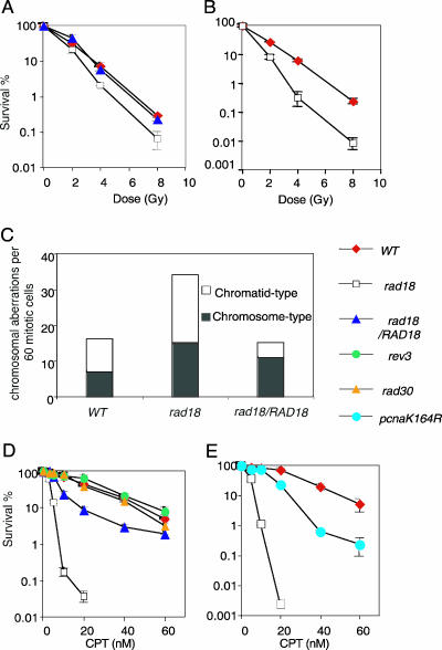
We previously showed that asynchronous populations of rad18-deficient DT40 cells are moderately sensitive to killing by IR in a colony formation assay (Fig. 1A) (63). This phenotype may result from two nonexclusive mechanisms: diminished bypass of base lesions and reduced capacity for homology-directed DSB repair. To investigate whether Rad18 is involved in DSB repair, we exposed cells to IR during the G2 phase, measured colony survival (Fig. 1B), and counted the chromosomal breaks in the subsequent M phase (Fig. 1C), as previously described (41, 53). Interestingly, rad18 cells showed increased sensitivity to γ rays compared with wild-type cells (Fig. 1B). Moreover, the level of unrepaired chromosomal breaks scored in mitotic spreads following irradiation in the G2 phase was on average 2.5-fold higher in rad18 than in wild-type cells (Fig. 1C). These observations suggest that Rad18 can affect DSB repair in the G2 phase even after completion of DNA replication.
FIG. 1.
Increased sensitivity of rad18-deficient cells to killing by camptothecin, as well as γ-irradiation. (A) The fractions of colonies surviving after γ-irradiation compared with untreated controls of the same genotype are shown on the y axis on a logarithmic scale. The error bars show the mean ± standard deviation for at least three separate experiments. (B) IR sensitivities of cells synchronized in the G2 phase. (C) Deficient DSB repair during the G2 phase in rad18 cells. The data shown in the histogram indicate the types and numbers of chromosomal breaks in 60 analyzed mitotic cells. Two breaks at the same site of both sister chromatids are defined as chromosome-type breaks, while breaks at either sister chromatid are chromatid type. Mitotic cells were enriched by Colcemid treatment for 3 hours before cell harvest. Colcemid was added immediately after γ-irradiation. (D and E) Fractions of colonies arising from CPT-treated cells relative to non-CPT-treated controls. The cells were continuously exposed to CPT during clonal expansion in methylcellulose-containing culture media. The data are shown as in panel A. WT, wild type.
To assess the repair of DSBs that arise during replication, we measured the ability of rad18 cells to form colonies in methyl cellulose containing increasing amounts of CPT. Interestingly, rad18 cells were extremely sensitive to CPT in comparison with wild-type cells, and the sensitivity was reversed by complementation with a RAD18 transgene (Fig. 1D). This finding is in marked contrast to findings in budding yeast, where the rad18 mutant does not display CPT sensitivity (5, 51). In contrast to rad18 DT40 cells, TLS polymerase-deficient mutants, including rad30 (polη) or rev3 cells (30, 53, 65), exhibited no increase in sensitivity to CPT (Fig. 1D). Conceivably, if topoisomerase I is covalently linked to DNA, the resulting broken template strands could not be bypassed by any TLS polymerases.
Furthermore, pcnaK164R cells were far more CPT resistant than were rad18 cells (Fig. 1E), indicating that Rad18 can contribute to CPT tolerance independently of the modification of PCNA. Taken together, these data suggest that Rad18 may contribute to the repair of DSB arising during, as well as after the completion of, replication.
The increased sensitivities of rad18-deficient cells to IR and camptothecin are reversed by disruption of Ku70.
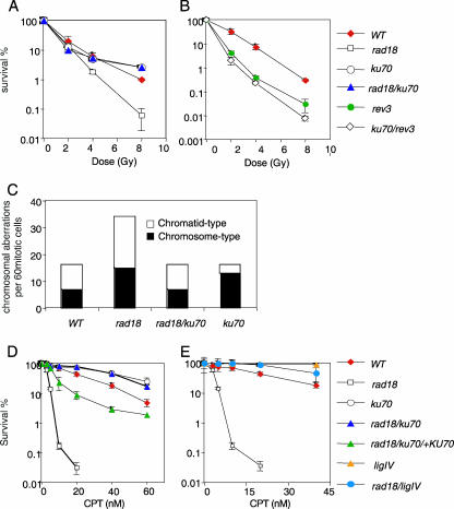
To understand which major DSB repair pathway is regulated by Rad18, we abolished an essential early NHEJ factor, Ku70, in rad18 cells. As a control, we also deleted Ku70 in the rev3 background. The resulting double-mutant cells were viable and did not show any further growth retardation (data not shown). Surprisingly, the γ-ray sensitivity of rad18 cells, but not that of rev3 cells, was completely reversed by the additional disruption of the KU70 gene (Fig. 2A and B). Accordingly, the patterns of IR sensitivity in ku70 and rad18/ku70 cells were indistinguishable (Fig. 2A). Likewise, the deletion of KU70 in rad18 cells normalized induced chromosomal breaks at 3 hours post-IR exposure (Fig. 2C). These observations indicate that IR sensitivity of rad18 cells depends on the Ku protein. This is in marked contrast with the findings in budding yeast, where the IR sensitivity of rad18 is not dependent on ku70 (9). Alternatively, this phenotype could arise as a result of an increase in recombinogenic lesions due to a PRR defect in rad18 cells. Additionally, these lesions could be repaired more efficiently by HR, particularly in the absence of Ku70. If this scenario is true, deletion of Ku70 should also suppress other PRR mutants, such as rev3 mutants. However, deletion of KU70 in rev3 cells slightly increased their IR sensitivity (Fig. 2B), as opposed to the suppression of IR sensitivity in rad18/ku70 cells. These data suggest that Ku70-dependent NHEJ and rev3-dependent TLS contribute to cellular tolerance of IR independently of each other and that the predominant role of Rad18 in tolerance of IR is distinct from that of Rev3.
FIG. 2.
Deletion of KU70 reversed the increased sensitivity of rad18-deficient cells to γ-irradiation and CPT. (A and B) Fractions of colonies surviving after γ-irradiation compared with untreated controls. The data are shown as in Fig. 1A. (C) Deficient DSB repair during the G2 phase in rad18 cells was restored by inactivation of Ku70. The data are shown as in Fig. 1C. (D) Fractions of colonies surviving after continuous exposure to CPT. The data are shown as in Fig. 1D. WT, wild type.
We next measured CPT sensitivity and found that rad18 cells were highly sensitive to CPT only in the presence of Ku70 (Fig. 2D). This toxic effect of Ku70 on rad18 cells might be attributed to the tight association of the Ku70 protein with CPT-induced DSB ends, which could directly interfere with HR-mediated DSB repair. Alternatively, the process of end joining itself might be toxic, as loss of NHEJ in ligaseIV (LigIV)-deficient cells also confers CPT resistance on wild-type cells (1). To distinguish between these two possibilities, we created rad18/LigIV double-mutant cells and examined their sensitivities to CPT. Like rad18/ku70 cells, rad18/LigIV cells displayed higher tolerance of CPT than did rad18 cells (Fig. 2E). These observations suggest that NHEJ-dependent ligation, rather than simple occlusion of DNA ends by Ku70, is toxic in repairing CPT-induced DSBs in the absence of Rad18.
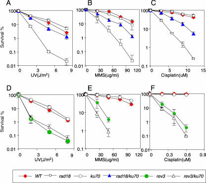
The reversion of rad18 cell hypersensitivity to IR and CPT by additional inactivation of Ku70 led us to measure cellular sensitivity to UV, MMS, and cisplatin by comparing rad18 and rad18/ku70 cells. The deletion of KU70 reversed the hypersensitivity of rad18 cells to UV, MMS, and cisplatin by more than 50% (Fig. 3A, B, and C). This phenotype is in marked contrast to the additive effect of KU70 deletion on cells deficient in Rev3, which plays a critical role in TLS past a wide variety of DNA damage (Fig. 3D, E, and F). As discussed above, this observation suggests that Ku70 does not necessarily interfere with PRR. In conclusion, Rad18 contributes to cellular tolerance of a wide range of DNA damage in the following two distinct manners: avoidance of the toxic effect of NHEJ on HR and facilitation of PRR.
FIG. 3.
Effect of KU70 deletion on rad18- and rev3-deficient cells. Cells of the indicated genotypes were exposed to UV (A and D), MMS (B and E), and cisplatin (C and F). The data are shown as in Fig. 1. WT, wild type.
Rad18 promotes homology-directed I-SceI-induced DSB repair when proficient NHEJ is present.
Our observation that the number of chromosomal breaks following IR exposure in G2 cells was reduced in rad18/ku70 cells compared to rad18 cells (Fig. 2C) suggested that Ku70 could interfere with HR-mediated DSB repair in the absence of Rad18. To test this hypothesis, we measured HR-dependent repair of the DSBs that are generated by the I-SceI restriction enzyme. To this end, we inserted the SCneo substrate, including a rare-cutting endonuclease site, I-SceI, into the OVALBUMIN locus of the wild-type, rad18, ku70, and rad18/ku70 genotypes (17, 26). This construct carries two mutant neomycin resistance genes (S2neo and 3′-neo), which are localized in tandem and complementary to each other (Fig. 4A). Following transient expression of I-SceI, intragenic or sister chromatid recombination repairs the induced DSB in S2neo, with 3′-neo serving as a donor for gene conversion. This leads to the restoration of a functional neomycin resistance gene. Thus, the HR frequency can be evaluated by counting the neomycin-resistant (Neor) colonies following transient expression of I-SceI. The number of Neor colonies was reduced by 22-fold in rad18 cells in comparison to wild-type cells (Fig. 4B). This reduction was reversed when rad18 cells were transfected with the I-SceI expression plasmid, together with a RAD18 transgene. We therefore concluded that Rad18 can contribute to HR-mediated DSB repair. We next examined I-SceI-induced HR in rad18/ku70 cells and those transiently expressing Ku70. The frequency of gene conversion in rad18/ku70 cells was restored to wild-type levels, while transient expression of Ku70 in rad18/ku70 cells reduced the frequency of HR-mediated repair (Fig. 4B). These results indicate that the HR defect in rad18 cells depends on the presence of Ku70. We therefore suggest that Rad18 facilitates HR by suppressing NHEJ factors.
To gain an insight into the role of Rad18 in HR, we examined HR products recovered from 24 neomycin-resistant clones from each genotype, using Southern hybridization analysis with a probe shown in Fig. 4A. The majority of the repair products were associated with STGC (Fig. 4C), as previously reported (28, 50). To evaluate the accuracy of DSB repair, HR products were PCR amplified using the primers indicated in Fig. 4A (P1 and P2), and 24 fragments from each genotype were subjected to base sequence determination. Interestingly, three clones derived from rad18 cells, but not any from cells of other genotypes, contained 4- and 8-base pair deletions (see Fig. S1 in the supplemental material). In order to further evaluate the accuracy of DSB repair, we examined DSB repair products 24 h after transient expression of I-SceI without neomycin selection. To exclude the repair substrate in the PCR amplification products, genomic DNA was digested with I-SceI prior to the amplification. Accurate HR would replace the I-SceI site with an NcoI site, while inaccurate DSB repair, either HR or NHEJ, might result in loss of both I-SceI and NcoI sites. A higher percentage of the amplified fragments from rad18 cells were resistant to both NcoI and I-SceI digestion compared with those from wild-type and rad18/ku70 cells (Fig. 4D, bottom panel). Collectively, deletion of Rad18 resulted in an increase in the percentage of inaccurate DSB repair events. Conceivably, Rad18 may be directly associated with DSBs and protect them from inaccurate repair by NHEJ, thereby facilitating HR.
Human Rad18 facilitates DSB repair.
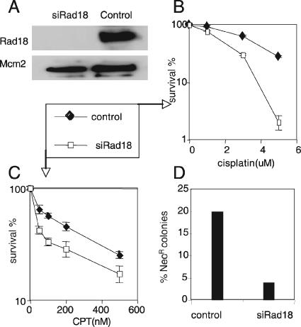
We wished to know whether the findings from DT40 cells presented here are also relevant to human cells. To this end, we depleted Rad18 in human SW480 cells, a mismatch repair-proficient cell line from a colon cancer (36), using siRNA and evaluated DSB repair. SW480 cells were transfected with rad18 siRNA or mock transfected (control) (see Materials and Methods). The level of Rad18 protein was reduced by more than 80% at 72 h after siRNA transfection (Fig. 5A), while the growth rate of the cells was unaffected (data not shown). As seen in rad18 mutant DT40 cells, the SW480 Rad18-depleted cells exhibited a slight increase in sensitivity to CPT, as well as cisplatin (Fig. 5B and C). Increased sensitivities to both CPT and cisplatin were also observed in a RAD18−/− HCT116 human cell line (personal communication from T. Shiomi, National Institute of Radiological Sciences, Japan). The less prominent phenotype of the human cell line (Fig. 5) in comparison with DT40 cells may be attributed to the duration of CPT exposure, only 48 h for the siRNA-treated cells but continuous during clonal expansion for DT40 cells. To assess HR, we used an assay based on the SCneo substrate (26), a single copy of which is integrated into SW480sn3 cells (36). At 72 h after the addition of either control or Rad18 siRNA to the cells, they were transfected with an I-SceI expression plasmid and were subsequently subjected to selection with neomycin. Rad18-depleted cells produced Neor recombinants at a frequency fivefold lower than the control (mock-treated) cells (Fig. 5D). These observations indicate that human Rad18 is also required for optimal execution of HR.
FIG. 5.
Rad18 plays a role in DSB repair in human cells. (A) Immunoblot assay 72 h after siRNA transfection. Control, mock transfection of oligofectamine alone; siRad18, rad18 siRNA. Cell lysate was prepared from control and siRNA-treated cells and immunoblotted with anti-Rad18 antiserum. An immunoblot with antibody against Mcm2 was used as a loading control. (B and C) Fractions of colonies surviving after exposure of cells to cisplatin for 1 hour (B) and CPT for 48 h (C), relative to untreated controls, comparing cells treated with 100 nM siRNA with untreated cells (see Materials and Methods). Shown are means ± standard deviations for at least three separate experiments. (D) Decreased HR-mediated DSB repair in siRad18-treated cells. The experiments were done as for Fig. 4. The frequency of Neo-resistant colonies was calculated as the percentage of neomycin-resistant colonies relative to the number of colonies in G418-free medium.
Parallel actions of Rad18 and poly(ADP-ribose) polymerase in suppressing the toxic effect of NHEJ on DSB repair at a replication block.
We previously reported that the deletion of Ku70 suppresses the sensitivity of parp1 to IR, CPT, MMS, and cisplatin (20). This phenotypic similarity between rad18- and parp1-deficient DT40 cells led us to analyze cells deficient in both the RAD18 and PARP1 genes to understand their functional relationship. The resulting rad18/parp1 cells showed a slight retardation of cellular proliferation (Fig. 6A). Interestingly, the concomitant deletion of these two genes caused a synergistic increase in sensitivity to CPT (Fig. 6B and C), as well as cisplatin and MMS (Fig. 6D and E). Furthermore, deletion of the KU70 gene normalized the CPT sensitivity of the rad18/parp1-deficient cells (Fig. 6B, low-dose treatment, and C, higher-dose treatment). Cisplatin and MMS sensitivities were also partially restored in the triple-mutant cells. Thus, Rad18 can partially substitute for a lack of PARP1 in reducing the toxic effect of NHEJ in DSB repair at a replication block. Likewise, PARP1 can also partially substitute for a lack of Rad18. These data suggest that Rad18 and PARP1 suppress the access of NHEJ factors to DSBs caused by replication blocks independently of each other.
FIG. 6.
Synergistic increase in sensitivity to DNA-damaging agents by simultaneous inactivation of RAD18 and PARP1 genes. (A) Relative growth rates plotted for the indicated genotypes. (B to E) Colony survival to evaluate the sensitivities of the indicated cells to killing by CPT at a low dose (B) and at a high dose (C), cisplatin (D), MMS (E), and UV (F). Each value represents the mean (± standard deviation) of the results from three independent experiments.
DISCUSSION
Rad18 is a key player in controlling PRR in yeast, and its action is carried out mainly by ubiquitylation of PCNA at replication blocks (22, 55, 60). Mammalian Rad18 can also control the PRR pathway through ubiquitylation of PCNA (29, 62). In the present study, we have provided genetic evidence that Rad18 has another function, the facilitation of HR-dependent DSB repair in chicken and human cells. In chicken DT40 cells, this function depends on intact NHEJ, because concomitant suppression of NHEJ normalized cellular sensitivity to CPT in rad18 cells. This observation sheds light on a novel role of Rad18, the suppression of the toxic effect of NHEJ on HR, in addition to release of replication blocks by conventional PRR. Furthermore, we demonstrated that Rad18 and PARP1 collaboratively contribute to cellular tolerance of CPT and cisplatin, which are widely used for chemotherapeutic treatment of cancer and leukemia (38, 51).
Rad18 plays a role in HR independently of its contribution to PRR.
In this study, we presented evidence that the CPT sensitivity of rad18 cells is mainly caused by a defect in HR-mediated DSB repair, but not by impaired PRR. We derived this conclusion from the following results. First, in contrast to yeast rad18 mutant cells (5, 51) or DT40 cells deficient in TLS polymerases, rad18 cells displayed a strong increase in CPT sensitivity (Fig. 1D). This phenotype cannot be explained by defective TLS, because the attachment of topoisomerase I to the backbone of template strands would prevent DNA synthesis by polymerases. Second, in contrast with rad18 cells, DT40 cells carrying the PCNA K164R mutation displayed only a modest increase in sensitivity to CPT (Fig. 1E). Thus, Rad18 may contribute to cellular tolerance of CPT independently of PCNA's posttranslational modification, which seems to play a key role in PRR in mammalian cells (2, 29, 52, 62). Third, the tolerance of CPT by rad18 cells was restored by deletion of KU70 and LigIV, which act only in DSB repair and not in PRR (Fig. 2D and E). Fourth, rad18 DT40 cells, as well as Rad18-depleted human cells, showed reductions in the rate of HR-mediated repair of I-SceI-induced DSBs (Fig. 4B and 5D). Furthermore, the accuracy of DSB repair was moderately compromised in rad18 cells (Fig. 4D; see Fig. S1 in the supplemental material). This result agrees with a recent report that in rad18 DT40 cells immunoglobulin gene conversion is frequently associated with deletions and disruptions (56). These observations provide compelling evidence for the dual roles of Rad18 in HR-dependent DSB repair and PRR in vertebrate cells.
In the cases of UV, MMS, and cisplatin treatments, deletion of Ku partly suppressed the sensitivity of rad18 cells (Fig. 3A to C). Clearly, this suppression does not depend on the PRR function of Rad18, because Ku does not interact in a similar manner with other PRR factors, such as Rev3 (Fig. 3D to F). A comparison between the rad18 and rad18/ku70 phenotypes as shown in Fig. 3 allows a clear dissection of the HR and PRR branches of Rad18.
It is postulated that HR does not require Rad18 in budding yeast, as the Rad18 and Rad52 epistasis groups constitute different repair pathways, PRR and HR, respectively (reviewed in reference 21). However, a recent study showed that although the yeast rad18 mutant is very sensitive to IR, the pol30 (pcnaK164R) mutant exhibits only slight sensitivity to IR (9), indicating that Rad18 can promote HR-mediated repair of IR-induced DSBs independently of the PCNA ubiquitylation. Moreover, hypersensitivity to both CPT and topoisomerase II inhibitors is observed in fission yeast defective in the ortholog gene of RAD18 (A. M. Carr and K. Furuya, Sussex, United Kingdom, personal communication). Taken together, these observations may uncover a role of Rad18 in DSB repair in lower- as well as higher-eukaryotic cells.
Functional interaction between Rad18 and Parp1 in preventing NHEJ.
The present data indicate that the hypersensitivity of rad18 cells to CPT may require proficient NHEJ, including both the Ku proteins and LigIV (Fig. 2D and E). Thus, the entire NHEJ pathway is unfavorable under these circumstances. Presumably, when a large number of DSBs arise within a replication factory following CPT treatment, active NHEJ makes inappropriate joins between DSB ends, which could eventually result in chromosomal translocation and deletion. Our findings postulate a role for Rad18 in suppressing such inappropriate NHEJ at stalled replication forks. We have recently found that cells deficient in PARP1, an enzyme that poly(ADP-ribose)ylates proteins at DNA breaks, shares a strikingly similar phenotype with Rad18-deficient cells with regard to their interactions with NHEJ (20). Furthermore, in the case of PARP, biochemical studies have already shown that the attachment of PAR to the Ku proteins reduces affinity to DSBs in vitro (33). Thus, both biochemical data and our genetic results consistently support the idea that PARP1 suppresses the access of the Ku proteins to DSB ends. Finding substrates of Rad18-mediated ubiquitylation in this novel pathway will be crucial for understanding the molecular mechanism of Rad18 function in DSB repair.
In this study, we showed that the sensitivity of a rad18/parp1 double mutant to CPT was dramatically increased compared to those of the relevant single mutants and that cellular tolerance of CPT was restored by deletion of KU70 in rad18/parp1 cells (Fig. 6). Based on these genetic findings, a model emerges in which Rad18-mediated ubiquitylation and PARP-mediated poly(ADP-ribose)ylation separately limit the access of NHEJ factors to DNA ends at stalled replication forks. Conceivably, higher eukaryotes have evolved a novel regulatory mechanism to keep their highly active NHEJ pathways in check. This may be necessary in order to avoid the unwanted ligation of DSB breaks at replication forks and to allow access of HR factors when this reaction is more favorable for cellular survival.
Multiple functions of DNA repair factors in vertebrates.
Genetic studies in yeast have clearly established defined functions for the genes in the RAD3, RAD52, and RAD6 epistasis groups (23, 47). Vertebrate cells are faced with a more complex task in safeguarding their DNA, due to the sheer size of their genomes. As the vertebrate genetics of DNA repair become more accessible, it appears that, besides evolving new genes, the functions of old players have shifted during metazoan evolution to help cope with the more complex tasks of ensuring chromosome stability. This seems to be particularly true for PRR genes, which overlap much more with HR (30, 41, 53) than appears to be the case in yeast. The present study indicated that another example of such a PRR gene is the gene encoding Rad18, which may inhibit access of NHEJ to DSBs while at the same time losing its dominant role in controlling every PRR subpathway in the more complex system in vertebrates. These crossovers of DNA repair pathways in vertebrates have to be kept in mind when specific factors are targeted for therapeutic purposes. Multifunctional effectors, such as Rad18, could thus be ideal candidates for inhibitor screens or gene therapy in order to maximize the effects of DNA-damaging agents, such as CPT, during chemotherapy.
Supplementary Material
Acknowledgments
We thank R. Ohta, Y. Sato, and M. Nagao for their technical assistance. We also thank H. D. Ulrich (Clare Hall Laboratories, Cancer Research, United Kingdom), and A. R. Lehmann (University of Sussex) for critical reading and discussion.
Financial support was provided in part by CREST.JST (Saitama, Japan) and the center of excellence (COE) grant for Scientific Research from the Ministry of Education, Culture, Sports and Technology. A. Saberi has a scholarship from the Health Ministry of Medical Education of Iran. D.S. was supported by the Leukemia Research Fund.
Footnotes
Published ahead of print on 22 January 2007.
Supplemental material for this article may be found at http://mcb.asm.org/.
REFERENCES
- 1.Adachi, N., S. So, and H. Koyama. 2004. Loss of nonhomologous end joining confers camptothecin resistance in DT40 cells. Implications for the repair of topoisomerase I-mediated DNA damage. J. Biol. Chem. 279:37343-37348. [DOI] [PubMed] [Google Scholar]
- 2.Arakawa, H., G. L. Moldovan, H. Saribasak, N. N. Saribasak, S. Jentsch, and J. M. Buetstedde. 2006. A role for PCNA ubiquitination in immunoglobulin hypermutation. PloS Biol. 4:e366. [DOI] [PMC free article] [PubMed] [Google Scholar]
- 3.Bailly, V., J. Lamb, P. Sung, S. Prakash, and L. Prakash. 1994. Specific complex formation between yeast RAD6 and RAD18 proteins: a potential mechanism for targeting RAD6 ubiquitin-conjugating activity to DNA damage sites. Genes Dev. 8:811-820. [DOI] [PubMed] [Google Scholar]
- 4.Bailly, V., S. Prakash, and L. Prakash. 1997. Domains required for dimerization of yeast Rad6 ubiquitin-conjugating enzyme and Rad18 DNA binding protein. Mol. Cell. Biol. 17:4536-4543. [DOI] [PMC free article] [PubMed] [Google Scholar]
- 5.Bennett, C. B., L. K. Lewis, G. Karthikeyan, K. S. Lobachev, Y. H. Jin, J. F. Sterling, J. R. Snipe, and M. A. Resnick. 2001. Genes required for ionizing radiation resistance in yeast. Nat. Genet. 29:426-434. [DOI] [PubMed] [Google Scholar]
- 6.Broomfield, S., T. Hryciw, and W. Xiao. 2001. DNA postreplication repair and mutagenesis in Saccharomyces cerevisiae. Mutat. Res. 486:167-184. [DOI] [PubMed] [Google Scholar]
- 7.Burkle, A. 2005. Poly(ADP-ribose). The most elaborate metabolite of NAD+. FEBS J. 272:4576-4589. [DOI] [PubMed] [Google Scholar]
- 8.Chang, I. Y., M. H. Kim, H. B. Kim, Y. L. Do, S. H. Kim, H. Y. Kim, and H. J. You. 2005. Small interfering RNA-induced suppression of ERCC1 enhances sensitivity of human cancer cells to cisplatin. Biochem. Biophys. Res. Commun. 327:225-233. [DOI] [PubMed] [Google Scholar]
- 9.Chen, S., A. A. Davies, D. Sagan, and H. D. Ulrich. 2005. The RING finger ATPase Rad5p of Saccharomyces cerevisiae contributes to DNA double-strand break repair in a ubiquitin-independent manner. Nucleic Acids Res. 33:5878-5886. [DOI] [PMC free article] [PubMed] [Google Scholar]
- 10.Connelly, J. C., and D. R. Leach. 2004. Repair of DNA covalently linked to protein. Mol. Cell 13:307-316. [DOI] [PubMed] [Google Scholar]
- 11.Couedel, C., K. D. Mills, M. Barchi, L. Shen, A. Olshen, R. D. Johnson, A. Nussenzweig, J. Essers, R. Kanaar, G. C. Li, F. W. Alt, and M. Jasin. 2004. Collaboration of homologous recombination and nonhomologous end-joining factors for the survival and integrity of mice and cells. Genes Dev. 18:1293-1304. [DOI] [PMC free article] [PubMed] [Google Scholar]
- 12.Doherty, A. J., and S. P. Jackson. 2001. DNA repair: how Ku makes ends meet. Curr. Biol. 11:R920-R924. [DOI] [PubMed] [Google Scholar]
- 13.Dracopoli, N. C., G. A. Bruns, G. M. Brodeur, G. M. Landes, T. C. Matise, M. F. Seldin, J. M. Vance, and A. Weith. 1994. Report and abstracts of the First International Workshop on Human Chromosome 1 Mapping 1994. Bethesda, Maryland, March 25-27, 1994. Cytogenet. Cell Genet. 67:144-165. [PubMed] [Google Scholar]
- 14.Frank-Vaillant, M., and S. Marcand. 2002. Transient stability of DNA ends allows nonhomologous end joining to precede homologous recombination. Mol. Cell 10:1189-1199. [DOI] [PubMed] [Google Scholar]
- 15.Friedberg, E. C. 2005. Suffering in silence: the tolerance of DNA damage. Nat. Rev. Mol. Cell. Biol. 6:943-953. [DOI] [PubMed] [Google Scholar]
- 16.Friedberg, E. C., A. J. Bardwell, L. Bardwell, W. J. Feaver, R. D. Kornberg, J. Q. Svejstrup, A. E. Tomkinson, and Z. Wang. 1995. Nucleotide excision repair in the yeast Saccharomyces cerevisiae: its relationship to specialized mitotic recombination and RNA polymerase II basal transcription. Phil. Trans. R. Soc. Lond. B 347:63-68. [DOI] [PubMed] [Google Scholar]
- 17.Fukushima, T., M. Takata, C. Morrison, R. Araki, A. Fujimori, M. Abe, K. Tatsumi, M. Jasin, P. K. Dhar, E. Sonoda, T. Chiba, and S. Takeda. 2001. Genetic analysis of the DNA-dependent protein kinase reveals an inhibitory role of Ku in late S-G2 phase DNA double-strand break repair. J. Biol. Chem. 276:44413-44418. [DOI] [PubMed] [Google Scholar]
- 18.Haber, J. E. 1999. DNA recombination: the replication connection. Trends Biochem. Sci. 24:271-275. [DOI] [PubMed] [Google Scholar]
- 19.Higgins, N. P., K. Kato, and B. Strauss. 1976. A model for replication repair in mammalian cells. J. Mol. Biol. 101:417-425. [DOI] [PubMed] [Google Scholar]
- 20.Hochegger, H., D. Dejsuphong, T. Fukushima, C. Morrison, E. Sonoda, V. Schreiber, G. Y. Zhao, A. Saberi, M. Masutani, N. Adachi, H. Koyama, G. de Murcia, and S. Takeda. 2006. Parp-1 protects homologous recombination from interference by Ku and Ligase IV in vertebrate cells. EMBO J. 25:1305-1314. [DOI] [PMC free article] [PubMed] [Google Scholar]
- 21.Hochegger, H., E. Sonoda, and S. Takeda. 2004. Post-replication repair in DT40 cells: translesion polymerases versus recombinases. Bioessays 26:151-158. [DOI] [PubMed] [Google Scholar]
- 22.Hoege, C., B. Pfander, G. L. Moldovan, G. Pyrowolakis, and S. Jentsch. 2002. RAD6-dependent DNA repair is linked to modification of PCNA by ubiquitin and SUMO. Nature 419:135-141. [DOI] [PubMed] [Google Scholar]
- 23.Hoeijmakers, J. H. 2001. Genome maintenance mechanisms for preventing cancer. Nature 411:366-374. [DOI] [PubMed] [Google Scholar]
- 24.Jeggo, P. A. 1998. DNA breakage and repair. Adv. Genet. 38:185-218. [DOI] [PubMed] [Google Scholar]
- 25.Jentsch, S., J. P. McGrath, and A. Varshavsky. 1987. The yeast DNA repair gene RAD6 encodes a ubiquitin-conjugating enzyme. Nature 329:131-134. [DOI] [PubMed] [Google Scholar]
- 26.Johnson, R. D., and M. Jasin. 2001. Double-strand-break-induced homologous recombination in mammalian cells. Biochem. Soc. Trans. 29:196-201. [DOI] [PubMed] [Google Scholar]
- 27.Johnson, R. D., and M. Jasin. 2000. Sister chromatid gene conversion is a prominent double-strand break repair pathway in mammalian cells. EMBO J. 19:3398-3407. [DOI] [PMC free article] [PubMed] [Google Scholar]
- 28.Johnson, R. D., N. Liu, and M. Jasin. 1999. Mammalian XRCC2 promotes the repair of DNA double-strand breaks by homologous recombination. Nature 401:397-399. [DOI] [PubMed] [Google Scholar]
- 29.Kannouche, P. L., and A. R. Lehmann. 2004. Ubiquitination of PCNA and the polymerase switch in human cells. Cell Cycle 3:1011-1013. [PubMed] [Google Scholar]
- 30.Kawamoto, T., K. Araki, E. Sonoda, Y. M. Yamashita, K. Harada, K. Kikuchi, C. Masutani, F. Hanaoka, K. Nozaki, N. Hashimoto, and S. Takeda. 2005. Dual roles for DNA polymerase eta in homologous DNA recombination and translesion DNA synthesis. Mol. Cell 20:793-799. [DOI] [PubMed] [Google Scholar]
- 31.Lawrence, C. 1994. The RAD6 DNA repair pathway in Saccharomyces cerevisiae: what does it do, and how does it do it? Bioessays 16:253-258. [DOI] [PubMed] [Google Scholar]
- 32.Lehmann, A. R. 2002. Replication of damaged DNA in mammalian cells: new solutions to an old problem. Mutat. Res. 509:23-34. [DOI] [PubMed] [Google Scholar]
- 33.Li, B., S. Navarro, N. Kasahara, and L. Comai. 2004. Identification and biochemical characterization of a Werner's syndrome protein complex with Ku70/80 and poly(ADP-ribose) polymerase-1. J. Biol. Chem. 279:13659-13667. [DOI] [PubMed] [Google Scholar]
- 34.Li, Z., W. Xiao, J. J. McCormick, and V. M. Maher. 2002. Identification of a protein essential for a major pathway used by human cells to avoid UV-induced DNA damage. Proc. Natl. Acad. Sci. USA 99:4459-4464. [DOI] [PMC free article] [PubMed] [Google Scholar]
- 35.Liang, F., P. J. Romanienko, D. T. Weaver, P. A. Jeggo, and M. Jasin. 1996. Chromosomal double-strand break repair in Ku80-deficient cells. Proc. Natl. Acad. Sci. USA 93:8929-8933. [DOI] [PMC free article] [PubMed] [Google Scholar]
- 36.Mohindra, A., L. E. Hays, E. N. Phillips, B. D. Preston, T. Helleday, and M. Meuth. 2002. Defects in homologous recombination repair in mismatch-repair-deficient tumour cell lines. Hum. Mol. Genet. 11:2189-2200. [DOI] [PubMed] [Google Scholar]
- 37.Nelson, J. R., C. W. Lawrence, and D. C. Hinkle. 1996. Thymine-thymine dimer bypass by yeast DNA polymerase zeta. Science 272:1646-1649. [DOI] [PubMed] [Google Scholar]
- 38.Nojima, K., H. Hochegger, A. Saberi, T. Fukushima, K. Kikuchi, M. Yoshimura, B. J. Orelli, D. K. Bishop, S. Hirano, M. Ohzeki, M. Ishiai, K. Yamamoto, M. Takata, H. Arakawa, J. M. Buerstedde, M. Yamazoe, T. Kawamoto, K. Araki, J. A. Takahashi, N. Hashimoto, S. Takeda, and E. Sonoda. 2005. Multiple repair pathways mediate tolerance to chemotherapeutic cross-linking agents in vertebrate cells. Cancer Res. 65:11704-11711. [DOI] [PubMed] [Google Scholar]
- 39.Ohmori, H., E. C. Friedberg, R. P. Fuchs, M. F. Goodman, F. Hanaoka, D. Hinkle, T. A. Kunkel, C. W. Lawrence, Z. Livneh, T. Nohmi, L. Prakash, S. Prakash, T. Todo, G. C. Walker, Z. Wang, and R. Woodgate. 2001. The Y-family of DNA polymerases. Mol. Cell 8:7-8. [DOI] [PubMed] [Google Scholar]
- 40.Okada, T., E. Sonoda, Y. M. Yamashita, S. Koyoshi, S. Tateishi, M. Yamaizumi, M. Takata, O. Ogawa, and S. Takeda. 2002. Involvement of vertebrate polkappa in Rad18-independent postreplication repair of UV damage. J. Biol. Chem. 277:48690-48695. [DOI] [PubMed] [Google Scholar]
- 41.Okada, T., E. Sonoda, M. Yoshimura, Y. Kawano, H. Saya, M. Kohzaki, and S. Takeda. 2005. Multiple roles of vertebrate REV genes in DNA repair and recombination. Mol. Cell. Biol. 25:6103-6111. [DOI] [PMC free article] [PubMed] [Google Scholar]
- 42.O-Wang, J., K. Kajiwara, K. Kawamura, M. Kimura, H. Miyagishima, H. Koseki, and M. Tagawa. 2002. An essential role for REV3 in mammalian cell survival: absence of REV3 induces p53-independent embryonic death. Biochem. Biophys. Res. Commun. 293:1132-1137. [DOI] [PubMed] [Google Scholar]
- 43.Paques, F., and J. E. Haber. 1999. Multiple pathways of recombination induced by double-strand breaks in Saccharomyces cerevisiae. Microbiol. Mol. Biol. Rev. 63:349-404. [DOI] [PMC free article] [PubMed] [Google Scholar]
- 44.Pierce, A. J., P. Hu, M. Han, N. Ellis, and M. Jasin. 2001. Ku DNA end-binding protein modulates homologous repair of double-strand breaks in mammalian cells. Genes Dev. 15:3237-3242. [DOI] [PMC free article] [PubMed] [Google Scholar]
- 45.Pommier, Y., C. Redon, V. A. Rao, J. A. Seiler, O. Sordet, H. Takemura, S. Antony, L. Meng, Z. Liao, G. Kohlhagen, H. Zhang, and K. W. Kohn. 2003. Repair of and checkpoint response to topoisomerase I-mediated DNA damage. Mutat. Res. 532:173-203. [DOI] [PubMed] [Google Scholar]
- 46.Prakash, S., R. E. Johnson, and L. Prakash. 2005. Eukaryotic translesion synthesis DNA polymerases: specificity of structure and function. Annu. Rev. Biochem. 74:317-353. [DOI] [PubMed] [Google Scholar]
- 47.Prakash, S., P. Sung, and L. Prakash. 1993. DNA repair genes and proteins of Saccharomyces cerevisiae. Annu. Rev. Genet. 27:33-70. [DOI] [PubMed] [Google Scholar]
- 48.Ross, A. L., L. J. Simpson, and J. E. Sale. 2005. Vertebrate DNA damage tolerance requires the C-terminus but not BRCT or transferase domains of REV1. Nucleic Acids Res. 33:1280-1289. [DOI] [PMC free article] [PubMed] [Google Scholar]
- 49.Rouet, P., F. Smih, and M. Jasin. 1994. Introduction of double-strand breaks into the genome of mouse cells by expression of a rare-cutting endonuclease. Mol. Cell. Biol. 14:8096-8106. [DOI] [PMC free article] [PubMed] [Google Scholar]
- 50.Saleh-Gohari, N., H. E. Bryant, N. Schultz, K. M. Parker, T. N. Cassel, and T. Helleday. 2005. Spontaneous homologous recombination is induced by collapsed replication forks that are caused by endogenous DNA single-strand breaks. Mol. Cell. Biol. 25:7158-7169. [DOI] [PMC free article] [PubMed] [Google Scholar]
- 51.Simon, J. A., P. Szankasi, D. K. Nguyen, C. Ludlow, H. M. Dunstan, C. J. Roberts, E. L. Jensen, L. H. Hartwell, and S. H. Friend. 2000. Differential toxicities of anticancer agents among DNA repair and checkpoint mutants of Saccharomyces cerevisiae. Cancer Res. 60:328-333. [PubMed] [Google Scholar]
- 52.Simpson, L. J., A. L. Ross, D. Szuts, C. A. Alviani, V. H. Oestergaard, K. J. Patel, and J. E. Sale. 2006. RAD18-independent ubiquitination of proliferating-cell nuclear antigen in the avian cell line DT40. EMBO. Rep. 7:927-932. [DOI] [PMC free article] [PubMed] [Google Scholar]
- 53.Sonoda, E., T. Okada, G. Y. Zhao, S. Tateishi, K. Araki, M. Yamaizumi, T. Yagi, N. S. Verkaik, D. C. van Gent, M. Takata, and S. Takeda. 2003. Multiple roles of Rev3, the catalytic subunit of polzeta in maintaining genome stability in vertebrates. EMBO J. 22:3188-3197. [DOI] [PMC free article] [PubMed] [Google Scholar]
- 54.Sonoda, E., M. S. Sasaki, J. M. Buerstedde, O. Bezzubova, A. Shinohara, H. Ogawa, M. Takata, Y. Yamaguchi-Iwai, and S. Takeda. 1998. Rad51-deficient vertebrate cells accumulate chromosomal breaks prior to cell death. EMBO J. 17:598-608. [DOI] [PMC free article] [PubMed] [Google Scholar]
- 55.Stelter, P., and H. D. Ulrich. 2003. Control of spontaneous and damage-induced mutagenesis by SUMO and ubiquitin conjugation. Nature 425:188-191. [DOI] [PubMed] [Google Scholar]
- 56.Szuts, D., L. J. Simpson, S. Kabani, M. Yamazoe, and J. E. Sale. 2006. Role for RAD18 in homologous recombination in DT40. Mol. Cell. Biol. 26:8032-8041. [DOI] [PMC free article] [PubMed] [Google Scholar]
- 57.Takata, M., M. S. Sasaki, E. Sonoda, C. Morrison, M. Hashimoto, H. Utsumi, Y. Yamaguchi-Iwai, A. Shinohara, and S. Takeda. 1998. Homologous recombination and non-homologous end-joining pathways of DNA double-strand break repair have overlapping roles in the maintenance of chromosomal integrity in vertebrate cells. EMBO J. 17:5497-5508. [DOI] [PMC free article] [PubMed] [Google Scholar]
- 58.Tateishi, S., H. Niwa, J. Miyazaki, S. Fujimoto, H. Inoue, and M. Yamaizumi. 2003. Enhanced genomic instability and defective postreplication repair in RAD18 knockout mouse embryonic stem cells. Mol. Cell. Biol. 23:474-481. [DOI] [PMC free article] [PubMed] [Google Scholar]
- 59.Tauchi, H., J. Kobayashi, K. Morishima, D. C. van Gent, T. Shiraishi, N. S. Verkaik, D. vanHeems, E. Ito, A. Nakamura, E. Sonoda, M. Takata, S. Takeda, S. Matsuura, and K. Komatsu. 2002. Nbs1 is essential for DNA repair by homologous recombination in higher vertebrate cells. Nature 420:93-98. [DOI] [PubMed] [Google Scholar]
- 60.Ulrich, H. D. 2004. How to activate a damage-tolerant polymerase: consequences of PCNA modifications by ubiquitin and SUMO. Cell Cycle 3:15-18. [PubMed] [Google Scholar]
- 61.Van Sloun, P. P., I. Varlet, E. Sonneveld, J. J. Boei, R. J. Romeijn, J. C. Eeken, and N. De Wind. 2002. Involvement of mouse Rev3 in tolerance of endogenous and exogenous DNA damage. Mol. Cell. Biol. 22:2159-2169. [DOI] [PMC free article] [PubMed] [Google Scholar]
- 62.Watanabe, K., S. Tateishi, M. Kawasuji, T. Tsurimoto, H. Inoue, and M. Yamaizumi. 2004. Rad18 guides poleta to replication stalling sites through physical interaction and PCNA monoubiquitination. EMBO J. 23:3886-3896. [DOI] [PMC free article] [PubMed] [Google Scholar]
- 63.Yamashita, Y. M., T. Okada, T. Matsusaka, E. Sonoda, G. Y. Zhao, K. Araki, S. Tateishi, M. Yamaizumi, and S. Takeda. 2002. RAD18 and RAD54 cooperatively contribute to maintenance of genomic stability in vertebrate cells. EMBO J. 21:5558-5566. [DOI] [PMC free article] [PubMed] [Google Scholar]
- 64.Yamazoe, M., E. Sonoda, H. Hochegger, and S. Takeda. 2004. Reverse genetic studies of the DNA damage response in the chicken B lymphocyte line DT40. DNA Repair 3:1175-1185. [DOI] [PubMed] [Google Scholar]
- 65.Yoshimura, A., K. Nishino, J. Takezawa, S. Tada, T. Kobayashi, E. Sonoda, T. Kawamoto, S. Takeda, Y. Ishii, K. Yamada, T. Enomoto, and M. Seki. 2006. A novel Rad18 function involved in protection of the vertebrate genome after exposure to camptothecin. DNA Repair 5:1307-1316. [DOI] [PubMed] [Google Scholar]
Associated Data
This section collects any data citations, data availability statements, or supplementary materials included in this article.


