

Abstract
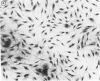
Rat myofibroblasts from granulating wound biopsies (RGW) were successfully cultured and compared in terms of growth and morphology with fibroblasts from uninjured rat dermis (RD). Populations of early passage (P-3) RGW myofibroblasts grew significantly more slowly than RD fibroblasts. Logarithmic growth was nearly the same in late passage (P-30) populations of both cell types. Early passage RGW myofibroblasts were similar to those in vivo, as shown by well-defined microfilament bundles. RD fibroblasts contained less well defined microfilaments. Both late passage RGW myofibroblasts and RD fibroblasts displayed evidence of morphologic dedifferentiation. These data show that morphologic features of myofibroblasts and fibroblasts in vivo are maintained in vitro. Evidence is presented that cultured animal myofibroblasts maintain differentiation in early passage, whereas late passage cells suggest that these differences disappear with time.
Full text
PDF


Images in this article


Selected References
These references are in PubMed. This may not be the complete list of references from this article.
- Adelstein R. S., Conti M. A., Johnson G. S., Pastan I., Pollard T. D. Isolation and characterization of myosin from cloned mouse fibroblasts. Proc Natl Acad Sci U S A. 1972 Dec;69(12):3693–3697. doi: 10.1073/pnas.69.12.3693. [DOI] [PMC free article] [PubMed] [Google Scholar]
- Bell E., Marek L. F., Levinstone D. S., Merrill C., Sher S., Young I. T., Eden M. Loss of division potential in vitro: aging or differentiation? Science. 1978 Dec 15;202(4373):1158–1163. doi: 10.1126/science.725592. [DOI] [PubMed] [Google Scholar]
- Gabbiani G., Ryan G. B., Lamelin J. P., Vassalli P., Majno G., Bouvier C. A., Cruchaud A., Lüscher E. F. Human smooth muscle autoantibody. Its identification as antiactin antibody and a study of its binding to "nonmuscular" cells. Am J Pathol. 1973 Sep;72(3):473–488. [PMC free article] [PubMed] [Google Scholar]
- Gabbiani G., Ryan G. B., Majne G. Presence of modified fibroblasts in granulation tissue and their possible role in wound contraction. Experientia. 1971 May 15;27(5):549–550. doi: 10.1007/BF02147594. [DOI] [PubMed] [Google Scholar]
- Hirschel B. J., Gabbiani G., Ryan G. B., Majno G. Fibroblasts of granulation tissue: immunofluorescent staining with antismooth muscle serum. Proc Soc Exp Biol Med. 1971 Nov;138(2):466–469. doi: 10.3181/00379727-138-35920. [DOI] [PubMed] [Google Scholar]
- Johnson G. D., Holborow E. J., Glynn L. E. Antibody to smooth muscle in patients with liver disease. Lancet. 1965 Oct 30;2(7418):878–879. doi: 10.1016/s0140-6736(65)92505-5. [DOI] [PubMed] [Google Scholar]
- Rudolph R. Location of the force of wound contraction. Surg Gynecol Obstet. 1979 Apr;148(4):547–551. [PubMed] [Google Scholar]
- Rudolph R., Woodward M. Spatial orientation of microtubules in contractile fibroblasts in vivo. Anat Rec. 1978 Jun;191(2):169–181. doi: 10.1002/ar.1091910204. [DOI] [PubMed] [Google Scholar]
- Taylor J. F. Changes in nuclear dimensions and orientation during contraction of a cultured fibroblast sheet. J Anat. 1971 Apr;108(Pt 3):509–517. [PMC free article] [PubMed] [Google Scholar]
- Van Winkle W., Jr Wound contraction. Surg Gynecol Obstet. 1967 Jul;125(1):131–142. [PubMed] [Google Scholar]