Abstract
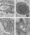
This study represents a systematic analysis of the fine-structural characteristics of atherosclerotic lesions of the superficial femoral artery in man together with the growth characteristics in culture of the smooth muscle cells derived from these lesions. Occlusive fibrous atherosclerotic plaques were obtained from 29 male patients at the time of bypass surgery for occlusion of the superficial femoral artery and were studied by light and transmission electron microscopy. The occluded segment of each artery was obtained immediately after removal from the patient and examined with sterile techniques, and representative segments were fixed for light- and electron-microscopic study. Adjacent segments were used for dissection of the lesion away from the underlying media, and smooth muscle cells were cultured from lesion and nonlesion areas and compared in terms of their growth responses to increasing concentrations of a pool of human whole blood serum. The majority of the lesions were fibroproliferative and contained relatively little lipid. The fibrous cap that covered each lesion consisted of a special form of dense connective tissue that contained flat, pancake-shaped smooth muscle cells in a lacunalike space. This space consisted of concentric layers of basement membrane, collagen fibrils, and proteoglycan. The majority of the cells beneath the fibrous cap were smooth muscle cells mixed with small but varying numbers of macrophages. Most of the lesions were occluded by a thrombus, which had undergone organization and recanalization. A small number of the lesions had deep lipid deposits together with foci of degeneration and calcification. The occluded thrombi contained smooth muscle cells and a larger proportion of macrophages than the lesions themselves. The in vitro growth properties of the smooth muscle cells isolated from the lesion and the underlying media suggested that the lesion cells had senesced, compared with the medial smooth muscle cells derived from the same artery.
Full text
PDF


Images in this article
Selected References
These references are in PubMed. This may not be the complete list of references from this article.
- BALIS J. U., HAUST M. D., MORE R. H. ELECTRON-MICROSCOPIC STUDIES IN HUMAN ATHEROSCLEROSIS; CELLULAR ELEMENTS IN AORTIC FATTY STREAKS. Exp Mol Pathol. 1964 Oct;90:511–525. doi: 10.1016/0014-4800(64)90031-0. [DOI] [PubMed] [Google Scholar]
- Burke J. M., Ross R. Synthesis of connective tissue macromolecules by smooth muscle. Int Rev Connect Tissue Res. 1979;8:119–157. doi: 10.1016/b978-0-12-363708-6.50010-2. [DOI] [PubMed] [Google Scholar]
- Eskin S. G., Sybers H. D., Lester J. W., Navarro L. T., Gotto A. M., Jr, DeBakey M. E. Human smooth muscle cells cultured from atherosclerotic plaques and uninvolved vessel wall. In Vitro. 1981 Aug;17(8):713–718. doi: 10.1007/BF02628408. [DOI] [PubMed] [Google Scholar]
- GEER J. C., McGILL H. C., Jr, STRONG J. P. The fine structure of human atherosclerotic lesions. Am J Pathol. 1961 Mar;38:263–287. [PMC free article] [PubMed] [Google Scholar]
- Geer J. C. Fine structure of human aortic intimal thickening and fatty streaks. Lab Invest. 1965 Oct;14(10):1764–1783. [PubMed] [Google Scholar]
- Ghidoni J. J., O'Neal R. M. Recent advances in molecular pathology: a review ultrastructure of human atheroma. Exp Mol Pathol. 1967 Dec;7(3):378–400. doi: 10.1016/0014-4800(67)90049-4. [DOI] [PubMed] [Google Scholar]
- HAYFLICK L. THE LIMITED IN VITRO LIFETIME OF HUMAN DIPLOID CELL STRAINS. Exp Cell Res. 1965 Mar;37:614–636. doi: 10.1016/0014-4827(65)90211-9. [DOI] [PubMed] [Google Scholar]
- Hoff H. F., Heideman C. L., Gaubatz J. W., Scott D. W., Titus J. L., Gotto A. M., Jr Correlation of apolipoprotein B retention with the structure of atherosclerotic plaques from human aortas. Lab Invest. 1978 May;38(5):560–567. [PubMed] [Google Scholar]
- Hoff H. H. Human intracranial atherosclerosis. A histochemical and ultrastructural study of gross fatty steak lesions. Am J Pathol. 1972 Dec;69(3):421–438. [PMC free article] [PubMed] [Google Scholar]
- LUFT J. H. Improvements in epoxy resin embedding methods. J Biophys Biochem Cytol. 1961 Feb;9:409–414. doi: 10.1083/jcb.9.2.409. [DOI] [PMC free article] [PubMed] [Google Scholar]
- PARKER F. An electron microscope study of coronary arteries. Am J Anat. 1958 Sep;103(2):247–273. doi: 10.1002/aja.1001030206. [DOI] [PubMed] [Google Scholar]
- Parker F., Odland G. F. A correlative histochemical, biochemical and electron microscopic study of experimental atherosclerosis in the rabbit aorta with special reference to the myo-intimal cell. Am J Pathol. 1966 Feb;48(2):197–239. [PMC free article] [PubMed] [Google Scholar]
- Pietilä K., Nikkari T. Enhanced growth of smooth muscle cells from atherosclerotic rabbit aortas in culture. Atherosclerosis. 1980 Jun;36(2):241–248. doi: 10.1016/0021-9150(80)90232-4. [DOI] [PubMed] [Google Scholar]
- Ross R. George Lyman Duff Memorial Lecture. Atherosclerosis: a problem of the biology of arterial wall cells and their interactions with blood components. Arteriosclerosis. 1981 Sep-Oct;1(5):293–311. doi: 10.1161/01.atv.1.5.293. [DOI] [PubMed] [Google Scholar]
- Ross R., Glomset J. A. The pathogenesis of atherosclerosis (first of two parts). N Engl J Med. 1976 Aug 12;295(7):369–377. doi: 10.1056/NEJM197608122950707. [DOI] [PubMed] [Google Scholar]
- Ross R. The smooth muscle cell. II. Growth of smooth muscle in culture and formation of elastic fibers. J Cell Biol. 1971 Jul;50(1):172–186. doi: 10.1083/jcb.50.1.172. [DOI] [PMC free article] [PubMed] [Google Scholar]
- Takebayashi S., Kamio A. Ultrastructural studies on human arteriosclerosis-comparison between hypertensive and normotensive groups. Acta Pathol Jpn. 1969 Nov;19(4):479–499. doi: 10.1111/j.1440-1827.1969.tb00091.x. [DOI] [PubMed] [Google Scholar]
- Thomas W. A., Kim D. N. Biology of disease. Atherosclerosis as a hyperplastic and/or neoplastic process. Lab Invest. 1983 Mar;48(3):245–255. [PubMed] [Google Scholar]
- Wight T. N., Ross R. Proteoglycans in primate arteries. I. Ultrastructural localization and distribution in the intima. J Cell Biol. 1975 Dec;67(3):660–674. doi: 10.1083/jcb.67.3.660. [DOI] [PMC free article] [PubMed] [Google Scholar]


