Abstract
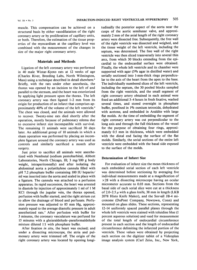
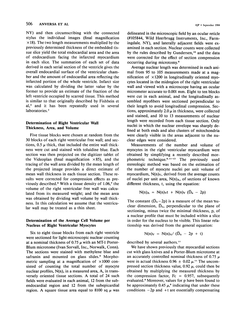
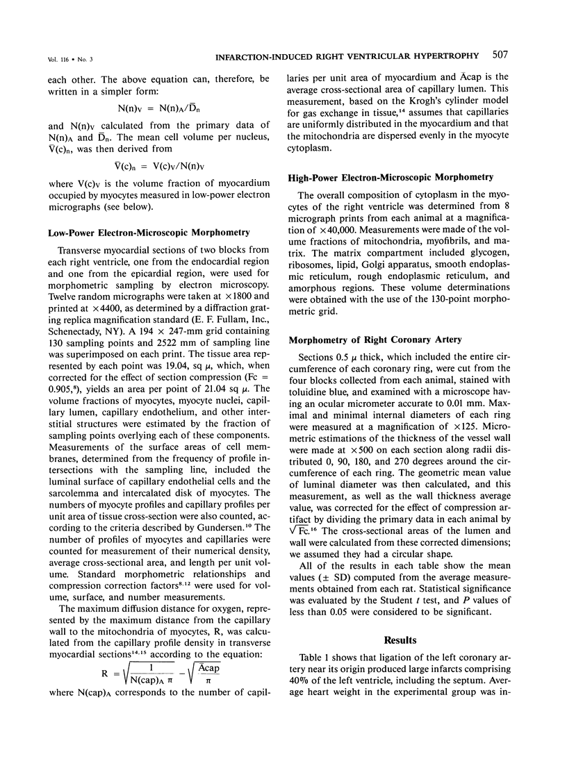
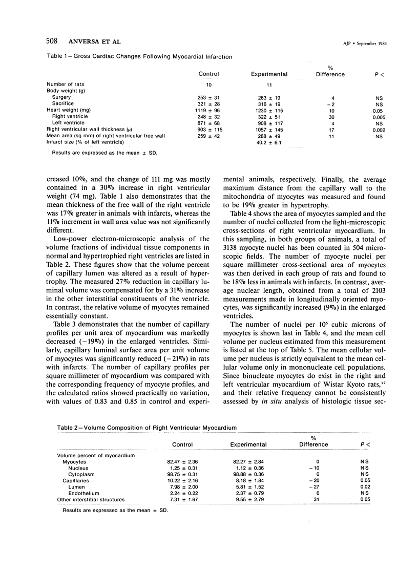
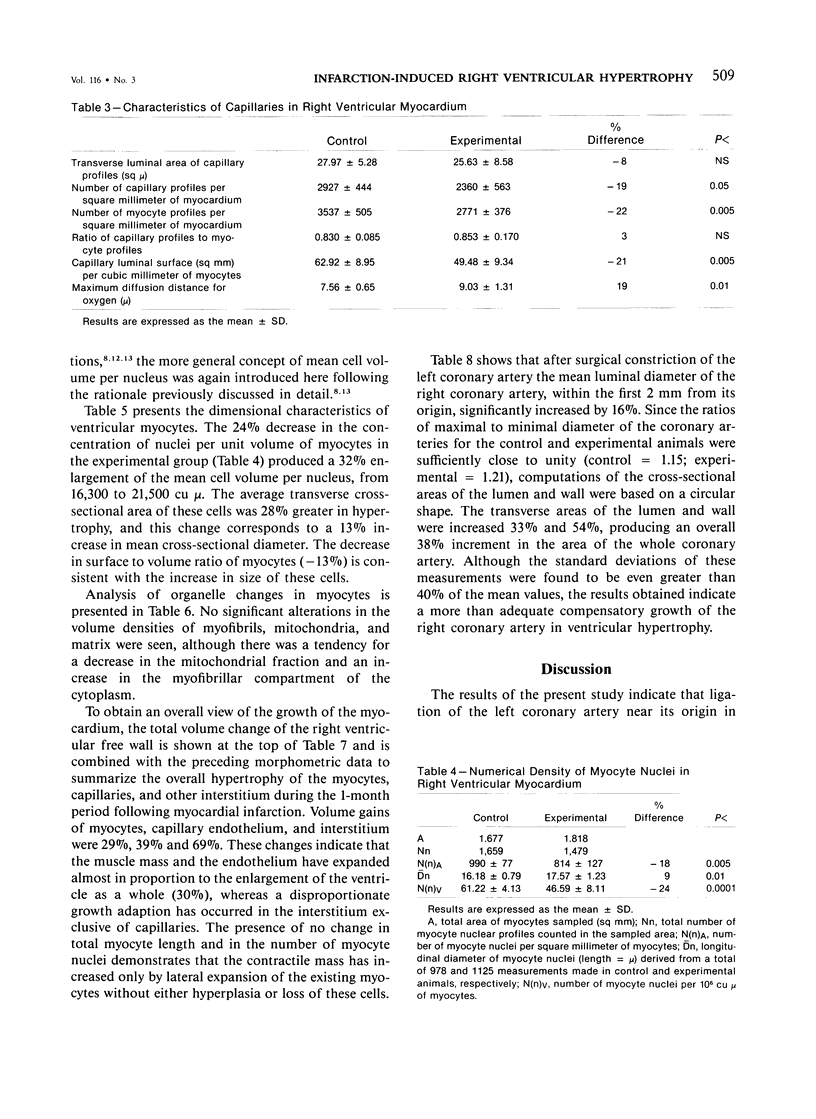
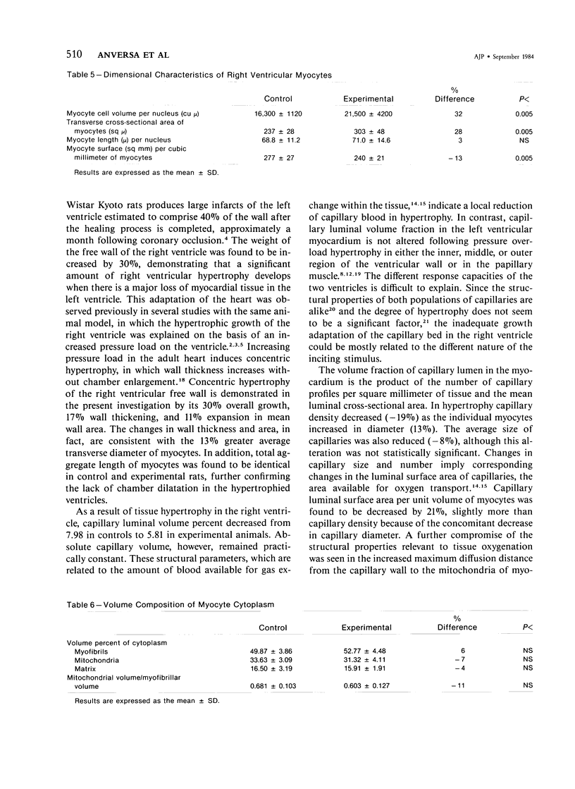
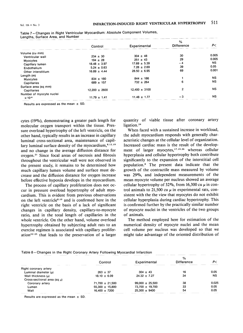
The growth response of the right ventricle was studied in rats following ligation of the left coronary artery, which produced infarcts comprising approximately 40% of the left ventricle. A month after surgery the weight of the right ventricle was increased 30%, and this hypertrophic change was characterized by a 17% wall thickening, consistent with the 13% greater diameter of myocytes. Myocardial hypertrophy was accompanied by an inadequate growth of the microvasculature that supports tissue oxygenation. This was seen by relative decreases in capillary luminal volume density (-27%) and capillary luminal surface density (-21%) and by an increase in the average maximum distance from the capillary wall to the mitochondria of myocytes (19%). In contrast, measurements of the mean myocyte volume per nucleus showed a proportional enlargement of these cells (32%), from 16,300 cu mu in control animals to 21,500 cu mu in experimental rats. Quantitative analysis of the right coronary artery revealed a 33% increase in its luminal area, commensurate with the magnitude of ventricular hypertrophy.
Full text
PDF
Selected References
These references are in PubMed. This may not be the complete list of references from this article.
- Anversa P., Giacomelli F., Wiener J. Regional variation in capillary permeability of ventricular myocardium. Microvasc Res. 1973 Nov;6(3):273–285. doi: 10.1016/0026-2862(73)90076-9. [DOI] [PubMed] [Google Scholar]
- Anversa P., Levicky V., Beghi C., McDonald S. L., Kikkawa Y. Morphometry of exercise-induced right ventricular hypertrophy in the rat. Circ Res. 1983 Jan;52(1):57–64. doi: 10.1161/01.res.52.1.57. [DOI] [PubMed] [Google Scholar]
- Anversa P., Loud A. V., Giacomelli F., Wiener J. Absolute morphometric study of myocardial hypertrophy in experimental hypertension. II. Ultrastructure of myocytes and interstitium. Lab Invest. 1978 May;38(5):597–609. [PubMed] [Google Scholar]
- Anversa P., Olivetti G., Loud A. V. Morphometric study of early postnatal development in the left and right ventricular myocardium of the rat. I. Hypertrophy, hyperplasia, and binucleation of myocytes. Circ Res. 1980 Apr;46(4):495–502. doi: 10.1161/01.res.46.4.495. [DOI] [PubMed] [Google Scholar]
- Anversa P., Olivetti G., Melissari M., Loud A. V. Morphometric study of myocardial hypertrophy induced by abdominal aortic stenosis. Lab Invest. 1979 Mar;40(3):341–349. [PubMed] [Google Scholar]
- Anversa P., Olivetti G., Melissari M., Loud A. V. Stereological measurement of cellular and subcellular hypertrophy and hyperplasia in the papillary muscle of adult rat. J Mol Cell Cardiol. 1980 Aug;12(8):781–795. doi: 10.1016/0022-2828(80)90080-2. [DOI] [PubMed] [Google Scholar]
- Bishop S. P., Oparil S., Reynolds R. H., Drummond J. L. Regional myocyte size in normotensive and spontaneously hypertensive rats. Hypertension. 1979 Jul-Aug;1(4):378–383. doi: 10.1161/01.hyp.1.4.378. [DOI] [PubMed] [Google Scholar]
- Driscol T. E., Eckstein R. W. Coronary inflow and outflow responses to coronary artery occlusion. Circ Res. 1967 May;20(5):485–495. doi: 10.1161/01.res.20.5.485. [DOI] [PubMed] [Google Scholar]
- Fishbein M. C., Maclean D., Maroko P. R. Experimental myocardial infarction in the rat: qualitative and quantitative changes during pathologic evolution. Am J Pathol. 1978 Jan;90(1):57–70. [PMC free article] [PubMed] [Google Scholar]
- HORT W., DACANALIS S., JUST H. UNTERSUCHUNGEN BEI CHRONISCHEM EXPERIMENTELLEN HERZINFARKT DER RATTE. Arch Kreislaufforsch. 1964 Oct;44:288–299. doi: 10.1007/BF02119542. [DOI] [PubMed] [Google Scholar]
- Herzberg R. M., Rubio R., Berne R. M. Coronary occlusion and embolization: effect on blood flow in adjacent arteries. Am J Physiol. 1966 Jan;210(1):169–175. doi: 10.1152/ajplegacy.1966.210.1.169. [DOI] [PubMed] [Google Scholar]
- Hoppeler H., Mathieu O., Krauer R., Claassen H., Armstrong R. B., Weibel E. R. Design of the mammalian respiratory system. VI Distribution of mitochondria and capillaries in various muscles. Respir Physiol. 1981 Apr;44(1):87–111. doi: 10.1016/0034-5687(81)90078-5. [DOI] [PubMed] [Google Scholar]
- Hoppeler H., Mathieu O., Weibel E. R., Krauer R., Lindstedt S. L., Taylor C. R. Design of the mammalian respiratory system. VIII Capillaries in skeletal muscles. Respir Physiol. 1981 Apr;44(1):129–150. doi: 10.1016/0034-5687(81)90080-3. [DOI] [PubMed] [Google Scholar]
- Korecky B., Rakusan K. Normal and hypertrophic growth of the rat heart: changes in cell dimensions and number. Am J Physiol. 1978 Feb;234(2):H123–H128. doi: 10.1152/ajpheart.1978.234.2.H123. [DOI] [PubMed] [Google Scholar]
- Lais L. T., Rios L. L., Boutelle S., DiBona G. F., Brody M. J. Arterial pressure development in neonatal and young spontaneously hypertensive rats. Blood Vessels. 1977;14(5):277–284. doi: 10.1159/000158134. [DOI] [PubMed] [Google Scholar]
- Leon A. S., Bloor C. M. The effect of complete and partial deconditioning on exercise-induced cardiovascular changes in the rat. Adv Cardiol. 1976;18(0):81–92. doi: 10.1159/000399514. [DOI] [PubMed] [Google Scholar]
- Lin H. L., Katele K. V., Grimm A. F. Functional morphology of the pressure- and the volume-hypertrophied rat heart. Circ Res. 1977 Dec;41(6):830–836. doi: 10.1161/01.res.41.6.830. [DOI] [PubMed] [Google Scholar]
- Loud A. V., Anversa P., Giacomelli F., Wiener J. Absolute morphometric study of myocardial hypertrophy in experimental hypertension. I. Determination of myocyte size. Lab Invest. 1978 May;38(5):586–596. [PubMed] [Google Scholar]
- McElroy C. L., Gissen S. A., Fishbein M. C. Exercise-induced reduction in myocardial infarct size after coronary artery occlusion in the rat. Circulation. 1978 May;57(5):958–962. doi: 10.1161/01.cir.57.5.958. [DOI] [PubMed] [Google Scholar]
- NORMAN T. D., COERS C. R. Cardiac hypertrophy after coronary artery ligation in rats. Arch Pathol. 1960 Feb;69:181–184. [PubMed] [Google Scholar]
- Olivetti G., Anversa P., Melissari M., Loud A. V. Morphometric study of early postnatal development of the thoracic aorta in the rat. Circ Res. 1980 Sep;47(3):417–424. doi: 10.1161/01.res.47.3.417. [DOI] [PubMed] [Google Scholar]
- Page E., McCallister L. P. Quantitative electron microscopic description of heart muscle cells. Application to normal, hypertrophied and thyroxin-stimulated hearts. Am J Cardiol. 1973 Feb;31(2):172–181. doi: 10.1016/0002-9149(73)91030-8. [DOI] [PubMed] [Google Scholar]
- Pfeffer M. A., Pfeffer J. M., Fishbein M. C., Fletcher P. J., Spadaro J., Kloner R. A., Braunwald E. Myocardial infarct size and ventricular function in rats. Circ Res. 1979 Apr;44(4):503–512. doi: 10.1161/01.res.44.4.503. [DOI] [PubMed] [Google Scholar]
- Polimeni P. I., Al-Sadir J. Expansion of extracellular space in the nonischemic zone of the infarcted heart and concomitant changes in tissue electrolyte contents in the rat. Circ Res. 1975 Dec;37(6):725–732. doi: 10.1161/01.res.37.6.725. [DOI] [PubMed] [Google Scholar]
- SELYE H., BAJUSZ E., GRASSO S., MENDELL P. Simple techniques for the surgical occlusion of coronary vessels in the rat. Angiology. 1960 Oct;11:398–407. doi: 10.1177/000331976001100505. [DOI] [PubMed] [Google Scholar]
- Tomanek R. J. Effects of age and exercise on the extent of the myocardial capillary bed. Anat Rec. 1970 May;167(1):55–62. doi: 10.1002/ar.1091670106. [DOI] [PubMed] [Google Scholar]
- Turek Z., Grandtner M., Kubát K., Ringnalda B. E., Kreuzer F. Arterial blood gases, muscle fiber diameter and intercapillary distance in cardiac hypertrophy of rats with an old myocardial infarction. Pflugers Arch. 1978 Sep 29;376(3):209–215. doi: 10.1007/BF00584952. [DOI] [PubMed] [Google Scholar]


