

Abstract
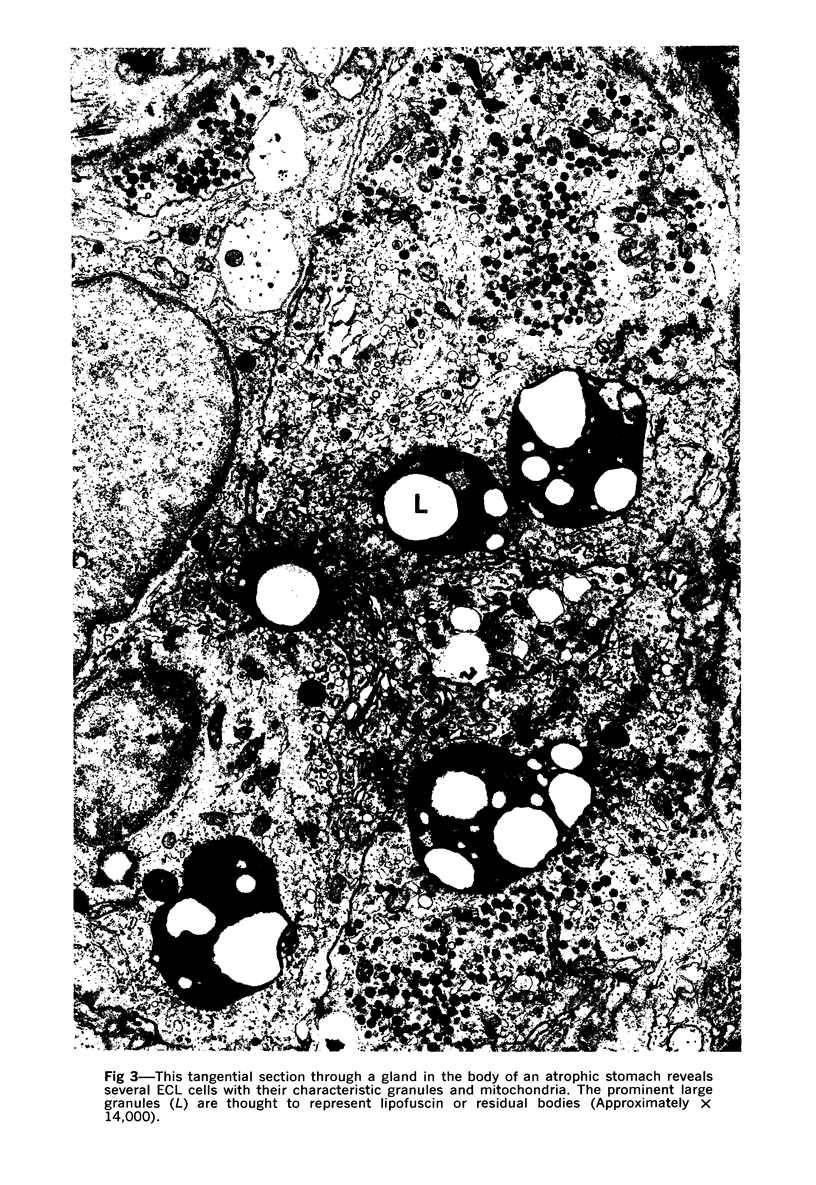
This study shows that most of the proliferated endocrine cells in nonintestinalized epithelium in the body of stomachs in patients with pernicious anemia have the fine structure of ECL (enterochromaffin-like) cells, the principal endocrine cell in the body of normal stomachs, and notes an absence of G (gastrin) cells, the principal endocrine cell in the normal pylorus. Since gastrin has been identified in many of these proliferated cells by immunofluorescence, these findings question the position that gastrin synthesis in the stomach is only associated with G cells.
Full text
PDF
Images in this article
Selected References
These references are in PubMed. This may not be the complete list of references from this article.
- Bussolati G., Pearse A. G. Immunofluorescent localization of the gastrin-secreting G cells in the pyloric antrum of the pig. Histochemie. 1970;21(1):1–4. doi: 10.1007/BF00304798. [DOI] [PubMed] [Google Scholar]
- Capella C., Solcia E., Vassallo G. Identification of six types of endocrine cells in the gastrointestinal mucosa of the rabbit. Arch Histol Jpn. 1969 Aug;30(5):479–495. doi: 10.1679/aohc1950.30.479. [DOI] [PubMed] [Google Scholar]
- Carvalheira A. F., Welsch U., Pearse A. G. Cytochemical and ultrastructural observations on the argentaffin and argyrophil cells of the gastro-intestinal tract in mammals, and their place in the APUD series of polypeptide-secreting cells. Histochemie. 1968;14(1):33–46. doi: 10.1007/BF00268031. [DOI] [PubMed] [Google Scholar]
- FLICK A. L., QUINTON W. E., RUBIN C. E. A peroral hydraulic biopsy tube for multiple sampling at any level of the gastro-intestinal tract. Gastroenterology. 1961 Jan;40:120–126. [PubMed] [Google Scholar]
- Forssmann W. G., Orci L., Pictet R., Renold A. E., Rouiller C. The endocrine cells in the epithelium of the gastrointestinal mucosa of the rat. An electron microscope study. J Cell Biol. 1969 Mar;40(3):692–715. doi: 10.1083/jcb.40.3.692. [DOI] [PMC free article] [PubMed] [Google Scholar]
- Ganguli P. C., Cullen D. R., Irvine W. J. Radioimmunoassay of plasmagastrin in pernicious anaemia, achlorhydria without pernicious anaemia, hypochlorhydria, and in controls. Lancet. 1971 Jan 23;1(7691):155–158. doi: 10.1016/s0140-6736(71)91932-5. [DOI] [PubMed] [Google Scholar]
- Kobayashi S., Fujita T., Sasagawa T. Electron microscope studies on the endocrine cells of the human gastric fundus. Arch Histol Jpn. 1971 Feb;32(5):429–444. doi: 10.1679/aohc1950.32.429. [DOI] [PubMed] [Google Scholar]
- LUFT J. H. Improvements in epoxy resin embedding methods. J Biophys Biochem Cytol. 1961 Feb;9:409–414. doi: 10.1083/jcb.9.2.409. [DOI] [PMC free article] [PubMed] [Google Scholar]
- McGuigan J. E., Trudeau W. L. Serum gastrin concentrations in pernicious anemia. N Engl J Med. 1970 Feb 12;282(7):358–361. doi: 10.1056/NEJM197002122820703. [DOI] [PubMed] [Google Scholar]
- Orci L., Pictet R., Forssmann W. G., Renold A. E., Rouiller C. Structural evidence for glucagon producing cells in the intestinal mucosa of the rat. Diabetologia. 1968 Jan;4(1):56–67. doi: 10.1007/BF01241034. [DOI] [PubMed] [Google Scholar]
- Pearse A. G., Bussolati G. Immunofluorescence studies on the distribution of gastrin cells in different clinical states. Gut. 1970 Aug;11(8):646–648. doi: 10.1136/gut.11.8.646. [DOI] [PMC free article] [PubMed] [Google Scholar]
- Pearse A. G., Coulling I., Weavers B., Friesen S. The endocrine polypeptide cells of the human stomach, duodenum, and jejunum. Gut. 1970 Aug;11(8):649–658. doi: 10.1136/gut.11.8.649. [DOI] [PMC free article] [PubMed] [Google Scholar]
- Polak J. M., Bloom S., Coulling I., Pearse A. G. Immunofluorescent localization of enteroglucagon cells in the gastrointestinal tract of the dog. Gut. 1971 Apr;12(4):311–318. doi: 10.1136/gut.12.4.311. [DOI] [PMC free article] [PubMed] [Google Scholar]
- Polak J. M., Doe W., Coulling I., Pearse A. G. The G cells in pernicious anaemia. Gut. 1971 Apr;12(4):319–323. doi: 10.1136/gut.12.4.319. [DOI] [PMC free article] [PubMed] [Google Scholar]
- REYNOLDS E. S. The use of lead citrate at high pH as an electron-opaque stain in electron microscopy. J Cell Biol. 1963 Apr;17:208–212. doi: 10.1083/jcb.17.1.208. [DOI] [PMC free article] [PubMed] [Google Scholar]
- Rbin W. Proliferation of endocrine-like enterochromaffin) cells in atrophic gastri mucosa. Gastroenterology. 1969 Dec;57(6):641–648. [PubMed] [Google Scholar]
- Rubin W. An unusual intimate relationship between endocrine cells and other types of epithelial cells in the human stomach. J Cell Biol. 1972 Jan;52(1):219–227. doi: 10.1083/jcb.52.1.219. [DOI] [PMC free article] [PubMed] [Google Scholar]
- Rubin W., Gershon M. D., Ross L. L. Electron microscope radioautographic identification of serotonin-synthesizing cells in the mouse gastric mucosa. J Cell Biol. 1971 Aug;50(2):399–415. doi: 10.1083/jcb.50.2.399. [DOI] [PMC free article] [PubMed] [Google Scholar]
- Rubin W., Ross L. L., Jeffries G. H., Sleisenger M. H. Intestinal heterotopia. A fine structural study. Lab Invest. 1966 Jun;15(6):1024–1049. [PubMed] [Google Scholar]
- SABATINI D. D., BENSCH K., BARRNETT R. J. Cytochemistry and electron microscopy. The preservation of cellular ultrastructure and enzymatic activity by aldehyde fixation. J Cell Biol. 1963 Apr;17:19–58. doi: 10.1083/jcb.17.1.19. [DOI] [PMC free article] [PubMed] [Google Scholar]
- Sasagawa T., Kobayashi S., Fujita T. The endocrine cells in the human pyloric antrum. An electron microscope study of biopsy materials. Arch Histol Jpn. 1970 Sep;32(3):275–288. doi: 10.1679/aohc1950.32.275. [DOI] [PubMed] [Google Scholar]
- Solcia E., Sampietro R. Cytologic observations on the pancreatic islets with reference to some endocrine-like cells of the gastrointestinal mucosa. Z Zellforsch Mikrosk Anat. 1965 Dec 10;68(5):689–698. doi: 10.1007/BF00340094. [DOI] [PubMed] [Google Scholar]
- Solcia E., Vassallo G., Capella C. Studies on the G cells of the pyloric mucosa, the probable site of gastrin secretion. Gut. 1969 May;10(5):379–388. doi: 10.1136/gut.10.5.379. [DOI] [PMC free article] [PubMed] [Google Scholar]
- Solcia E., Vassallo G., Sampietro R. Endocrine cells in the antro-pyloric mucosa of the stomach. Z Zellforsch Mikrosk Anat. 1967;81(4):474–486. doi: 10.1007/BF00541009. [DOI] [PubMed] [Google Scholar]
- TRUMP B. F., SMUCKLER E. A., BENDITT E. P. A method for staining epoxy sections for light microscopy. J Ultrastruct Res. 1961 Aug;5:343–348. doi: 10.1016/s0022-5320(61)80011-7. [DOI] [PubMed] [Google Scholar]
- VENABLE J. H., COGGESHALL R. A SIMPLIFIED LEAD CITRATE STAIN FOR USE IN ELECTRON MICROSCOPY. J Cell Biol. 1965 May;25:407–408. doi: 10.1083/jcb.25.2.407. [DOI] [PMC free article] [PubMed] [Google Scholar]
- Vassallo G., Capella C., Solcia E. Endocrine cells of the human gastric mucosa. Z Zellforsch Mikrosk Anat. 1971;118(1):49–67. doi: 10.1007/BF00331766. [DOI] [PubMed] [Google Scholar]
- Vassallo G., Solcia E., Capella C. Light and electron microscopic identification of several types of endocrine cells in the gastrointestinal mucosa of the cat. Z Zellforsch Mikrosk Anat. 1969;98(3):333–356. doi: 10.1007/BF00346679. [DOI] [PubMed] [Google Scholar]
- WATSON M. L. Staining of tissue sections for electron microscopy with heavy metals. II. Application of solutions containing lead and barium. J Biophys Biochem Cytol. 1958 Nov 25;4(6):727–730. doi: 10.1083/jcb.4.6.727. [DOI] [PMC free article] [PubMed] [Google Scholar]
- Yalow R. S., Berson S. A. Further studies on the nature of immunoreactive gastrin in human plasma. Gastroenterology. 1971 Feb;60(2):203–214. [PubMed] [Google Scholar]
- Yalow R. S., Berson S. A. Radioimmunoassay of gastrin. Gastroenterology. 1970 Jan;58(1):1–14. [PubMed] [Google Scholar]