Abstract
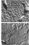
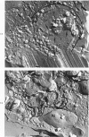
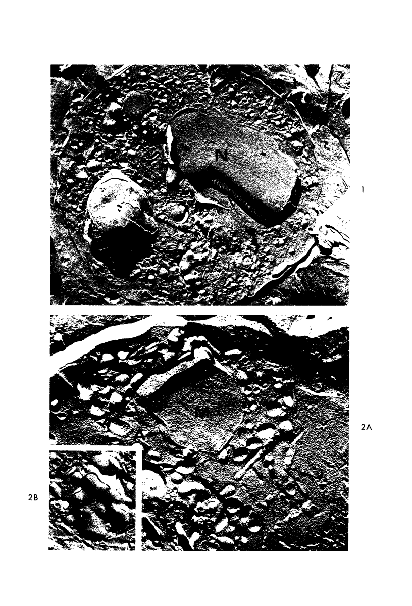
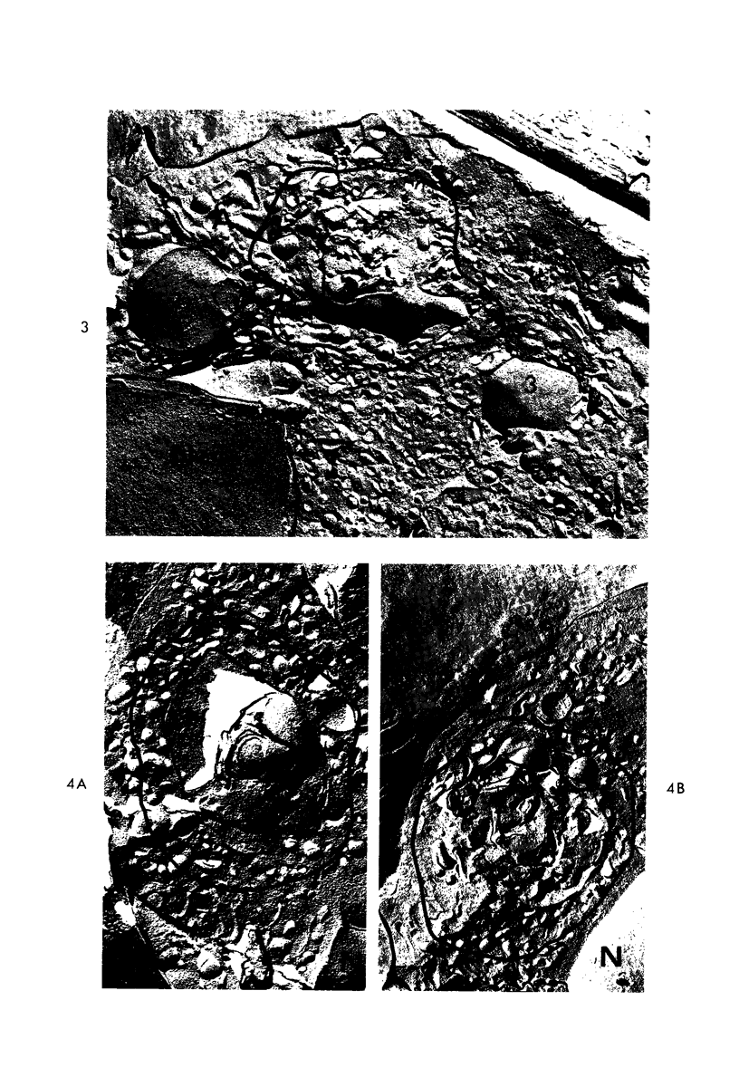
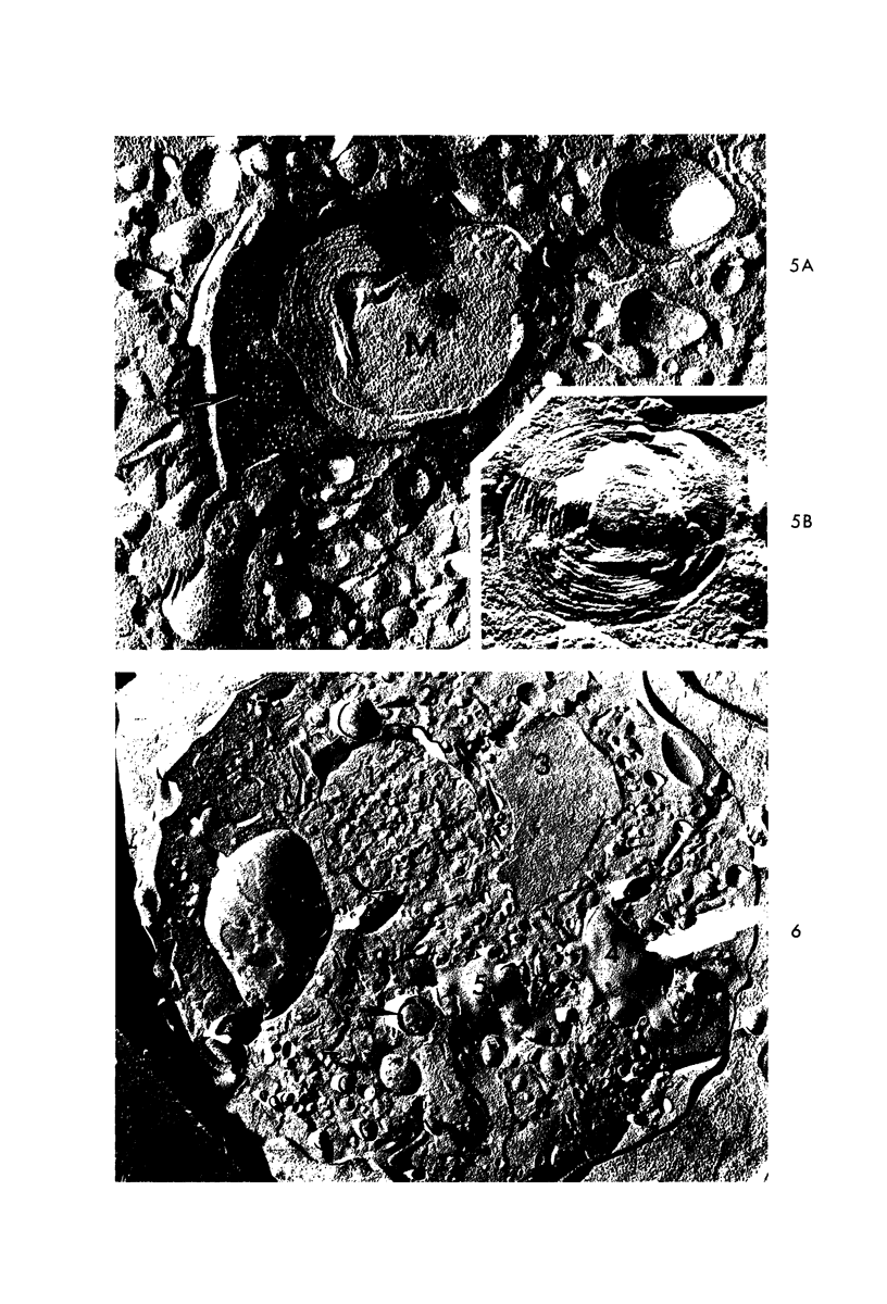
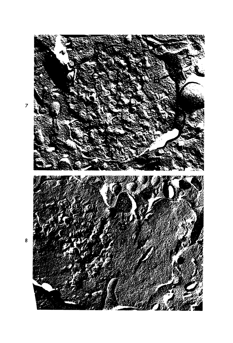
The technic of freeze-fracture and etching has been used in the present study to examine the fine structure of giant inclusions in circulating leukocytes from a patient with the Chediak-Higashi syndrome (CHS). The surface granularity of the membranes enclosing the giant inclusions differed slightly from that of normal sized organelles. Two types of giant granules were distinguished in replicas of freeze-fractured CHS neutrophils. The difference in fine structure suggests that one variety is a massively enlarged, but essentially unaltered primary lysosome, while the other develops as a result of continued fusion of small organelles with huge inclusions throughout the stages of polymorphonuclear leukocyte maturation.
Full text
PDF
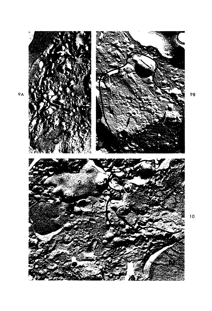
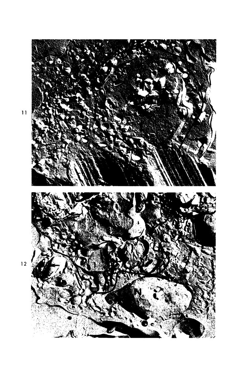
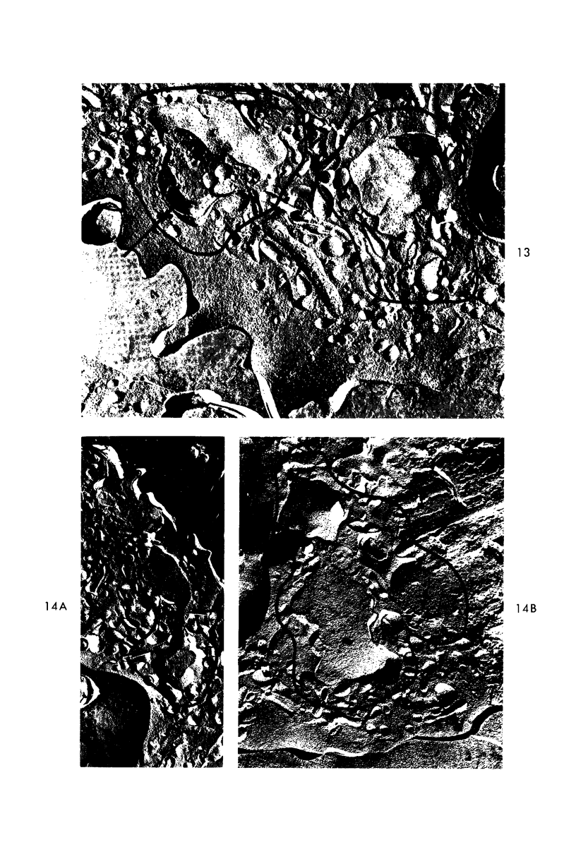
Images in this article
Selected References
These references are in PubMed. This may not be the complete list of references from this article.
- BERNARD J., BESSIS M., SELIGMANN M., CHASSIGNEUX J., CHOME J. [A case of Chediak-Steinbrinck-Higashi disease. Clinical and cytological study]. Presse Med. 1960 Mar 23;68:563–566. [PubMed] [Google Scholar]
- Bainton D. F., Farquhar M. G. Origin of granules in polymorphonuclear leukocytes. Two types derived from opposite faces of the Golgi complex in developing granulocytes. J Cell Biol. 1966 Feb;28(2):277–301. doi: 10.1083/jcb.28.2.277. [DOI] [PMC free article] [PubMed] [Google Scholar]
- Bedoya V., Grimley P. M., Duque O. Chediak-Higashi syndrome. Arch Pathol. 1969 Oct;88(4):340–349. [PubMed] [Google Scholar]
- Blume R. S., Glade P. R., Gralnick H. R., Chessin L. N., Haase A. T., Wolff S. M. The Chediak-Higashi syndrome: continuous suspension cultures derived from peripheral blood. Blood. 1969 Jun;33(6):821–832. [PubMed] [Google Scholar]
- Blume R. S., Wolff S. M. The Chediak-Higashi syndrome: studies in four patients and a review of the literature. Medicine (Baltimore) 1972 Jul;51(4):247–280. [PubMed] [Google Scholar]
- Branton D. Fracture faces of frozen membranes. Proc Natl Acad Sci U S A. 1966 May;55(5):1048–1056. doi: 10.1073/pnas.55.5.1048. [DOI] [PMC free article] [PubMed] [Google Scholar]
- CHEDIAK M. M. Nouvelle anomalie leucocytaire de caractère constitutionnel et familial. Rev Hematol. 1952;7(3):362–367. [PubMed] [Google Scholar]
- Chalcroft J. P., Bullivant S. An interpretation of liver cell membrane and junction structure based on observation of freeze-fracture replicas of both sides of the fracture. J Cell Biol. 1970 Oct;47(1):49–60. doi: 10.1083/jcb.47.1.49. [DOI] [PMC free article] [PubMed] [Google Scholar]
- Clark R. A., Kimball H. R. Defective granulocyte chemotaxis in the Chediak-Higashi syndrome. J Clin Invest. 1971 Dec;50(12):2645–2652. doi: 10.1172/JCI106765. [DOI] [PMC free article] [PubMed] [Google Scholar]
- Clawson C. C., White J. G. Platelet interaction with bacteria. I. Reaction phases and effects of inhibitors. Am J Pathol. 1971 Nov;65(2):367–380. [PMC free article] [PubMed] [Google Scholar]
- Davis W. C., Spicer S. S., Greene W. B., Padgett G. A. Ultrastructure of bone marrow granulocytes in normal mink and mink with the homolog of the Chediak-Higashi trait of humans. I. Origin of the abnormal granules present in the neutrophils of mink with the C-HS trait. Lab Invest. 1971 Apr;24(4):303–317. [PubMed] [Google Scholar]
- Davis W. C., Spicer S. S., Greene W. B., Padgett G. A. Ultrastructure of cells in bone marrow and peripheral blood of normal mink and mink with the homologue of the Chediak-Higashi trait of humans. II. Cytoplasmic granules in eosinophils, basophils, mononuclear cells and platelets. Am J Pathol. 1971 Jun;63(3):411–432. [PMC free article] [PubMed] [Google Scholar]
- Deamer D. W., Branton D. Fracture planes in an ice-bilayer model membrane system. Science. 1967 Nov 3;158(3801):655–657. doi: 10.1126/science.158.3801.655. [DOI] [PubMed] [Google Scholar]
- Douglas S. D., Blume R. S., Glade P. R., Chessin L. N., Wolff S. M. Fine structure of continuous long term lymphoid cell cultures from a Chediak-Higashi patient and heterozygote. Lab Invest. 1969 Sep;21(3):225–229. [PubMed] [Google Scholar]
- Douglas S. D., Blume R. S., Wolff S. M. Fine structural studies of leukocytes from patients and heterozygotes with the Chediak-Higashi syndrome. Blood. 1969 Apr;33(4):527–540. [PubMed] [Google Scholar]
- Efrati P., Danon D. Electron-microscopical study of bone marrow cells in a case of Chediak-Higashi-Steinbrinck syndrome. Br J Haematol. 1968 Aug;15(2):173–176. doi: 10.1111/j.1365-2141.1968.tb01526.x. [DOI] [PubMed] [Google Scholar]
- Friend D. S., Gilula N. B. Variations in tight and gap junctions in mammalian tissues. J Cell Biol. 1972 Jun;53(3):758–776. doi: 10.1083/jcb.53.3.758. [DOI] [PMC free article] [PubMed] [Google Scholar]
- HIGASHI O. Congenital gigantism of peroxidase granules; the first case ever reported of qualitative abnormity of peroxidase. Tohoku J Exp Med. 1954 Feb 25;59(3):315–332. doi: 10.1620/tjem.59.315. [DOI] [PubMed] [Google Scholar]
- Lutzner M. A., Tierney J. H., Benditt E. P. Giant granules and widespread cytoplasmic inclusions in a genetic syndrome of Aleutian mink. An electron microscopic study. Lab Invest. 1965 Dec;14(12):2063–2079. [PubMed] [Google Scholar]
- Moran T. J., Estevez J. M. Chediak-Higashi disease. Morphologic studies of a patient and her family. Arch Pathol. 1969 Oct;88(4):329–339. [PubMed] [Google Scholar]
- Padgett G. A. Neutrophilic function in animals with the Chediak-Higashi syndrome. Blood. 1967 Jun;29(6):906–915. [PubMed] [Google Scholar]
- Root R. K., Rosenthal A. S., Balestra D. J. Abnormal bactericidal, metabolic, and lysosomal functions of Chediak-Higashi Syndrome leukocytes. J Clin Invest. 1972 Mar;51(3):649–665. doi: 10.1172/JCI106854. [DOI] [PMC free article] [PubMed] [Google Scholar]
- Rosa F. Estudio electromicroscópico de la anomalía de Chediak-Higashi-Steinbrick. Acta Cient Venez. 1967;18(3):73–78. [PubMed] [Google Scholar]
- STEERE R. L. Electron microscopy of structural detail in frozen biological specimens. J Biophys Biochem Cytol. 1957 Jan 25;3(1):45–60. doi: 10.1083/jcb.3.1.45. [DOI] [PMC free article] [PubMed] [Google Scholar]
- Staehelin L. A., Chlapowski F. J., Bonneville M. A. Lumenal plasma membrane of the urinary bladder. I. Three-dimensional reconstruction from freeze-etch images. J Cell Biol. 1972 Apr;53(1):73–91. doi: 10.1083/jcb.53.1.73. [DOI] [PMC free article] [PubMed] [Google Scholar]
- Stossel T. P., Root R. K., Vaughan M. Phagocytosis in chronic granulomatous disease and the Chediak-Higashi syndrome. N Engl J Med. 1972 Jan 20;286(3):120–123. doi: 10.1056/NEJM197201202860302. [DOI] [PubMed] [Google Scholar]
- Tillack T. W., Marchesi V. T. Demonstration of the outer surface of freeze-etched red blood cell membranes. J Cell Biol. 1970 Jun;45(3):649–653. doi: 10.1083/jcb.45.3.649. [DOI] [PMC free article] [PubMed] [Google Scholar]
- White J. G., Conard W. J. The fine structure of freeze-fractured blood platelets. Am J Pathol. 1973 Jan;70(1):45–56. [PMC free article] [PubMed] [Google Scholar]
- White J. G. Giant organelles containing tubules in Chediak-Higashi lymphocytes. Am J Pathol. 1972 Nov;69(2):225–238. [PMC free article] [PubMed] [Google Scholar]
- White J. G. The Chediak-Higashi syndrome: a possible lysosomal disease. Blood. 1966 Aug;28(2):143–156. [PubMed] [Google Scholar]
- White J. G. The Chediak-Higashi syndrome: cytoplasmic sequestration in circulating leukocytes. Blood. 1967 Apr;29(4):435–451. [PubMed] [Google Scholar]
- Wolff S. M. The Chediak-Higashi syndrome: studies of host defenses. Ann Intern Med. 1972 Feb;76(2):293–306. doi: 10.7326/0003-4819-76-2-293. [DOI] [PubMed] [Google Scholar]


