Abstract
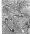
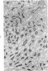
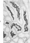
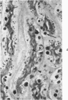
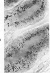
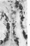
The beige mouse is considered to be a homologue of Chediak-Higashi syndrome (CHS). Cytochemical and electron microscopic studies have revealed an inherited lesion in the kidneys of these mice. The alteration was confined to the distal segments (S3) of the proximal tubules and was characterized by the accumulation of numerous massive polysaccharide-rich granules. These granules were reactive for acid phosphatase and β-glucuronidase activities and were therefore considered to be lysosomes. Small numbers of lymphocytes were also observed in the perivascular spaces and within the tubules of the S3 segment. The fine structure of S3 cells was also markedly altered. In addition to the massive lysosomes, dense material was found associated with the brush border and was present in pinocytotic vesicles at the base of the brush border and between basal invaginations of the plasma membranes. Despite these changes, reabsorption of exogeneous peroxidase by S3 cells appeared to be normal. The presence of a congenital defect in the kidney of the beige mouse appears to provide a useful model for studying the morphology and function of the S3 region of the nephron.
Full text
PDF


Images in this article
Selected References
These references are in PubMed. This may not be the complete list of references from this article.
- Beard M. E., Novikoff A. B. Distribution of peroxisomes (microbodies) in the nephron of the rat: a cytochemical study. J Cell Biol. 1969 Aug;42(2):501–518. doi: 10.1083/jcb.42.2.501. [DOI] [PMC free article] [PubMed] [Google Scholar]
- Bennett J. M., Blume R. S., Wolff S. M. Characterization and significance of abnormal leukocyte granules in the beige mouse: a possible homologue for Chediak-Higashi Aleutian trait. J Lab Clin Med. 1969 Feb;73(2):235–243. [PubMed] [Google Scholar]
- Blume R. S., Wolff S. M. The Chediak-Higashi syndrome: studies in four patients and a review of the literature. Medicine (Baltimore) 1972 Jul;51(4):247–280. [PubMed] [Google Scholar]
- DEITCH A. D. An improved Sakaguchi reaction for microspectrophotometric use. J Histochem Cytochem. 1961 Sep;9:477–483. doi: 10.1177/9.5.477. [DOI] [PubMed] [Google Scholar]
- Davis W. C., Douglas S. D. Defective granule formation and function in the Chediak-Higashi syndrome in man and animals. Semin Hematol. 1972 Oct;9(4):431–450. [PubMed] [Google Scholar]
- Davis W. C., Spicer S. S., Greene W. B., Padgett G. A. Ultrastructure of cells in bone marrow and peripheral blood of normal mink and mink with the homologue of the Chediak-Higashi trait of humans. II. Cytoplasmic granules in eosinophils, basophils, mononuclear cells and platelets. Am J Pathol. 1971 Jun;63(3):411–432. [PMC free article] [PubMed] [Google Scholar]
- Fahimi H. D. The fine structural localization of endogenous and exogenous peroxidase activity in Kupffer cells of rat liver. J Cell Biol. 1970 Oct;47(1):247–262. doi: 10.1083/jcb.47.1.247. [DOI] [PMC free article] [PubMed] [Google Scholar]
- HAYASHI M., NAKAJIMA Y., FISHMAN W. H. THE CYTOLOGIC DEMONSTRATION OF BETA-GLUCURONIDASE EMPLOYING NAPHTHOL AS-BI GLUCURONIDE AND HEXAZONIUM PARAROSANILIN; A PRELIMINARY REPORT. J Histochem Cytochem. 1964 Apr;12:293–297. doi: 10.1177/12.4.293. [DOI] [PubMed] [Google Scholar]
- LEADER R. W., WAGNER B. M., HENSON J. B., GORHAM J. R. Structural and histochemical observations of liver and kidney in Aleutian disease of mink. Am J Pathol. 1963 Jul;43:33–53. [PMC free article] [PubMed] [Google Scholar]
- LONGLEY J. B., FISHER E. R. Alkaline phosphatase and the periodic acid Schiff reaction in the proximal tubule of the vertebrate kidney; a study in segmental differentiation. Anat Rec. 1954 Sep;120(1):1–21. doi: 10.1002/ar.1091200102. [DOI] [PubMed] [Google Scholar]
- Lutzner M. A., Lowrie C. T., Jordan H. W. Giant granules in leukocytes of the beige mouse. J Hered. 1967 Nov-Dec;58(6):299–300. doi: 10.1093/oxfordjournals.jhered.a107620. [DOI] [PubMed] [Google Scholar]
- Lutzner M. A., Tierney J. H., Benditt E. P. Giant granules and widespread cytoplasmic inclusions in a genetic syndrome of Aleutian mink. An electron microscopic study. Lab Invest. 1965 Dec;14(12):2063–2079. [PubMed] [Google Scholar]
- Maunsbach A. B. Observations on the segmentation of the proximal tubule in the rat kidney. Comparison of results from phase contrast, fluorescence and electron microscopy. J Ultrastruct Res. 1966 Oct;16(3):239–258. doi: 10.1016/s0022-5320(66)80060-6. [DOI] [PubMed] [Google Scholar]
- Maunsbach A. B. The influence of different fixatives and fixation methods on the ultrastructure of rat kidney proximal tubule cells. I. Comparison of different perfusion fixation methods and of glutaraldehyde, formaldehyde and osmium tetroxide fixatives. J Ultrastruct Res. 1966 Jun;15(3):242–282. doi: 10.1016/s0022-5320(66)80109-0. [DOI] [PubMed] [Google Scholar]
- Oliver C., Essner E. Distribution of anomalous lysosomes in the beige mouse: a homologue of Chediak-Higashi syndrome. J Histochem Cytochem. 1973 Mar;21(3):218–228. doi: 10.1177/21.3.218. [DOI] [PubMed] [Google Scholar]
- PADGETT G. A., LEADER R. W., GORHAM J. R., O'MARY C. C. THE FAMILIAL OCCURRENCE OF THE CHEDIAK-HIGASHI SYNDROME IN MINK AND CATTLE. Genetics. 1964 Mar;49:505–512. doi: 10.1093/genetics/49.3.505. [DOI] [PMC free article] [PubMed] [Google Scholar]
- PIERRO L. J. PIGMENT GRANULE FORMATION IN SLATE, A COAT COLOR MUTANT IN THE MOUSE. Anat Rec. 1963 Aug;146:365–372. [PubMed] [Google Scholar]
- Prieur D. J., Davis W. C., Padgett G. A. Defective function of renal lysosomes in mice with the Chediak-Higashi syndrome. Am J Pathol. 1972 May;67(2):227–236. [PMC free article] [PubMed] [Google Scholar]
- REYNOLDS E. S. The use of lead citrate at high pH as an electron-opaque stain in electron microscopy. J Cell Biol. 1963 Apr;17:208–212. doi: 10.1083/jcb.17.1.208. [DOI] [PMC free article] [PubMed] [Google Scholar]
- SABATINI D. D., BENSCH K., BARRNETT R. J. Cytochemistry and electron microscopy. The preservation of cellular ultrastructure and enzymatic activity by aldehyde fixation. J Cell Biol. 1963 Apr;17:19–58. doi: 10.1083/jcb.17.1.19. [DOI] [PMC free article] [PubMed] [Google Scholar]
- SPICER S. S. A correlative study of the histochemical properties of rodent acid mucopolysaccharides. J Histochem Cytochem. 1960 Jan;8:18–35. doi: 10.1177/8.1.18. [DOI] [PubMed] [Google Scholar]
- SPICER S. S. DIAMINE METHODS FOR DIFFERENTIALING MUCOSUBSTANCES HISTOCHEMICALLY. J Histochem Cytochem. 1965 Mar;13:211–234. doi: 10.1177/13.3.211. [DOI] [PubMed] [Google Scholar]


