

Abstract
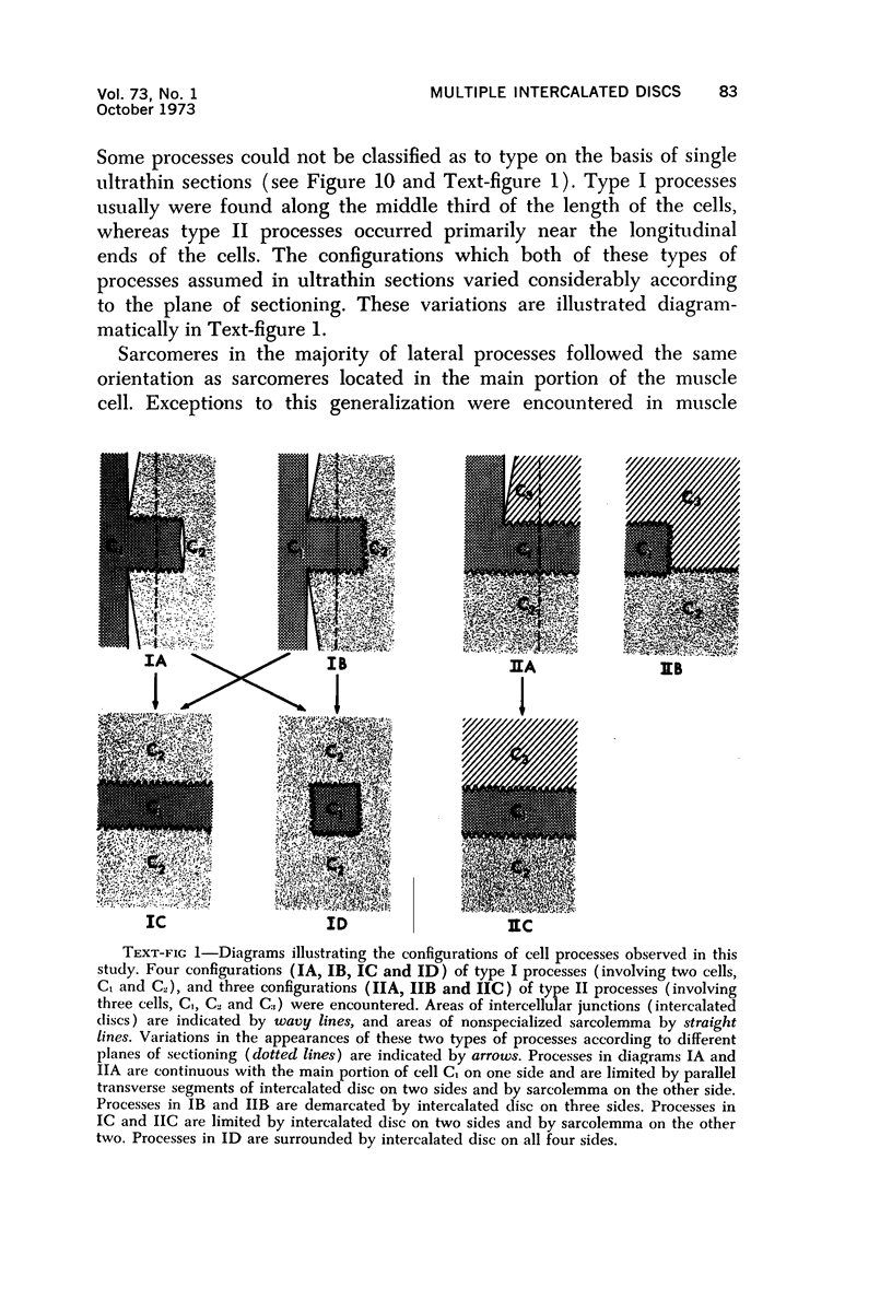
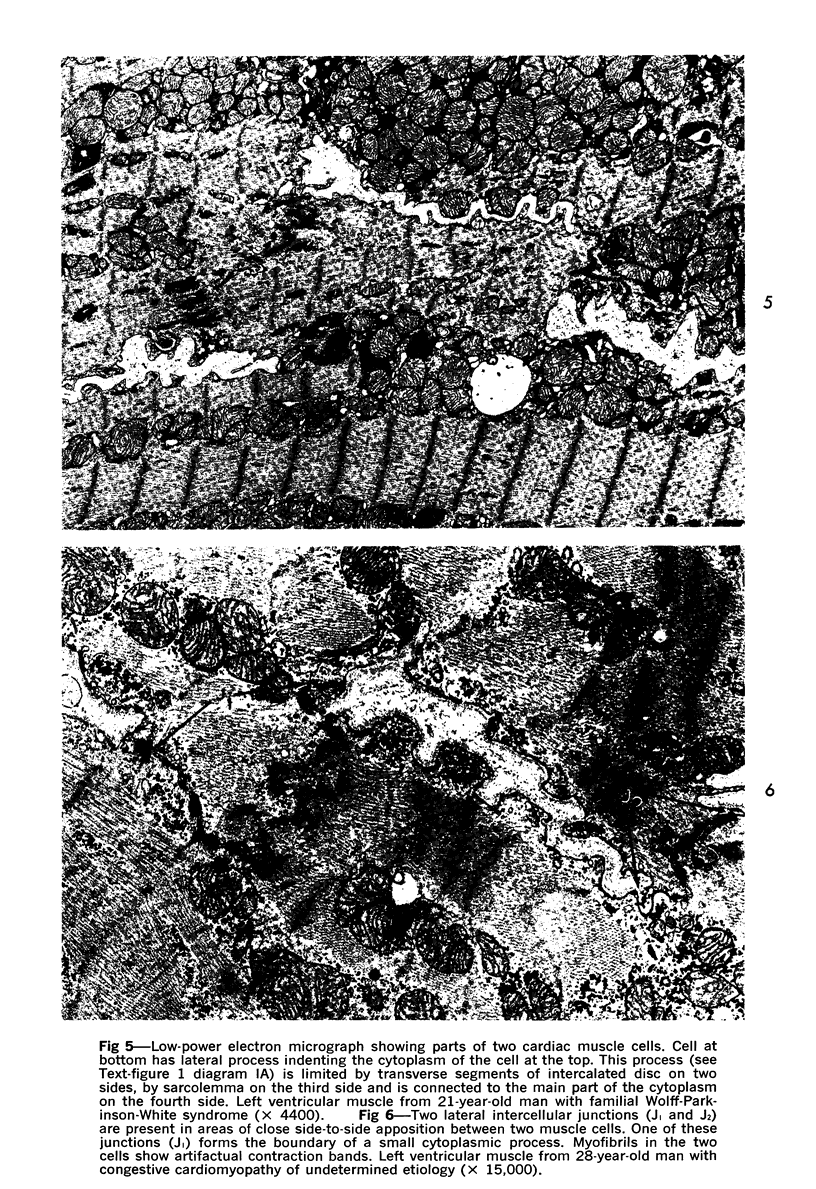
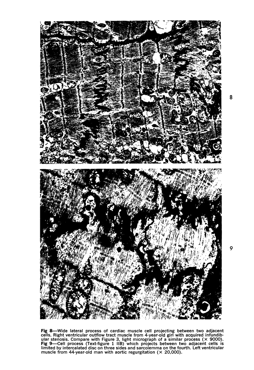
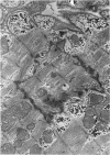
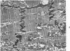
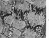
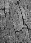
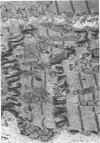
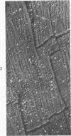
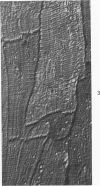
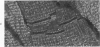
Multiple intercalated discs were frequently observed in muscle cells from patients with cardiac hypertrophy of various causes. Evidence is presented that these structures are the intercellular junctions of lateral processes of muscle cells. The sizes and shapes of these processes showed marked variations, depending in part on the plane of sectioning. It is postulated that lateral processes develop in certain cells which have side-to-side intercellular junctions with adjacent cells, and that localized mechanical tensions induce their growth. These tensions are due to shearing forces exerted at side-to-side junctions when contraction occurs at different rates or magnitudes in adjacent cells. These forces lead to asymmetric and complementary growth of sarcomeres along the two sides of the lateral junctions, to reorientation of these junctions and to the eventual formation of lateral processes.
Full text
PDF


Images in this article
Selected References
These references are in PubMed. This may not be the complete list of references from this article.
- FREEMAN J. A., SPURLOCK B. O. A new epoxy embedment for electron microscopy. J Cell Biol. 1962 Jun;13:437–443. doi: 10.1083/jcb.13.3.437. [DOI] [PMC free article] [PubMed] [Google Scholar]
- Fawcett D. W., McNutt N. S. The ultrastructure of the cat myocardium. I. Ventricular papillary muscle. J Cell Biol. 1969 Jul;42(1):1–45. doi: 10.1083/jcb.42.1.1. [DOI] [PMC free article] [PubMed] [Google Scholar]
- Ferrans V. J., Morrow A. G., Roberts W. C. Myocardial ultrastructure in idiopathic hypertrophic subaortic stenosis. A study of operatively excised left ventricular outflow tract muscle in 14 patients. Circulation. 1972 Apr;45(4):769–792. doi: 10.1161/01.cir.45.4.769. [DOI] [PubMed] [Google Scholar]
- Hibbs R. G., Ferrans V. J. An ultrastructural and histochemical study of rat atrial myocardium. Am J Anat. 1969 Mar;124(3):251–270. doi: 10.1002/aja.1001240302. [DOI] [PubMed] [Google Scholar]
- Kawamura K., James T. N. Comparative ultrastructure of cellular junctions in working myocardium and the conduction system under normal and pathologic conditions. J Mol Cell Cardiol. 1971 Sep;3(1):31–60. doi: 10.1016/0022-2828(71)90031-9. [DOI] [PubMed] [Google Scholar]
- Laks M. M., Morady F., Adomian G. E., Swan H. J. Presence of widened and multiple intercalated discs in the hypertrophied canine heart. Circ Res. 1970 Sep;27(3):391–402. doi: 10.1161/01.res.27.3.391. [DOI] [PubMed] [Google Scholar]
- Legato M. J. Sarcomerogenesis in human myocardium. J Mol Cell Cardiol. 1970 Dec;1(4):425–437. doi: 10.1016/0022-2828(70)90039-8. [DOI] [PubMed] [Google Scholar]
- Manasek F. J. Histogenesis of the embryonic myocardium. Am J Cardiol. 1970 Feb;25(2):149–168. doi: 10.1016/0002-9149(70)90576-x. [DOI] [PubMed] [Google Scholar]
- McNutt N. S., Fawcett D. W. The ultrastructure of the cat myocardium. II. Atrial muscle. J Cell Biol. 1969 Jul;42(1):46–67. doi: 10.1083/jcb.42.1.46. [DOI] [PMC free article] [PubMed] [Google Scholar]
- REYNOLDS E. S. The use of lead citrate at high pH as an electron-opaque stain in electron microscopy. J Cell Biol. 1963 Apr;17:208–212. doi: 10.1083/jcb.17.1.208. [DOI] [PMC free article] [PubMed] [Google Scholar]