

Full text
PDF

Images in this article
Selected References
These references are in PubMed. This may not be the complete list of references from this article.
- CASLEY-SMITH J. R. The identification of chylomicra and lipoproteins in tissue sections and their passage into jejunal lacteals. J Cell Biol. 1962 Nov;15:259–277. doi: 10.1083/jcb.15.2.259. [DOI] [PMC free article] [PubMed] [Google Scholar]
- CAULFIELD J. B. Effects of varying the vehicle for OsO4 in tissue fixation. J Biophys Biochem Cytol. 1957 Sep 25;3(5):827–830. doi: 10.1083/jcb.3.5.827. [DOI] [PMC free article] [PubMed] [Google Scholar]
- COHEN A. S., WEISS L., CALKINS E. Electron microscopic observations of the spleen during the induction of experimental amyloidosis in the rabbit. Am J Pathol. 1960 Oct;37:413–431. [PMC free article] [PubMed] [Google Scholar]
- DAVISON P. F., TAYLOR E. W. Physical-chemical studies of proteins of squid nerve axoplasm, with special reference to the axon fibrous protein. J Gen Physiol. 1960 Mar;43:801–823. doi: 10.1085/jgp.43.4.801. [DOI] [PMC free article] [PubMed] [Google Scholar]
- GONATAS N. K., TERRY R. D., WINKLER R., KOREY S. R., GOMEZ C. J., STEIN A. A CASE OF JUVENILE LIPIDOSIS: THE SIGNIFICANCE OF ELECTRON MICROSCOPIC AND BIOCHEMICAL OBSERVATIONS OF A CEREBRAL BIOPSY. J Neuropathol Exp Neurol. 1963 Oct;22:557–580. [PubMed] [Google Scholar]
- HEEFNER W. A., SORENSON G. D. Experimental amyloidosis. I. Light and electron microscopic observation of spleen and lymph nodes. Lab Invest. 1962 Aug;11:585–593. [PubMed] [Google Scholar]
- HUXLEY H. E., ZUBAY G. Preferential staining of nucleic acid-containing structures for electron microscopy. J Biophys Biochem Cytol. 1961 Nov;11:273–296. doi: 10.1083/jcb.11.2.273. [DOI] [PMC free article] [PubMed] [Google Scholar]
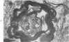
- KIDD M. Paired helical filaments in electron microscopy of Alzheimer's disease. Nature. 1963 Jan 12;197:192–193. doi: 10.1038/197192b0. [DOI] [PubMed] [Google Scholar]
- KOREY S. R., GOMEZ C. J., STEIN A., GONATAS J., SUZUKI K., TERRY R. D., WEISS M. Studies in Tay-Sachs disease. I. A. methods. J Neuropathol Exp Neurol. 1963 Jan;22:2–9. [PubMed] [Google Scholar]
- LUFT J. H. Improvements in epoxy resin embedding methods. J Biophys Biochem Cytol. 1961 Feb;9:409–414. doi: 10.1083/jcb.9.2.409. [DOI] [PMC free article] [PubMed] [Google Scholar]
- MAJNO G., PALADE G. E. Studies on inflammation. 1. The effect of histamine and serotonin on vascular permeability: an electron microscopic study. J Biophys Biochem Cytol. 1961 Dec;11:571–605. doi: 10.1083/jcb.11.3.571. [DOI] [PMC free article] [PubMed] [Google Scholar]
- MARGOLIS G. Senile cerebral disease: a critical survey of traditional concepts based upon observations with newer technics. Lab Invest. 1959 Mar-Apr;8(2):335–370. [PubMed] [Google Scholar]
- MILLONIG G. A modified procedure for lead staining of thin sections. J Biophys Biochem Cytol. 1961 Dec;11:736–739. doi: 10.1083/jcb.11.3.736. [DOI] [PMC free article] [PubMed] [Google Scholar]
- MOVAT H. Z., FERNANDO N. V. Allergic inflammation. I. The earliest fine structural changes at the blood-tissue barrier during antigen-antibody interaction. Am J Pathol. 1963 Jan;42:41–59. [PMC free article] [PubMed] [Google Scholar]
- NOVIKOFF A. B., GOLDFISCHER S. Nucleosidediphosphatase activity in the Golgi apparatus and its usefulness for cytological studies. Proc Natl Acad Sci U S A. 1961 Jun 15;47:802–810. doi: 10.1073/pnas.47.6.802. [DOI] [PMC free article] [PubMed] [Google Scholar]
- PALADE G. E. A study of fixation for electron microscopy. J Exp Med. 1952 Mar;95(3):285–298. doi: 10.1084/jem.95.3.285. [DOI] [PMC free article] [PubMed] [Google Scholar]
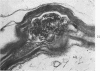
- TERRY R. D. THE FINE STRUCTURE OF NEUROFIBRILLARY TANGLES IN ALZHEIMER'S DISEASE. J Neuropathol Exp Neurol. 1963 Oct;22:629–642. doi: 10.1097/00005072-196310000-00005. [DOI] [PubMed] [Google Scholar]
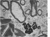
- TERRY R. D., WEISS M. Studies in Tay-Sachs disease. II. Ultrastructure of the cerebrum. J Neuropathol Exp Neurol. 1963 Jan;22:18–55. doi: 10.1097/00005072-196301000-00003. [DOI] [PubMed] [Google Scholar]
- TRUMP B. F., BENDITT E. P. Electron microscopic studies of human renal disease. Observations of normal visceral glomerular epithelium and its modification in disease. Lab Invest. 1962 Sep;11:753–781. [PubMed] [Google Scholar]
- VIAL J. D. The early changes in the axoplasm during wallerian degeneration. J Biophys Biochem Cytol. 1958 Sep 25;4(5):551–555. doi: 10.1083/jcb.4.5.551. [DOI] [PMC free article] [PubMed] [Google Scholar]
- WEBSTER H. D., SPIRO D., WAKSMAN B., ADAMS R. D. Phase and electron microscopic studies of experimental demyelination. II. Schwann cell changes in guinea pig sciatic nerves during experimental diphtheritic neuritis. J Neuropathol Exp Neurol. 1961 Jan;20:5–34. doi: 10.1097/00005072-196101000-00002. [DOI] [PubMed] [Google Scholar]
- WEBSTER H. D. Transient, focal accumulation of axonal mitochondria during the early stages of wallerian degeneration. J Cell Biol. 1962 Feb;12:361–383. doi: 10.1083/jcb.12.2.361. [DOI] [PMC free article] [PubMed] [Google Scholar]