

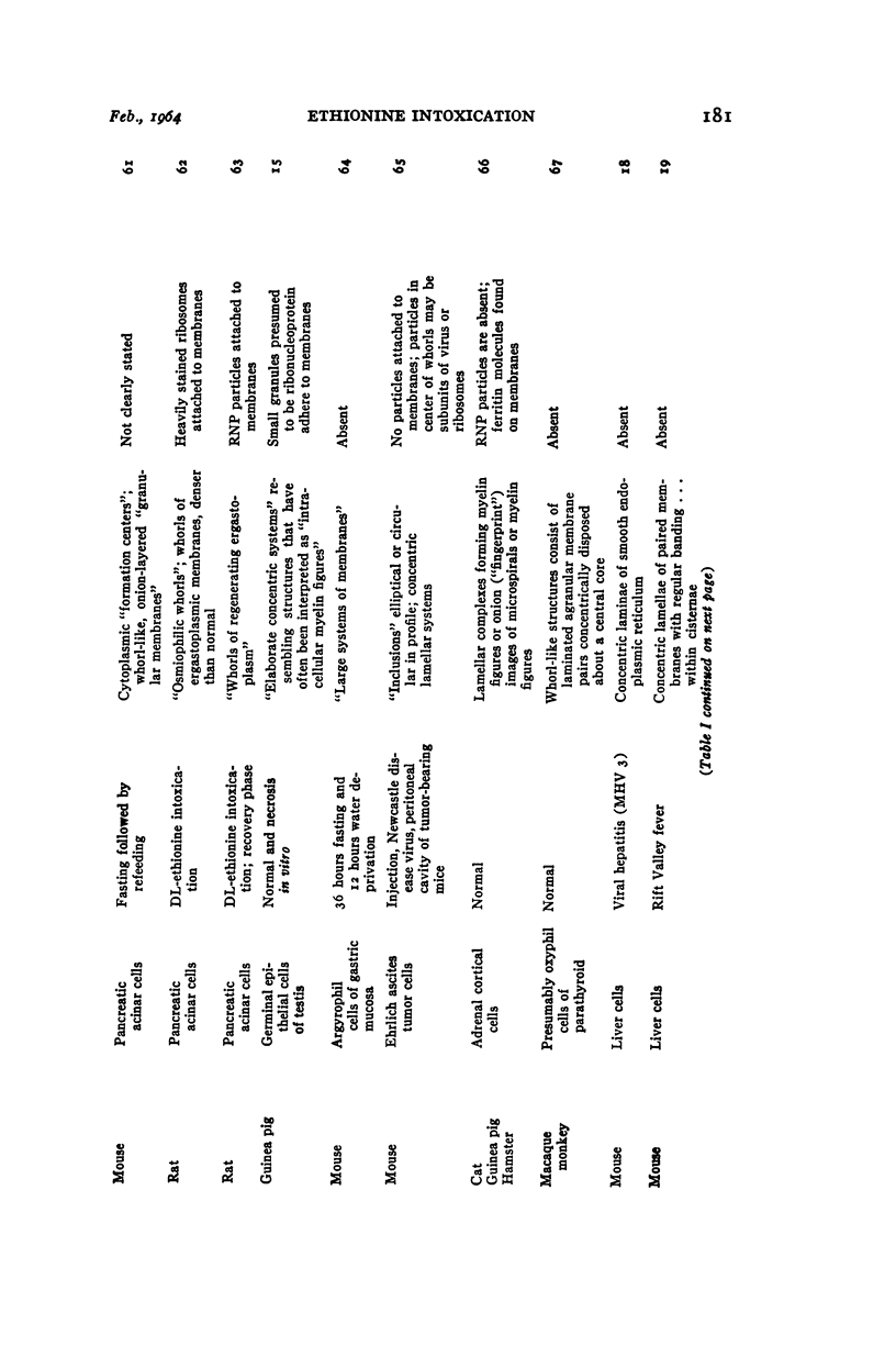
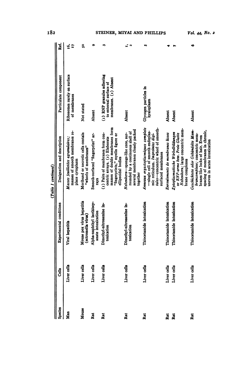
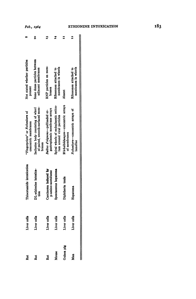
Full text
PDF
Images in this article
Selected References
These references are in PubMed. This may not be the complete list of references from this article.
- ADAMS W. R., PRINCE A. F. Cellular changes associated with infection of the Ehrlich ascites tumor with Newcastle disease virus. Ann N Y Acad Sci. 1959 Jul 21;81:89–100. doi: 10.1111/j.1749-6632.1959.tb49298.x. [DOI] [PubMed] [Google Scholar]
- ARHELGER R. B., DARLINGTON R. W., RANDALL C. C. An electron microscopic study of equine abortion virus infection in hamster liver. Am J Pathol. 1963 Jun;42:703–713. [PMC free article] [PubMed] [Google Scholar]
- BEARCROFT W. G. Electron-microscope studies on the liver in infective hepatitis. J Pathol Bacteriol. 1962 Apr;83:383–388. doi: 10.1002/path.1700830207. [DOI] [PubMed] [Google Scholar]
- BEARCROFT W. G. Electron-microscope studies on the livers of yellow-fever-infected African monkeys. J Pathol Bacteriol. 1962 Jan;83:59–64. doi: 10.1002/path.1700830108. [DOI] [PubMed] [Google Scholar]
- BRAUNSTEINER H., FELLINGER K., PAKESCH F., NEUMAYR A. Weitere elektronenmikroskopische Beobachtungen bei Virushepatitiden. Klin Wochenschr. 1958 Apr 15;36(8):379–382. doi: 10.1007/BF01477091. [DOI] [PubMed] [Google Scholar]
- BRUNI C. Hyaline degeneration of rat liver cells studied with the electron microscope. Lab Invest. 1960 Mar-Apr;9:209–215. [PubMed] [Google Scholar]
- COSSEL L. [Electron microscopic studies on the liver sinusoids in viral hepatitis]. Klin Wochenschr. 1959 Dec 15;37:1263–1278. doi: 10.1007/BF01488544. [DOI] [PubMed] [Google Scholar]
- COTTE G. Some problems posed by the ultrastructure of lipids of the adrenal cortex. J Ultrastruct Res. 1959 Dec;3:186–209. doi: 10.1016/s0022-5320(59)90014-0. [DOI] [PubMed] [Google Scholar]
- DROCHMANS P. [Demonstration of glycogen in liver cells by electron microscopy]. J Biophys Biochem Cytol. 1960 Oct;8:553–558. [PMC free article] [PubMed] [Google Scholar]
- EMMELOT P., BENEDETTI E. L. Changes in the fine structure of rat liver cells brought about by dimethylnitrosamine. J Biophys Biochem Cytol. 1960 Apr;7:393–396. doi: 10.1083/jcb.7.2.393. [DOI] [PMC free article] [PubMed] [Google Scholar]
- EPSTEIN M. A. The fine structural organisation of Rous tumour cells. J Biophys Biochem Cytol. 1957 Nov 25;3(6):851–858. doi: 10.1083/jcb.3.6.851. [DOI] [PMC free article] [PubMed] [Google Scholar]
- EPSTEIN M. A. The fine structure of the cells in mouse sarcoma 37 ascitic fluids. J Biophys Biochem Cytol. 1957 Jul 25;3(4):567–576. doi: 10.1083/jcb.3.4.567. [DOI] [PMC free article] [PubMed] [Google Scholar]
- FAVARD P. L'origine ergastoplasmique des granules protéiques dans les spermatocytes d'Ascaris. C R Hebd Seances Acad Sci. 1958 Jul 28;247(4):531–533. [PubMed] [Google Scholar]
- FAWCETT D. W. Observations on the cytology and electron microscopy of hepatic cells. J Natl Cancer Inst. 1955 Apr;15(5 Suppl):1475–1503. [PubMed] [Google Scholar]
- GUEFT B. Viral hepatitis under the electron microscope. Arch Pathol. 1961 Jul;72:61–69. [PubMed] [Google Scholar]
- HELANDER H. F. A preliminary note on the ultrastructure of the argyrophile cells of the mouse gastric mucosa. J Ultrastruct Res. 1961 Jun;5:257–262. doi: 10.1016/s0022-5320(61)90019-3. [DOI] [PubMed] [Google Scholar]
- HERMAN L., FITZGERALD P. J. Restitution of pancreatic acinar cells following ethionine. J Cell Biol. 1962 Feb;12:297–312. doi: 10.1083/jcb.12.2.297. [DOI] [PMC free article] [PubMed] [Google Scholar]
- HERMAN L., FITZGERALD P. J. The degenerative changes in pancreatic acinar cells caused by DL-ethionine. J Cell Biol. 1962 Feb;12:277–296. doi: 10.1083/jcb.12.2.277. [DOI] [PMC free article] [PubMed] [Google Scholar]
- KARRER H. E. Electron microscope observations on chick embryo liver. Glycogen, bile canaliculi, inclusion bodies and hematopoiesis. J Ultrastruct Res. 1961 Apr;5:116–141. doi: 10.1016/s0022-5320(61)90009-0. [DOI] [PubMed] [Google Scholar]
- KARRER H. E. Electron-microscopic study of glycogen in chick embryo liver. J Ultrastruct Res. 1960 Nov;4:191–212. doi: 10.1016/s0022-5320(60)90053-8. [DOI] [PubMed] [Google Scholar]
- LEDUC E. H., BERNHARD W. Electron microscope study of mouse liver infected by ectromelia virus. J Ultrastruct Res. 1962 Jun;6:466–488. doi: 10.1016/s0022-5320(62)80003-3. [DOI] [PubMed] [Google Scholar]
- PORTER K. R., BRUNI C. An electron microscope study of the early effects of 3'-Me-DAB on rat liver cells. Cancer Res. 1959 Nov;19:997–1009. [PubMed] [Google Scholar]
- ROUILLER C. Contribution de la microscopie électronique à l'étude du foie normal et pathologique. Ann Anat Pathol (Paris) 1957 Oct-Dec;2(4):548–562. [PubMed] [Google Scholar]
- RUTHMANN A. Basophilic lamellar systems in the crayfish spermatocyte. J Biophys Biochem Cytol. 1958 May 25;4(3):267–274. doi: 10.1083/jcb.4.3.267. [DOI] [PMC free article] [PubMed] [Google Scholar]
- SALOMON J. C. [Changes in liver parenchymal cells in the rat subjected to the effects of thioacetamide. Electron microscope study of lesions observed during the late phase of chronic poisoning]. J Ultrastruct Res. 1962 Oct;7:293–307. doi: 10.1016/s0022-5320(62)90025-4. [DOI] [PubMed] [Google Scholar]
- SVOBODA D., NIELSON A., WERBER A., HIGGINSON J. An electron microscopic study of viral hepatitis in mice. Am J Pathol. 1962 Aug;41:205–224. [PMC free article] [PubMed] [Google Scholar]
- THEMANN H. [On the electron microscope demonstration of glycogen and the relations of the cell organelles in glycogen synthesis and glycogenolysis]. Verh Dtsch Ges Pathol. 1961;45:291–297. [PubMed] [Google Scholar]
- THOENES W., BANNASCH P. [Electron and light microscopic studies on the cytoplasm of liver cells after acute and chronic thioacetamide poisoning]. Virchows Arch Pathol Anat Physiol Klin Med. 1962;335:556–583. [PubMed] [Google Scholar]
- THOENES W. [On information on the smooth endoplasmatic reticulum of the liver cells]. Verh Dtsch Ges Pathol. 1962;46:202–206. [PubMed] [Google Scholar]
- TRIER J. S. The fine structure of the parathyroid gland. J Biophys Biochem Cytol. 1958 Jan 25;4(1):13–22. doi: 10.1083/jcb.4.1.13. [DOI] [PMC free article] [PubMed] [Google Scholar]
- WATSON M. L. Staining of tissue sections for electron microscopy with heavy metals. II. Application of solutions containing lead and barium. J Biophys Biochem Cytol. 1958 Nov 25;4(6):727–730. doi: 10.1083/jcb.4.6.727. [DOI] [PMC free article] [PubMed] [Google Scholar]
- WEISS J. M. The ergastoplasm; its fine structure and relation to protein synthesis as studied with the electron microscope in the pancreas of the Swiss albino mouse. J Exp Med. 1953 Dec;98(6):607–618. doi: 10.1084/jem.98.6.607. [DOI] [PMC free article] [PubMed] [Google Scholar]