

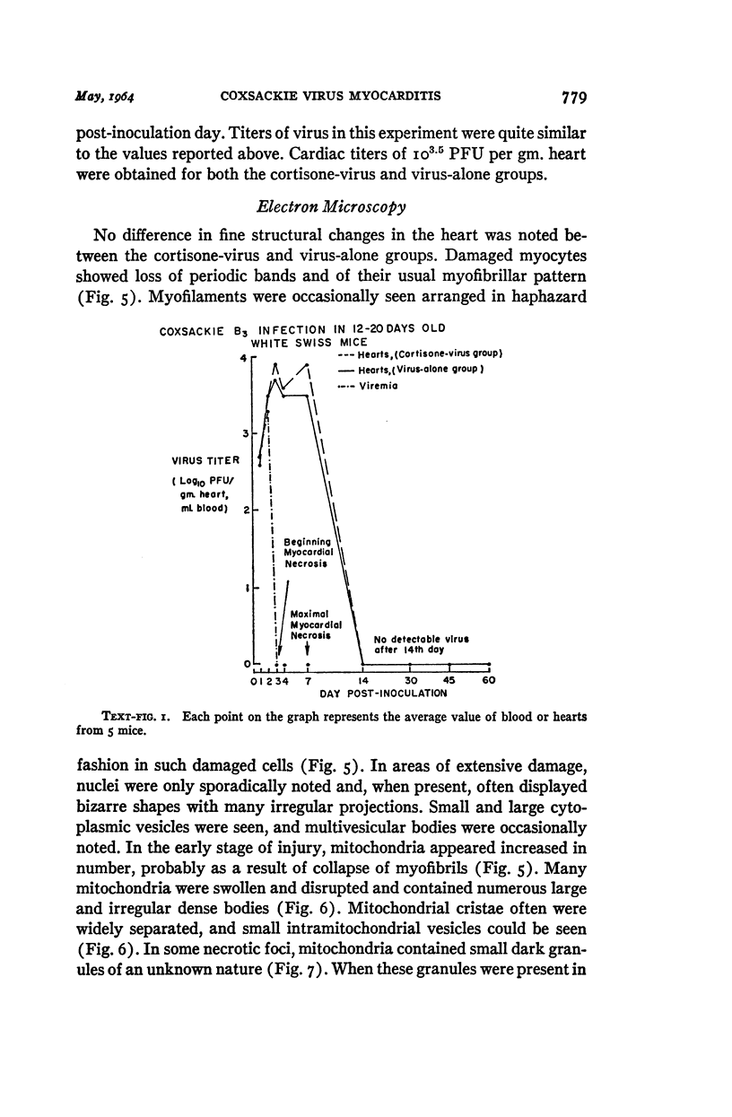
Full text
PDF
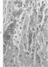
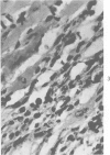
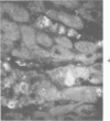
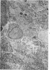
Images in this article
Selected References
These references are in PubMed. This may not be the complete list of references from this article.
- BABB J. M., STONEMAN M. E., STERN H. Myocarditis and croup caused by Coxsackie virus type B5. Arch Dis Child. 1961 Oct;36:551–556. doi: 10.1136/adc.36.189.551. [DOI] [PMC free article] [PubMed] [Google Scholar]
- BRYANT R. E., THOMAS W. A., O'NEAL R. M. An electron microscopic study of myocardial ischemia in the rat. Circ Res. 1958 Nov;6(6):699–709. doi: 10.1161/01.res.6.6.699. [DOI] [PubMed] [Google Scholar]
- COONS A. H., KAPLAN M. H. Localization of antigen in tissue cells; improvements in a method for the detection of antigen by means of fluorescent antibody. J Exp Med. 1950 Jan 1;91(1):1–13. doi: 10.1084/jem.91.1.1. [DOI] [PMC free article] [PubMed] [Google Scholar]
- FECHNER R. E., SMITH M. G., MIDDLEKAMP J. N. Coxsackie B virus infection of the newborn. Am J Pathol. 1963 Apr;42:493–505. [PMC free article] [PubMed] [Google Scholar]
- FOGH J. Filamentous organization of poliovirus particles. Virology. 1961 Aug;14:495–497. doi: 10.1016/0042-6822(61)90346-4. [DOI] [PubMed] [Google Scholar]
- GEORGIEV G. P., CHENTSOV J. S. On the structural organization of nucleolo-chromosomal ribonucleoproteins. Exp Cell Res. 1962 Sep;27:570–572. doi: 10.1016/0014-4827(62)90020-4. [DOI] [PubMed] [Google Scholar]
- GIFFORD R., DALLDORF G. The morbid anatomy of experimental Coxsackie virus infection. Am J Pathol. 1951 Nov-Dec;27(6):1047–1063. [PMC free article] [PubMed] [Google Scholar]
- GRIFFIN C. W., CARSKI T. R., WARNER G. S. Labeling procedures employing crystalline fluorescein isothiocyanate. J Bacteriol. 1961 Oct;82:534–537. doi: 10.1128/jb.82.4.534-537.1961. [DOI] [PMC free article] [PubMed] [Google Scholar]
- GRODUMS E. I., DEMPSTER G. Myocarditis in experimental Coxsackie B-3 infection. Can J Microbiol. 1959 Dec;5:605–615. doi: 10.1139/m59-074. [DOI] [PubMed] [Google Scholar]
- GRODUMS E. I., DEMPSTER G. The age factor in experimental Coxsackie B-3 infection. Can J Microbiol. 1959 Dec;5:595–604. doi: 10.1139/m59-073. [DOI] [PubMed] [Google Scholar]
- HOWES D. W., MELNICK J. L., REISSIG M. Sequence of morphological changes in epithelial cell cultures infected with poliovirus. J Exp Med. 1956 Sep 1;104(3):289–304. doi: 10.1084/jem.104.3.289. [DOI] [PMC free article] [PubMed] [Google Scholar]
- KILBOURNE E. D., WILSON C. B., PERRIER D. The induction of gross myocardial lesions by a Coxsackie (pleurodynia) virus and cortisone. J Clin Invest. 1956 Apr;35(4):362–370. doi: 10.1172/JCI103286. [DOI] [PMC free article] [PubMed] [Google Scholar]
- LERNER A. M., LEVIN H. S., FINLAND M. Age and susceptibility of mice to Coxsackie A viruses. J Exp Med. 1962 Apr 1;115:745–762. doi: 10.1084/jem.115.4.745. [DOI] [PMC free article] [PubMed] [Google Scholar]
- LOU T. Y., WENNER H. A., KAMITSUKA P. S. Experimental infections with Coxackie viruses. II. Myocarditis in Cynomolgus monkeys infected with B 4 virus. Arch Gesamte Virusforsch. 1961;10:451–464. [PubMed] [Google Scholar]
- LOVE R. Cytopathology of virus-infected tumor cells. Ann N Y Acad Sci. 1959 Jul 21;81:101–117. doi: 10.1111/j.1749-6632.1959.tb49299.x. [DOI] [PubMed] [Google Scholar]
- MATTERN C. F., CHI L. L. Studies on the sites and kinetics of Coxsackie A9 virus multiplication in the monkey kidney cell. Virology. 1962 Oct;18:257–265. doi: 10.1016/0042-6822(62)90012-0. [DOI] [PubMed] [Google Scholar]
- MATTERN C. F. Some physical and chemical properties of Coxsackie viruses A9 and A10. Virology. 1962 Aug;17:520–532. doi: 10.1016/0042-6822(62)90151-4. [DOI] [PubMed] [Google Scholar]
- MAYOR H. D., JORDAN L. E. Formation of poliovirus in monkey kidney tissue culture cells. Virology. 1962 Mar;16:325–333. doi: 10.1016/0042-6822(62)90254-4. [DOI] [PubMed] [Google Scholar]
- MELNICK J. L., GODMAN G. C. Pathogenesis of coxsackie virus infection; multiplication of virus and evolution of the muscle lesion in mice. J Exp Med. 1951 Mar;93(3):247–266. doi: 10.1084/jem.93.3.247. [DOI] [PMC free article] [PubMed] [Google Scholar]
- MOLNAR Z., LARSEN K., SPARGO B. Cardiac changes in the potassium-depleted rat. Arch Pathol. 1962 Oct;74:339–347. [PubMed] [Google Scholar]
- MORGAN C., HOWE C., ROSE H. M. Intracellular crystals of Coxsackie virus viewed in the electron microscope. Virology. 1959 Sep;9:145–149. doi: 10.1016/0042-6822(59)90110-2. [DOI] [PubMed] [Google Scholar]
- REVEL J. P., NAPOLITANO L., FAWCETT D. W. Identification of glycogen in electron micrographs of thin tissue sections. J Biophys Biochem Cytol. 1960 Dec;8:575–589. doi: 10.1083/jcb.8.3.575. [DOI] [PMC free article] [PubMed] [Google Scholar]
- RIFKIND R. A., GODMAN G. C., HOWE C., MORGAN C., ROSE H. M. Structure and development of viruses as observed in the electron microscope. VI. ECHO virus, type 9. J Exp Med. 1961 Jul 1;114:1–12. doi: 10.1084/jem.114.1.1. [DOI] [PMC free article] [PubMed] [Google Scholar]
- WATSON M. L. Further observations on the nuclear envelope of the animal cell. J Biophys Biochem Cytol. 1959 Oct;6:147–156. doi: 10.1083/jcb.6.2.147. [DOI] [PMC free article] [PubMed] [Google Scholar]
- Warner J. R., Rich A., Hall C. E. Electron Microscope Studies of Ribosomal Clusters Synthesizing Hemoglobin. Science. 1962 Dec 28;138(3548):1399–1403. doi: 10.1126/science.138.3548.1399. [DOI] [PubMed] [Google Scholar]