Abstract
Escherichia coli serogroup O26 consists of enterohemorrhagic E. coli (EHEC) and atypical enteropathogenic E. coli (aEPEC). The former produces Shiga toxins (Stx), major determinants of EHEC pathogenicity, encoded by bacteriophages; the latter is Stx negative. We have isolated EHEC O26 from patient stools early in illness and aEPEC O26 from stools later in illness, and vice versa. Intrapatient EHEC and aEPEC isolates had quite similar pulsed-field gel electrophoresis (PFGE) patterns, suggesting that they might have arisen by conversion between the EHEC and aEPEC pathotypes during infection. To test this hypothesis, we asked whether EHEC O26 can lose stx genes and whether aEPEC O26 can be lysogenized with Stx-encoding phages from EHEC O26 in vitro. The stx2 loss associated with the loss of Stx2-encoding phages occurred in 10% to 14% of colonies tested. Conversely, Stx2- and, to a lesser extent, Stx1-encoding bacteriophages from EHEC O26 lysogenized aEPEC O26 isolates, converting them to EHEC strains. In the lysogens and EHEC O26 donors, Stx2-converting bacteriophages integrated in yecE or wrbA. The loss and gain of Stx-converting bacteriophages diversifies PFGE patterns; this parallels findings of similar but not identical PFGE patterns in the intrapatient EHEC and aEPEC O26 isolates. EHEC O26 and aEPEC O26 thus exist as a dynamic system whose members undergo ephemeral interconversions via loss and gain of Stx-encoding phages to yield different pathotypes. The suggested occurrence of this process in the human intestine has diagnostic, clinical, epidemiological, and evolutionary implications.
Escherichia coli serogroup O26 has members classified as enterohemorrhagic E. coli (EHEC) or atypical enteropathogenic E. coli (aEPEC). EHEC O26 strains constitute the most common non-O157 EHEC group associated with diarrhea and hemolytic uremic syndrome (HUS) in Europe (16, 18, 19, 25, 48, 51). EHEC O26 is also the most common non-O157 EHEC serogroup in the United States, where, between 1983 and 2002, it accounted for 22% of non-O157 EHEC clinical isolates (10). In a recent prospective study from Montana, half of EHEC O26 isolates originated from patients with bloody diarrhea (23). Moreover, EHEC O26 has spread globally (24).
EHEC O26 strains produce Shiga toxin 1 (Stx1) and Stx2, either singly or together (10, 54). Indeed, phage H19B from a clinical EHEC O26 isolate that carries stx1 was one of the first Stx-converting phages described (45). Moreover, these strains contain the intimin-encoding eae gene (6, 54), an important characteristic of EHEC (33). EHEC O26 represents a highly dynamic group of organisms that rapidly engender new pathogenic clones (54). This is exemplified by emergence of a novel EHEC O26:H11 clonal subgroup in Germany in the 1990s that possessed stx2 as the sole stx gene, in contrast to stx1, exclusively identified in EHEC O26 previously. The pathogenicity of this clone was demonstrated by its strong association with HUS (29, 54) and its ability to spread rapidly (2, 54).
aEPEC O26 strains do not harbor stx genes (9, 20, 42) but share with EHEC the eae gene (20, 34) and the ability to produce attaching and effacing lesions in intestinal epithelial cells via actin rearrangement (9, 20, 42). Unlike typical EPEC strains (49), aEPEC O26 strains lack the EPEC adherence factor plasmid (6) encoding bundle-forming pili that mediate localized adherence on cultured epithelial cells. The absence of the EPEC adherence factor plasmid is a common feature of aEPEC strains, which cause gastroenteritis in children (12, 20, 49).
It has been hypothesized (15) that aEPEC O26 is ancestral to EHEC O26. According to this hypothesis, the acquisition of stx1 by aEPEC O26 gave rise to globally distributed toxigenic EHEC O26 (15). Furthermore, replacement of stx1 with stx2 has been postulated as the cause of the recent emergence of the new stx2-harboring EHEC O26 clonal subgroup in Europe (15, 54). A prerequisite for such an evolutionary process is that aEPEC O26 strains undergo lysogeny by Stx-encoding bacteriophages. However, this has not yet been systematically investigated. Moreover, it is not clear if the sequence of events proposed for the evolution of EHEC O26 is unidirectional, where aEPEC O26 strains are always progenitors of EHEC O26 strains, or bidirectional, with EHEC O26 also being converted to aEPEC by loss of an stx gene. Therefore, we investigated the role of Stx-encoding bacteriophages in the postulated transition between EHEC and aEPEC O26 to determine if (i) Stx-encoding phages originating from EHEC O26 lysogenize aEPEC O26 under laboratory conditions, (ii) stx genes and their encoding phages are lost by EHEC O26 in vitro, (iii) the loss and gain of Stx-encoding phages influence the genomic architecture, (iv) there is an identifiable site where these bacteriophages integrate into the genomes of EHEC O26 and the lysogens, and (v) there is bidirectional conversion between EHEC O26 and aEPEC O26 during human infection.
MATERIALS AND METHODS
Bacterial strains.
Three EHEC (stx2-positive, eae-positive, bfpA-negative) and three aEPEC (stx-negative, eae-positive, bfpA-negative) O26:H11 strains were isolated from initial stools (collected 5 days after the onset of diarrhea) and follow-up stools (collected 9 days after the initial samples), respectively, of three children (13, 16, and 17 months old) during an outbreak of HUS in Germany (29). The other EHEC and aEPEC O26:H11 strains used in transduction experiments were isolated from patients between 1971 and 1999; they were epidemiologically unrelated except for EHEC strain 46 and aEPEC strain 47 (Table 1), the latter being a spontaneous stx2-negative laboratory derivative of the former. The donors and recipients of Stx-encoding phages were selected from our strain collection to contain strains with related as well as dissimilar pulsed-field gel electrophoresis (PFGE) patterns. E. coli strain C600(φH19B), which contains Stx1-converting phage H19B from a clinical EHEC O26:H11 isolate H19 (44), was described previously (45).
TABLE 1.
Ability of Stx-encoding phages from EHEC O26:H11 to lysogenize aEPEC O26:H11 and E. coli K-12 strain C600
| EHEC O26 phage donora
|
Result (frequency) of transduction of aEPEC O26 phage recipientb,c,f
|
Result (frequency) of transduction of E. coli C600 recipientf | |||||||
|---|---|---|---|---|---|---|---|---|---|
| Strain (phage)e | Diagnosis or statusc,d | stx gene | 10 (D) | 15 (D) | 22 (D) | 47 (LD) | 32 (D) | 40 (HUS) | |
| 54(φ54) | D | stx1 | — | — | — | — | — | — | C600(φ54) (1 × 10⁻⁶) |
| 49(φ49) | D | stx1 | — | — | — | — | — | — | — |
| 51(φ51) | HUS | stx1 | — | — | — | — | — | — | C600(φ51) (2 × 10⁻⁶) |
| C600(φH19B) (φH19B) | LS | stx1 | 10(φH19B) (1 × 10⁻⁷) | — | — | — | — | — | C600(φH19B) (8 × 10⁻⁵) |
| 46(φ46) | HUS | stx2 | — | — | — | 47(φ46) (1 × 10⁻⁶) | 32(φ46) (6 ×10⁻⁶) | — | C600(φ46) (3 × 10⁻⁵) |
| 61(φ61) | HUS | stx2 | 10(φ61) (3 × 10⁻⁷) | — | — | — | 32(φ61) (1 × 10⁻⁷) | — | C600(φ61) (7 × 10⁻⁶) |
| 50(φ50) | HUS | stx2 | — | 15(φ50) (1 × 10⁻⁷) | 22(φ50) (3 × 10⁻⁷) | — | — | 40(φ50) (6 × 10⁻⁷) | C600(φ50) (4 × 10⁻⁶) |
Six phage donors are clinical EHEC O26:H11 isolates from our laboratory; E. coli strain C600(φH19B) (45) contains stx1-harboring phage φH19B from a clinical EHEC O26 isolate H19 (44).
Five phage recipients (strains 10, 15, 22, 32, and 40) are clinical aEPEC O26 isolates, all stx negative; aEPEC strain 40 was isolated from a follow-up stool of a HUS patient (patient A) whose initial stool had EHEC strain 50; strain 47 is an stx2-negative laboratory derivative of EHEC strain 46 that fits the definition of aEPEC.
Data are results of transduction of the respective recipient with the respective phage; lysogen designations are given as the number of the recipient strain (number of the transducing phage). Data in parentheses are rates of transduction (number of lysogens per recipient cell). —, no lysogens were identified.
E. coli K-12 strain C600 was a positive control for phage transduction.
stx-harboring bacteriophages were designated by numbers of the donor strains.
D, diarrhea; HUS, hemolytic uremic syndrome; LD, laboratory derivative; LS, laboratory strain.
PCR techniques.
PCRs were performed in an iCycler (version 1.259; Bio-Rad, München, Germany) or a Biometra TGradient 96 cycler (Biometra GmbH, Göttingen, Germany) (46) using reagents from PEQLAB Biotechnologie (Erlangen, Germany) and primers synthesized by Sigma Genosys (Haverhill, United Kingdom). stx1, stx2, eae, and bfpA (encoding the structural subunit of bundle-forming pili) were detected using published protocols (6, 18). The chromosomal loci that serve as integration sites for Stx-encoding phages in E. coli O157 were interrogated using primer pairs A-B (yehV) (43), wrbA1-wrbA2 (wrbA) (47), EC10-EC11 (yecE) (14), and sbcB1-sbcB2 (sbcB) (47). The linkage between yecE and the integrase gene (int) of stx2-harboring bacteriophage φ258320, which integrates in yecE in E. coli O157 (7), was tested using primers Int-258320 and EC11 (7). The linkage between wrbA and the int gene of stx2-harboring bacteriophage φ933W, which integrates in wrbA in E. coli O157:H7 strain EDL933 (35, 37), was investigated using primers WrbA (5′-CGCCATCCACTTTGCTTG-3′) and Int933W (5′-TATGCTACCGAGGCTTGG-3′); the PCR consisted of 30 cycles of denaturing (94°C, 30 s), annealing (55°C, 1 min), and extension (72°C, 90 s) followed by a final extension (72°C, 5 min). The specificity of PCR products was confirmed by analyzing the sequence of representative amplicons as described below.
PFGE and Southern hybridization.
PFGE was performed using the PulseNet protocol (22) and with XbaI-digested DNA of Salmonella enterica serovar Braenderup strain H9812 (22) as a standard. Restriction patterns were analyzed with BioNumerics, version 4.0 (Applied Maths BVBA, Sint-Martens-Latem, Belgium). XbaI-digested, PFGE-separated genomic DNAs were hybridized with a digoxigenin-11-dUTP-labeled (DIG High Prime kit; Roche Molecular Biochemicals, Mannheim, Germany) stxA2 probe (7).
MLST.
Internal fragments of seven housekeeping genes (adk, fumC, gyrB, icd, mdh, purA, and recA) were analyzed using a published multilocus sequence typing (MLST) scheme for E. coli (52), except for a newly designed forward primer for icd (5′-CCGATTATCCCTTACATTGAAG-3′), which is 79 bp downstream of the original primer. Because of optimized proximity to the analyzed region of icd, the sequence trace quality was substantially higher without any ambiguous base callings. After purifying the PCR products, we sequenced both strands in 10 μl containing 0.5 μl premix (ABI Prism BigDye Terminator v3.1 Ready Reaction cycle sequencing kit; Applied Biosystems, Darmstadt, Germany), 1.8 μl 400 mmol/liter Tris-HCl, 10 mmol/liter MgCl2, 10 pmol sequencing primer, and 2 μl PCR product. Sequencing products were purified (Centri-Sep spin columns; Princeton Separations, Adelphia, NJ) and analyzed with the ABI Prism 3100 Avant genetic analyzer (Applied Biosystems) according to the manufacturer's instructions. The alleles and sequence types (ST) were assigned in accordance with the E. coli MLST website (http://web.mpiib-berlin.mpg.de/mlst/dbs/Ecoli).
Induction of Stx-encoding phages and transduction experiments.
Stx-encoding bacteriophages were induced using mitomycin C (Sigma-Aldrich, Deisenhofen, Germany) (41) from six wild-type EHEC O26 isolates that contained stx1 or stx2 and from strain C600(φH19B) (Table 1). To isolate stx-harboring phages, sterile filtrates of induced bacterial cultures were subjected to a plaque assay using E. coli C600 as an indicator (41); plaques were PCR screened for stx1 or stx2 using primer pair KS7-KS8 or LP43-LP44 (18), respectively. stx-harboring phages were propagated from single PCR-positive plaques (40). The resulting lysates contained the phages at titers between 2 × 10⁷ and 3.1 × 10⁸ PFU/ml, as determined by plaque assay (41). In transduction experiments, 104 PFU of each phage was mixed with 100 μl of log-phase culture (107 CFU) of each aEPEC O26 recipient or E. coli C600 and 125 μl of 0.1 M CaCl2 solution and incubated for 2 h at 37°C without shaking. The mixtures were then transferred into 4 ml of Luria-Bertani (LB) broth and incubated at 37°C and 180 rpm for 24 h. The cultures (100 μl) were then streaked on LB agar, and overnight bacterial growths that had been harvested into 1 ml of saline were PCR screened for stx1 or stx2. Tenfold dilutions of PCR-positive cultures were tested for lysogens using an Stx immunoblot assay (Shiga toxin [verocytotoxin] immunoblot; Sifin, Berlin, Germany). To identify stable lysogens, Stx-producing colonies were subcultured three times on LB agar, and the presence of stx genes was confirmed by PCR after the third passage.
Loss of stx in vitro.
A single colony of an stx2-positive EHEC O26 strain was suspended in 50 μl of sterile saline, and 2.5 μl was used to confirm the presence of stx2 by PCR. Another 5 μl was inoculated into 5 ml of Trypticase soy broth and incubated overnight at 37°C. Tenfold dilutions of the liquid culture were then inoculated onto sorbitol MacConkey agar, and after overnight incubation, 30 to 60 colonies from plates with 150 to 200 well-separated colonies were PCR screened for stx2. The frequency of stx2 loss was expressed as the percent stx2-negative colonies among the total number of colonies tested.
Stx production.
Stx1 and Stx2 production was determined using a commercial latex agglutination assay (verotoxin-producing E. coli reverse passive latex agglutination; Denka Seiken Co., Tokyo, Japan). Stx cytotoxicity titers were assessed by the Vero cell assay (26).
RESULTS
EHEC and aEPEC O26:H11 strains in consecutive stools collected from patients.
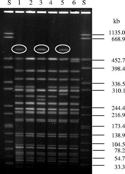
During an outbreak of HUS in Germany in 1999, stools from three infected children contained EHEC O26:H11 (stx2 positive, eae positive, bfpA negative) in their initial samples and aEPEC O26:H11 (stx negative, eae positive, bfpA negative) in follow-up samples. All six isolates belonged to ST 29 and had similar but not identical PFGE patterns (Fig. 1, lanes 1 to 6). Specifically, EHEC and aEPEC isolates from consecutive stools of individual patients differed by two to five bands; one of these variant bands was always a 550-kb XbaI fragment that contains stx2 in all EHEC isolates (Fig. 1, lanes 1, 3, and 5) but which is absent from all aEPEC isolates (Fig. 1, lanes 2, 4, and 6). The similarities in the PFGE patterns of the consecutive EHEC and aEPEC isolates from each patient, and the fact that one of the differences was the presence or absence of the genomic fragment containing stx2, suggested that aEPEC strains were derived from the EHEC strains by the loss of stx2 in these patients.
FIG. 1.
XbaI-digested genomic DNA from EHEC and aEPEC O26:H11 strains isolated from initial and follow-up stools, respectively, of three patients during a HUS outbreak. Lane 1, EHEC O26 (strain 50), patient A; lane 2, aEPEC O26 (strain 40), patient A; lane 3, EHEC O26 (strain 140), patient B; lane 4, aEPEC O26 (strain 41), patient B; lane 5, EHEC O26 (strain 141), patient C; lane 6, aEPEC O26 (strain 42), patient C; lanes S, molecular size standards (S. enterica serovar Braenderup strain H9812; Centers for Disease Control and Prevention, Atlanta, GA). XbaI fragments containing stx2 as demonstrated by hybridization with an stxA2 probe are circled.
Loss of stx2 in vitro.
To test this hypothesis, two EHEC O26 outbreak isolates (strain 50 from patient A and strain 140 from patient B) were tested for the stability of stx2 in vitro. Both lost stx2 in each of three independent experiments, with the frequency of the loss ranging from 10 to 14% of colonies tested on first subculture. The ease with which these EHEC strains lost stx2 in vitro makes it plausible that stx2 loss might have also occurred during infection in the human host, giving rise to the aEPEC O26 strains isolated from the follow-up stools.
Transduction of aEPEC O26 with Stx1- and Stx2-encoding phages from EHEC O26.
To determine if conversion between EHEC and aEPEC O26 is bidirectional, we investigated the ability of Stx-encoding phages from EHEC O26 to lysogenize aEPEC O26. High-titer phage lysates from three EHEC O26:H11 strains harboring stx2 only, three EHEC O26:H11 strains harboring stx1 only, and stx1-harboring E. coli strain C600(φH19B) were used to infect six aEPEC O26:H11 strains and E. coli C600. Stable lysogens were identified based on their ability to retain stx genes after three passages on LB agar (Table 1). Three of the four Stx1-encoding phages lysogenized E. coli C600, but only phage φH19B formed lysogens with one of the aEPEC O26 strains. In contrast, each of the three Stx2-encoding phages from EHEC O26 lysogenized, in addition to E. coli C600, at least two of the six aEPEC O26 recipients. Each of the aEPEC O26 recipients could be lysogenized with at least one of the Stx2-encoding phages (Table 1). These phages lysogenized aEPEC recipients with PFGE patterns related to those of the phage donors as well as aEPEC recipients with distant PFGE patterns. Notably, aEPEC strain 40, from the follow-up stool specimen of patient A, was lysogenized with an Stx2-encoding phage from EHEC strain 50, which was isolated from the initial stool of this patient [lysogen 40(φ50)] (Table 1). Similarly, aEPEC strain 47, an stx-negative laboratory derivative of EHEC strain 46, could be lysogenized with the Stx2-encoding phage originating in the parental EHEC O26 strain 46 [lysogen 47(φ46)] (Table 1). The rates of transduction of aEPEC strains with the three different stx2-harboring phages ranged from 1 × 10⁻⁷ to 6 × 10⁻⁶ per recipient cell; E. coli C600 was transduced with each respective phage at a rate that was approximately 10-fold greater (Table 1). Phage φH19B transduced aEPEC strain 10 and E. coli C600 at a rate similar to that of stx2-harboring phages (Table 1).
Stx production by the lysogens.
All lysogens from aEPEC O26 produced Stx1 or Stx2, depending on the donor. Moreover, supernatants of all lysogens were toxic to Vero cells at dilutions between 1:256 and 1:2,048; the lysogen Stx titers were comparable to those of the phage donors (1:512 to 1:2,048). Thus, aEPEC O26:H11 strains can be converted to EHEC O26:H11 strains that produce active Stx via transduction with Stx-encoding bacteriophages from EHEC O26:H11.
Genomic positions of stx2 genes in EHEC O26 donors and lysogens.
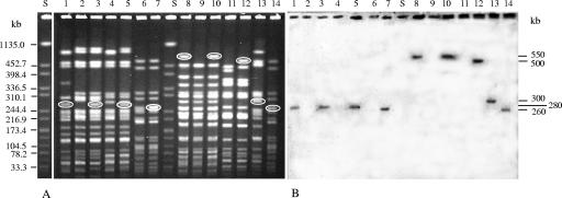
To compare genomic positions of stx2 genes in the EHEC donors and lysogens, XbaI-digested, PFGE-separated DNA was hybridized with an stxA2 probe (Fig. 2). In lysogens derived from aEPEC strains that had PFGE patterns related to that of the respective EHEC phage donor (Fig. 2A, lanes 1 to 5 and lanes 8 to 10), stx2 was located on the same XbaI fragment as in EHEC (Fig. 2B, lanes 1, 3, and 5 and lanes 8 and 10). In a lysogen (Fig. 2A, lane 12) derived from an aEPEC strain (Fig. 2A, lane 11) that differed in PFGE pattern from the EHEC donor (Fig. 2A, lane 8), the stx2 genomic position (Fig. 2B, lane 12) differed from the one in the donor (Fig. 2B, lane 8). In E. coli C600 transduced with phage φ46 [lysogen C600(φ46)] (Fig. 2B, lane 7) or with phage φ61 [lysogen C600(φ61)] (Fig. 2B, lane 14), stx2 was on a 260-kb XbaI fragment. In contrast, E. coli C600 transduced with phage φ50 contained stx2 on a 440-kb XbaI fragment (data not shown). Thus, stx2-harboring phages excised from their integration sites in the genomes of EHEC O26 donors integrate into the same locus in the aEPEC O26 transductants, and there are at least two different integration sites for stx2-harboring phages in the genomes of EHEC O26 and corresponding lysogens.
FIG. 2.
PFGE (A) and stxA2 hybridization (B) of XbaI-digested genomic DNA from EHEC O26 phage donors, aEPEC O26 recipients, and lysogens transduced with stx2-harboring bacteriophages from EHEC O26. Lanes 1, EHEC O26 strain 46 (donor of phage φ46); lanes 2, aEPEC O26 strain 47; lanes 3, lysogen 47(φ46); lanes 4, aEPEC O26 strain 32; lanes 5, lysogen 32(φ46); lanes 6, E. coli strain C600; lanes 7, lysogen C600(φ46); lanes 8, EHEC O26 strain 50 (donor of phage φ50); lanes 9, aEPEC O26 strain 40; lanes 10, lysogen 40(φ50); lanes 11, aEPEC O26 strain 22; lanes 12, lysogen 22(φ50); lanes 13, EHEC O26 strain 61 (donor of phage φ61); lanes 14, lysogen C600(φ61); lanes S, molecular size standards (S. enterica serovar Braenderup strain H9812; Centers for Disease Control and Prevention, Atlanta, GA). The XbaI fragments that hybridized with the stxA2 probe are circled in panel A, and their sizes are given in panel B.
Integration sites of Stx2-encoding phages in EHEC O26:H11.
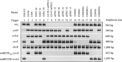
In EHEC O26 strains 46 and 61, yecE was occupied by foreign DNA (Fig. 3, lanes 4 and 5), but this locus was intact in an stx2-negative laboratory derivative of strain 46, strain 47 (Fig. 3, lane 8). Moreover, strains 46 and 61 produced an amplicon of 425 bp in PCR linking yecE with the int gene of φ258320 (Fig. 3, lanes 4 and 5), which integrates in yecE (7), demonstrating that the Stx2-encoding phages in these strains are integrated in yecE. In contrast, in EHEC strain 50, the wrbA insertion site was occupied (Fig. 3, lane 6), but wrbA was intact in its stx2-negative laboratory derivative, strain 50-1 (Fig. 3, lane 7). Strain 50 (Fig. 3, lane 6), but not strain 50-1 (Fig. 3, lane 7), produced an amplicon of 1,045 bp in PCR linking wrbA with the int gene of φ933W, which integrates in wrbA (35, 37). The Stx2-encoding phage in EHEC strain 50 is thus integrated in wrbA.
FIG. 3.
PCR analyses of phage integration sites in EHEC O26, aEPEC O26, and lysogens. Strains tested, loci examined, and lengths of resulting amplicons are listed across the top and to the left and right of the rows of amplicons, respectively. stx2-negative laboratory derivatives (LD) 47 and 50-1 fit the definition of aEPEC. Strains EDL933 (stx1- and stx2-harboring phages integrated in yehV and wrbA, respectively) (35, 37), 258/98 (stx2-harboring phage φ258320 integrated in yecE) (7), and E. coli K-12 C600 (all the genes investigated as putative phage integration sites intact) (8) were used as controls. In PCRs targeting yehV, wrbA, yecE, and sbcB, the presence of an amplicon indicates that the locus is intact, whereas the absence of an amplicon (or a very weak amplicon) indicates that the locus is occupied by foreign DNA. In PCRs connecting yecE with the int gene of phage φ258320 and wrbA with the int gene of phage φ933W (rows 6 and 7, respectively), the presence of an amplicon indicates that a phage with a homologous int gene is integrated in the respective locus; the absence of an amplicon indicates the absence of such a phage.
Integration sites of Stx2-converting phages in lysogens.
yecE and wrbA were intact in all aEPEC strains (Fig. 3, lanes 8 to 13) and a control E. coli C600 strain (Fig. 3, lane 3). The acquisition of phage φ46 or φ61 resulted in occupation of yecE and a positive signal in PCR linking yecE with int of φ258320 in lysogens arising from aEPEC (Fig. 3, lanes 14 and 15) and from E. coli C600 (Fig. 3, lanes 16 and 17). In contrast, phage φ50 integrated in wrbA in the aEPEC and E. coli C600 lysogens (Fig. 3, lanes 18 to 20). Thus, Stx2-encoding phages integrate in EHEC O26 in more than one locus, including yecE and wrbA. These loci are intact in aEPEC O26 and represent hot spots for the integration of such phages into the genomes and the conversion of aEPEC O26 to EHEC O26.
Impact of loss and gain of Stx-converting bacteriophages on PFGE patterns.
As demonstrated in Fig. 2A, loss and gain of Stx2-converting phages diversifies PFGE patterns, even in organisms that are epidemiologically related or are derivatives of the same isolate.
DISCUSSION
Several non-O157 EHEC serogroups are important causes of human disease, and the most common of these is O26 (10, 24, 25). We demonstrate that stx2- and stx1-harboring phages from EHEC O26 can transduce aEPEC O26 strains, converting them to stable lysogens that produce Stx in amounts comparable to that produced by wild-type EHEC O26. Conversely, EHEC O26 can lose stx genes at appreciable frequencies, thereby reverting to being aEPEC. EHEC O26 and aEPEC O26 represent a dynamic system in which bidirectional conversion yields different pathotypes; Stx-encoding bacteriophages are the major elements facilitating this conversion. Data from in vitro experiments correlate with observations obtained with patients infected with E. coli O26, in whom we observed the frequent loss (reference 28 and this study) as well as gain (though at considerably lower frequencies) (H. Karch, unpublished data) of stx2 genes.
EHEC O26:H11 and aEPEC O26:H11 belong to a common phylogenetic group, EHEC 2 (15). It has been hypothesized that aEPEC O26:H11 is ancestral to the EHEC 2 group (15). According to this hypothesis, the acquisition by the ancestral cell of stx genes, the EHEC plasmid, and a high-pathogenicity island that encodes an iron uptake system gave rise to EHEC O26:H11 (15). However, aEPEC O26:H11 already possesses a high-pathogenicity island and the EHEC plasmid (6) and differs from EHEC O26 most notably by the absence of stx genes (6). This observation and our data support the concept of interconversions between aEPEC O26 and EHEC O26 involving loss as well as gain of Stx-encoding bacteriophages. Such bidirectional conversion in nature is also evidenced by the fact that EHEC O26 and aEPEC O26 from different sources have closely related core genomes (3) and highly conserved housekeeping genes (39, 52) and share multiple non-Stx virulence and fitness genes (6). A proposed progenitor of these pathogenic E. coli O26 strains has yet to be identified.
Loss and acquisition of stx2-harboring bacteriophages in vitro alter the genomic architecture of E. coli O26, as reflected by changed PFGE patterns. This observation agrees with previous findings that the loss of stx genes is associated with variant PFGE patterns in EHEC O157:H7/NM (7, 17, 30). These data are also in keeping with the concept that bacteriophages are major drivers of genome diversity in E. coli O157:H7 (31). Therefore, we hypothesize that a diversification of the genome of the infecting EHEC O26 HUS outbreak strain via multiple losses and gains of Stx2-converting phages during human infection accounts for EHEC and aEPEC O26 strains with related, but not identical, PFGE patterns in the same individual and within the outbreak (Fig. 1). Such a possibility should be considered when PFGE is applied to epidemiological investigations of outbreaks caused by E. coli O26. It is noteworthy that variations in PFGE patterns caused by insertions or deletions of phage-associated DNA fragments have also been reported for Campylobacter jejuni (4).
The ability of Stx-encoding phages from clinical EHEC O26 and EHEC O157 isolates to transduce E. coli laboratory strain K-12, commensal E. coli, and EPEC of various origins within the gastrointestinal tract has been demonstrated in mice (1, 55), sheep (13), and house flies (Musca domestica) (36) in vivo and in porcine ligated ileal loops (47). These findings support the hypothesis that interconversion between aEPEC O26 and EHEC O26 can occur in the human intestine during an infection. However, conditions favoring lysogenic conversion or loss in vivo are poorly understood (47, 50, 55) and require further studies.
The ability of EHEC O26 strains to cause HUS indicates that such organisms are more virulent than aEPEC strains lacking the stx gene. If this is true, a change in the pathotype of the infecting E. coli O26 strain during an infection by the loss or acquisition of an Stx-encoding bacteriophage might have clinical implications. For example, using the paradigm that Stx causes HUS, the loss of an stx gene early in infection, before Stx is produced, might prevent the development of HUS in the infected patient. Furthermore, the stx loss by the infecting EHEC at a later stage in the infection, after Stx has already bound to the target organs and has injured the microvasculature, could confound diagnostic schemes dependent on toxin detection. Acquisition of an stx gene by an aEPEC O26 strain during an infection appears to be possible (Karch, unpublished) but is extremely rare. Therefore, the question of whether such an event could trigger HUS cannot be answered at present.
Three integration sites for Stx2-encoding bacteriophages have been identified in EHEC O157:H7, including wrbA (5, 21, 27, 35, 37, 43), sbcB (32), and yecE (14). This last locus is also a common integration site for Stx2-encoding phages in sorbitol-fermenting EHEC O157:NM strains (7), as well as an integration site for phage φP27, which encodes Stx2e in a non-O157 EHEC strain (38). In this study, we demonstrate for the first time that yecE is also an integration site for Stx2-encoding phages in EHEC O26:H11; the other is wrbA. Taken together, these data indicate that yecE and wrbA may be common integration sites for Stx2-encoding bacteriophages in EHEC strains. These loci were intact in all aEPEC O26:H11 strains we investigated and thus represent hot spots where stx2-harboring phages can enter the genomes of aEPEC O26, converting them to EHEC.
A remaining question is why all three Stx2-encoding phages, but only one of four Stx1-encoding phages (phage φH19B), lysogenized aEPEC O26. In general, the susceptibility of a wild-type strain to lysogenization by a phage depends on the presence of the phage receptor, the absence of a highly related bacteriophage within the host that confers immunity against the infecting phage, and the availability of a free integration site for the infecting phage in the genome of the recipient. One reason why wild-type aEPEC strains were not so easily lysogenized by Stx1-encoding phages might be their lack of receptors for such phages or the presence of cryptic prophages that mediate immunity (11). It is possible that such cryptic or truncated bacteriophages occupy a genomic integration site(s) for stx1-harboring phages in aEPEC O26, as has been recently shown in EHEC O157:H7 (5, 43). Although the integration site for stx1-harboring phages in EHEC O26 is unknown, it is noteworthy that in two of the six aEPEC recipients we examined, yehV, an integration site for stx1-harboring phages in EHEC O157:H7 (5, 35, 53), was already occupied by DNA of unknown origin. Future studies should examine the factors promoting transduction of aEPEC O26 by Stx1-converting phages from EHEC O26 and determine the nature of the phage integration site(s).
Acknowledgments
This work was supported by a grant from the EU Network of Excellence EuroPathoGenomics (number LSHB-CT-2005-512061), by a grant from the EU Network ERA-NET PathoGenoMics (project number 0313937C), and by NIH grant AI47499.
The skillful technical assistance of Dagmar Mense, Nadine Brandt (Münster), and Gerlinde Bartel and Evelyn Skiebe (Wernigerode) is greatly appreciated.
Footnotes
Published ahead of print on 30 March 2007.
REFERENCES
- 1.Acheson, D. W. K., J. Reidl, X. Zhang, G. T. Keusch, J. J. Mekalanos, and M. K. Waldor. 1998. In vivo transduction with Shiga toxin 1-encoding phage. Infect. Immun. 66:4496-4498. [DOI] [PMC free article] [PubMed] [Google Scholar]
- 2.Allerberger, F., A. W. Friedrich, K. Grif, M. P. Dierich, H. J. Dornbusch, C. J. Mache, E. Nachbaur, M. Freilinger, P. Rieck, M. Wagner, A. Caprioli, H. Karch, and L. B. Zimmerhackl. 2003. Hemolytic-uremic syndrome associated with enterohemorrhagic Escherichia coli O26:H− infection and consumption of unpasteurized cow's milk. Int. J. Infect. Dis. 7:42-45. [DOI] [PubMed] [Google Scholar]
- 3.Anjum, M. F., S. Lucchini, A. Thompson, J. C. Hinton, and M. J. Woodward. 2003. Comparative genomic indexing reveals the phylogenomics of Escherichia coli pathogens. Infect. Immun. 71:4674-4683. [DOI] [PMC free article] [PubMed] [Google Scholar]
- 4.Barton, C., L.-K. Ng, S. D. Tyler, and C. G. Clark. 2007. Temperate bacteriophages affect pulsed-field gel electrophoresis patterns of Campylobacter jejuni. J. Clin. Microbiol. 45:386-391. [DOI] [PMC free article] [PubMed] [Google Scholar]
- 5.Besser, T. E., N. Shaikh, N. J. Holt, P. I. Tarr, M. E. Konkel, P. Malik-Kale, C. W. Walsh, T. Whittam, and J. L. Bono. 2007. Greater diversity of Shiga toxin-encoding bacteriophage insertion sites among Escherichia coli O157:H7 isolates from cattle than in those from humans. Appl. Environ. Microbiol. 73:671-679. [DOI] [PMC free article] [PubMed] [Google Scholar]
- 6.Bielaszewska, M., W. Zhang, P. I. Tarr, A.-K. Sonntag, and H. Karch. 2005. Molecular profiling and phenotype analysis of Escherichia coli O26:H11 and O26:NM: secular and geographic consistency of enterohemorrhagic and enteropathogenic isolates. J. Clin. Microbiol. 43:4225-4228. [DOI] [PMC free article] [PubMed] [Google Scholar]
- 7.Bielaszewska, M., R. Prager, W. Zhang, A. W. Friedrich, A. Mellmann, H. Tschape, and H. Karch. 2006. Chromosomal dynamism in progeny of outbreak-related sorbitol-fermenting enterohemorrhagic Escherichia coli O157:NM. Appl. Environ. Microbiol. 72:1900-1909. [DOI] [PMC free article] [PubMed] [Google Scholar]
- 8.Blattner, F. R., G. Plunkett, C. A. Bloch, N. T. Perna, V. Burland, M. Riley, V. J. Collado, J. D. Glasner, C. K. Rode, G. F. Mayhew, J. Gregor, N. W. Davis, H. A. Kirkpatrick, M. A. Goeden, D. J. Rose, B. Mau, and Y. Shao. 1997. The complete genome sequence of Escherichia coli K-12. Science 277:1453-1474. [DOI] [PubMed] [Google Scholar]
- 9.Bokete, T. N., T. S. Whittam, R. S. Wilson, C. R. Clausen, C. M. O'Callaghan, S. L. Moseley, T. R. Fritsche, and P. I. Tarr. 1997. Genetic and phenotypic analysis of Escherichia coli with enteropathogenic characteristics isolated from Seattle children. J. Infect. Dis. 175:1382-1389. [DOI] [PubMed] [Google Scholar]
- 10.Brooks, J. T., E. G. Sowers, J. G. Wells, K. D. Greene, P. M. Griffin, R. M. Hoekstra, and N. A. Strockbine. 2005. Non-O157 Shiga toxin-producing Escherichia coli infections in the United States, 1983-2002. J. Infect. Dis. 192:1422-1429. [DOI] [PubMed] [Google Scholar]
- 11.Campbell, A. M. 1996. Cryptic prophages, p. 2041-2046. In F. C. Neidhardt, R. Curtiss III, J. L. Ingraham, E. C. Lin, K. B. Low, B. Magasanik, W. S. Reznikoff, M. Riley, M. Schaechter, and H. E. Umbarger (ed.), Escherichia coli and Salmonella: cellular and molecular biology, 2nd ed. ASM Press, Washington, DC.
- 12.Cohen, M. B., J. P. Nataro, D. I. Bernstein, J. Hawkins, N. Roberts, and M. A. Staat. 2005. Prevalence of diarrheagenic Escherichia coli in acute childhood enteritis: a prospective controlled study. J. Pediatr. 146:54-61. [DOI] [PubMed] [Google Scholar]
- 13.Cornick, N. A., A. F. Helgerson, V. Mai, J. M. Ritchie, and D. W. K. Acheson. 2006. In vivo transduction of an Stx-encoding phage in ruminants. Appl. Environ. Microbiol. 72:5086-5088. [DOI] [PMC free article] [PubMed] [Google Scholar]
- 14.De Greve, H., C. Qizhi, F. Deboeck, and J. P. Hernalsteens. 2002. The Shiga-toxin VT2-encoding bacteriophage Φ297 integrates at a distinct position in the Escherichia coli genome. Biochim. Biophys. Acta 1579:196-202. [DOI] [PubMed] [Google Scholar]
- 15.Donnenberg, M., and T. S. Whittam. 2001. Pathogenesis and evolution of virulence in enteropathogenic and enterohemorrhagic Escherichia coli. J. Clin. Investig. 107:539-548. [DOI] [PMC free article] [PubMed] [Google Scholar]
- 16.Eklund, M., F. Scheutz, and A. Siitonen. 2001. Clinical isolates of non-O157 Shiga toxin-producing Escherichia coli: serotypes, virulence characteristics, and molecular profiles of strains of the same serotype. J. Clin. Microbiol. 39:2829-2834. [DOI] [PMC free article] [PubMed] [Google Scholar]
- 17.Feng, P., M. Dey, A. Abe, and T. Takeda. 2001. Isogenic strain of Escherichia coli O157:H7 that has lost both Shiga toxin 1 and 2 genes. Clin. Diagn. Lab. Immunol. 8:711-717. [DOI] [PMC free article] [PubMed] [Google Scholar]
- 18.Friedrich, A. W., M. Bielaszewska, W.-L. Zhang, M. Pulz, T. Kuczius, A. Ammon, and H. Karch. 2002. Escherichia coli harboring Shiga toxin 2 gene variants: frequency and association with clinical symptoms. J. Infect. Dis. 185:74-84. [DOI] [PubMed] [Google Scholar]
- 19.Gerber, A., H. Karch, F. Allerberger, H. M. Verweyen, and L. B. Zimmerhackl. 2002. Clinical course and the role of Shiga toxin-producing Escherichia coli infection in the hemolytic-uremic syndrome in pediatric patients, 1997-2000, in Germany and Austria: a prospective study. J. Infect. Dis. 186:493-500. [DOI] [PubMed] [Google Scholar]
- 20.Giammanco, A., M. Maggio, G. Giammanco, R. Morelli, F. Minelli, F. Scheutz, and A. Caprioli. 1996. Characteristics of Escherichia coli strains belonging to enteropathogenic E. coli serogroups isolated in Italy from children with diarrhea. J. Clin. Microbiol. 34:689-694. [DOI] [PMC free article] [PubMed] [Google Scholar]
- 21.Hayashi, T., K. Makino, M. Ohnishi, K. Kurokawa, K. Ishii, K. Yokoyama, C. Han, E. Ohtsubo, K. Nakayama, T. Murata, M. Tanaka, T. Tobe, T. Iida, H. Takami, T. Honda, C. Sasakawa, N. Ogasawara, T. Yasunaga, S. Kuhara, T. Shiba, M. Hattori, and H. Shinagawa. 2001. Complete genome sequence of enterohemorrhagic Escherichia coli O157:H7 and genomic comparison with a laboratory strain K-12. DNA Res. 28:11-22. [DOI] [PubMed] [Google Scholar]
- 22.Hunter, S. B., P. Vauterin, M. A. Lambert-Fair, M. S. Van Duyne, K. Kubota, L. Graves, D. Wrigley, T. Barrett, and E. Ribot. 2005. Establishment of a universal size standard strain for use with the PulseNet standardized pulsed-field gel electrophoresis protocols: converting the national databases to the new size standard. J. Clin. Microbiol. 43:1045-1050. [DOI] [PMC free article] [PubMed] [Google Scholar]
- 23.Jelacic, J. K., T. Damrow, G. S. Chen, S. Jelacic, M. Bielaszewska, M. Ciol, H. M. Carvalho, A. R. Melton-Celsa, A. D. O'Brien, and P. I. Tarr. 2003. Shiga toxin-producing Escherichia coli in Montana: bacterial genotypes and clinical profiles. J. Infect. Dis. 188:719-729. [DOI] [PubMed] [Google Scholar]
- 24.Johnson, K. E., C. M. Thorpe, and C. L. Sears. 2006. The emerging clinical importance of non-O157 Shiga toxin-producing Escherichia coli. Clin. Infect. Dis. 43:1587-1595. [DOI] [PubMed] [Google Scholar]
- 25.Karch, H., P. I. Tarr, and M. Bielaszewska. 2005. Enterohaemorrhagic Escherichia coli in human medicine. Int. J. Med. Microbiol. 295:405-418. [DOI] [PubMed] [Google Scholar]
- 26.Karmali, M. A., M. Petric, C. Lim, P. C. Fleming, G. S. Arbus, and H. Lior. 1985. The association between idiopathic hemolytic uremic syndrome and infection by verotoxin-producing Escherichia coli. J. Infect. Dis. 151:775-782. [DOI] [PubMed] [Google Scholar]
- 27.Makino, K., K. Yokoyama, Y. Kubota, C. H. Yutsudo, S. Kimura, K. Kurokawa, K. Ishii, M. Hattori, I. Tatsuno, H. Abe, T. Iida, K. Yamamoto, M. Onishi, T. Hayashi, T. Yasunaga, T. Honda, C. Sasakawa, and H. Shinagawa. 1999. Complete nucleotide sequence of the prophage VT2-Sakai carrying the verotoxin 2 genes of the enterohemorrhagic Escherichia coli O157:H7 derived from the Sakai outbreak. Genes Genet. Syst. 74:227-239. [DOI] [PubMed] [Google Scholar]
- 28.Mellmann, A., M. Bielaszewska, L. B. Zimmerhackl, R. Prager, D. Harmsen, H. Tschape, and H. Karch. 2005. Enterohemorrhagic Escherichia coli in human infection: in vivo evolution of a bacterial pathogen. Clin. Infect. Dis. 41:785-792. [DOI] [PubMed] [Google Scholar]
- 29.Misselwitz, J., H. Karch, M. Bielaszewska, U. John, F. Ringelmann, G. Rönnefarth, and L. Patzer. 2003. Cluster of hemolytic-uremic syndrome caused by Shiga toxin-producing Escherichia coli O26:H11. Pediatr. Infect. Dis. J. 22:349-354. [DOI] [PubMed] [Google Scholar]
- 30.Murase, T., S. Yamai, and H. Watanabe. 1999. Changes in pulsed-field gel electrophoresis patterns in clinical isolates of enterohemorrhagic Escherichia coli O157:H7 associated with loss of Shiga toxin genes. Curr. Microbiol. 38:48-50. [DOI] [PubMed] [Google Scholar]
- 31.Ohnishi, M., K. Kurokawa, and T. Hayashi. 2001. Diversification of Escherichia coli genomes: are bacteriophages the major contributors? Trends Microbiol. 9:481-485. [DOI] [PubMed] [Google Scholar]
- 32.Ohnishi, M., J. Terajima, K. Kurokawa, K. Nakayama, T. Murata, K. Tamura, Y. Ogura, H. Watanabe, and T. Hayashi. 2002. Genomic diversity of enterohemorrhagic Escherichia coli O157 revealed by whole genome PCR scanning. Proc. Natl. Acad. Sci. USA 99:17043-17048. [DOI] [PMC free article] [PubMed] [Google Scholar]
- 33.Orth, D., and R. Würzner. 2006. What makes an enterohemorrhagic Escherichia coli? Clin. Infect. Dis. 43:1168-1169. [DOI] [PubMed] [Google Scholar]
- 34.Peixoto, J. C. C., S. Y. Bando, J. A. G. Ordonez, B. A. Botelho, L. R. Trabulsi, and C. A. Moreira-Filho. 2001. Genetic differences between Escherichia coli O26 strains isolated in Brazil and in other countries. FEMS Microbiol. Lett. 196:239-244. [DOI] [PubMed] [Google Scholar]
- 35.Perna, N. T., G. Plunkett III, V. Burland, B. Mau, J. D Glasner, D. J. Rose, G. F. Mayhew, P. S. Evans, J. Gregor, H. A. Kirkpatrick, G. Posfai, J. Hackett, S. Klink, A. Boutin, Y. Shao, L. Miller, E. J. Grotbeck, N. W. Davis, A. Lim, E. T. Dimalanta, K. D. Potamousis, J. Apodaca, T. S. Anantharaman, J. Lin, G. Yen, D. C. Schwartz, R. A. Welch, and F. R. Blattner. 2001. Genome sequence of enterohaemorrhagic Escherichia coli O157:H7. Nature 409:529-533. [DOI] [PubMed] [Google Scholar]
- 36.Petridis, M., M. Bagdasarian, M. K. Waldor, and E. Walker. 2006. Horizontal transfer of Shiga toxin and antibiotic resistance genes among Escherichia coli strains in house fly (Diptera: Muscidae) gut. J. Med. Entomol. 43:288-295. [DOI] [PubMed] [Google Scholar]
- 37.Plunkett, G., III, D. J. Rose, T. J. Durfee, and F. R. Blattner. 1999. Sequence of Shiga toxin 2 phage 933W from Escherichia coli O157:H7: Shiga toxin as a phage late-gene product. J. Bacteriol. 181:1767-1778. [DOI] [PMC free article] [PubMed] [Google Scholar]
- 38.Recktenwald, J., and H. Schmidt. 2002. The nucleotide sequence of Shiga toxin (Stx) 2e-encoding phage φP27 is not related to other Stx phage genomes, but the modular genetic structure is conserved. Infect. Immun. 70:1896-1908. [DOI] [PMC free article] [PubMed] [Google Scholar]
- 39.Reid, S. D., C. J. Herbelin, A. C. Bumbaugh, R. K. Selander, and T. S. Whittam. 2000. Parallel evolution of virulence in pathogenic Escherichia coli. Nature 406:64-67. [DOI] [PubMed] [Google Scholar]
- 40.Sambrook, J., E. F. Fritsch, and T. Maniatis. 1989. Molecular cloning: a laboratory manual, 2nd ed. Cold Spring Harbor Laboratory, Cold Spring Harbor, NY.
- 41.Schmidt, H., M. Bielaszewska, and H. Karch. 1999. Transduction of enteric Escherichia coli isolates with a derivative of Shiga toxin 2-encoding bacteriophage φ3585 isolated from Escherichia coli O157:H7. Appl. Environ. Microbiol. 65:3855-3861. [DOI] [PMC free article] [PubMed] [Google Scholar]
- 42.Scotland, S. M., G. A. Willshaw, H. R. Smith, and B. Rowe. 1990. Properties of strains of Escherichia coli O26:H11 in relation to their enteropathogenic or enterohemorrhagic classification. J. Infect. Dis. 162:1069-1074. [DOI] [PubMed] [Google Scholar]
- 43.Shaikh, N., and P. I. Tarr. 2003. Escherichia coli O157:H7 Shiga toxin-encoding bacteriophages: integrations, excisions, truncations, and evolutionary implications. J. Bacteriol. 185:3596-3605. [DOI] [PMC free article] [PubMed] [Google Scholar]
- 44.Smith, H. W., and M. A. Linggood. 1971. The transmissible nature of enterotoxin production in human enteropathogenic strain of Escherichia coli. J. Med. Microbiol. 4:301-305. [DOI] [PubMed] [Google Scholar]
- 45.Smith, H. W., P. Green, and Z. Parsell. 1983. Vero cell toxins in Escherichia coli and related bacteria: transfer by phage and conjugation and toxic action in laboratory animals, chickens and pigs. J. Gen. Microbiol. 129:3121-3137. [DOI] [PubMed] [Google Scholar]
- 46.Sonntag, A.-K., R. Prager, M. Bielaszewska, W. Zhang, A. Fruth, H. Tschape, and H. Karch. 2004. Phenotypic and genotypic analyses of enterohemorrhagic Escherichia coli O145 strains from patients in Germany. J. Clin. Microbiol. 42:954-962. [DOI] [PMC free article] [PubMed] [Google Scholar]
- 47.Tóth, I., H. Schmidt, M. Dow, A. Malik, E. Oswald, and B. Nagy. 2003. Transduction of porcine enteropathogenic Escherichia coli with a derivative of a Shiga toxin 2-encoding bacteriophage in a porcine ligated ileal loop system. Appl. Environ. Microbiol. 69:7242-7247. [DOI] [PMC free article] [PubMed] [Google Scholar]
- 48.Tozzi, A. E., A. Caprioli, F. Minelli, A. Gianviti, L. De Petris, A. Edefonti, G. Montini, A. Ferretti, T. De Palo, M. Gaido, G. Rizzoni, and the Hemolytic Uremic Syndrome Study Group. 2003. Shiga toxin-producing Escherichia coli infections associated with hemolytic uremic syndrome, Italy, 1988-2000. Emerg. Infect. Dis. 9:106-108. [DOI] [PMC free article] [PubMed] [Google Scholar]
- 49.Trabulsi, L. R., R. Keller, and T. A. T. Gomes. 2002. Typical and atypical enteropathogenic Escherichia coli. Emerg. Infect. Dis. 8:508-513. [DOI] [PMC free article] [PubMed] [Google Scholar]
- 50.Wagner, P. L., and M. K. Waldor. 2002. Bacteriophage control of bacterial virulence. Infect. Immun. 70:3985-3993. [DOI] [PMC free article] [PubMed] [Google Scholar]
- 51.Werber, D., A. Fruth, A. Liesegang, M. Littmann, U. Buchholz, R. Prager, H. Karch, T. Breuer, H. Tschäpe, and A. Ammon. 2002. A multistate outbreak of Shiga toxin-producing Escherichia coli O26:H11 infections in Germany, detected by molecular subtyping surveillance. J. Infect. Dis. 186:419-422. [DOI] [PubMed] [Google Scholar]
- 52.Wirth, T., D. Falush, R. Lan, F. Colles, P. Mensa, L. H. Wieler, H. Karch, P. R. Reeves, M. C. Maiden, H. Ochman, and M. Achtman. 2006. Sex and virulence in Escherichia coli: an evolutionary perspective. Mol. Microbiol. 60:1136-1151. [DOI] [PMC free article] [PubMed] [Google Scholar]
- 53.Yokoyama, K., K. Makino, Y. Kubota, M. Watanabe, S. Kimura, C. H. Yutsudo, K. Kurokawa, K. Ishii, M. Hattori, I. Tatsuno, H. Abe, M. Yoh, T. Iida, M. Ohnishi, T. Hayashi, T. Yasunaga, T. Honda, C. Sasakawa, and H. Shinagawa. 2000. Complete nucleotide sequence of the prophage VT1-Sakai carrying the Shiga toxin 1 genes of the enterohemorrhagic Escherichia coli O157:H7 strain derived from the Sakai outbreak. Gene 258:127-139. [DOI] [PubMed] [Google Scholar]
- 54.Zhang, W.-L., M. Bielaszewska, A. Liesegang, H. Tschäpe, H. Schmidt, M. Bitzan, and H. Karch. 2000. Molecular characteristics and epidemiological significance of Shiga toxin-producing Escherichia coli O26. J. Clin. Microbiol. 38:2134-2140. [DOI] [PMC free article] [PubMed] [Google Scholar]
- 55.Zhang, X., A. D. McDaniel, L. E. Wolf, G. T. Keusch, M. K. Waldor, and D. W. K. Acheson. 2000. Quinolone antibiotics induce Shiga toxin-encoding bacteriophages, toxin production and death in mice. J. Infect. Dis. 181:664-670. [DOI] [PubMed] [Google Scholar]


