

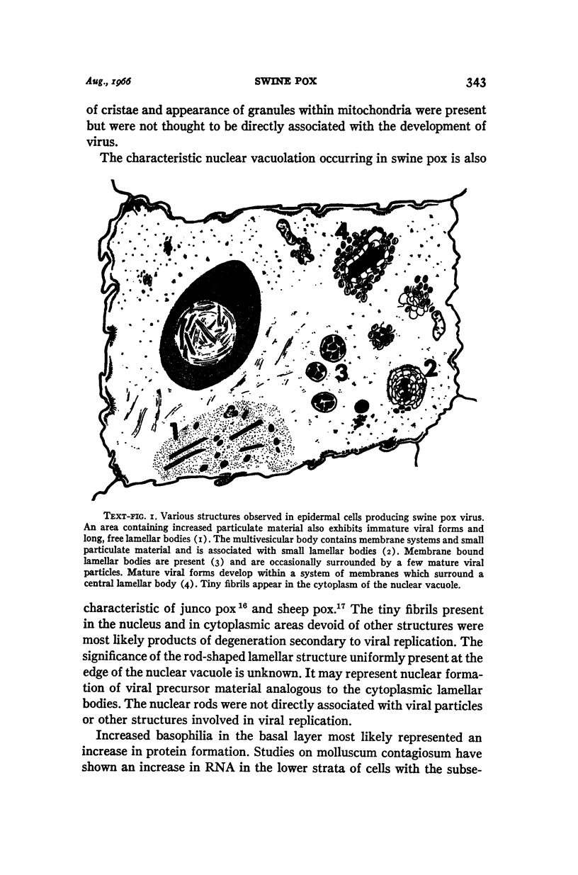
Full text
PDF
Images in this article
Selected References
These references are in PubMed. This may not be the complete list of references from this article.
- ABDUSSALAM M., BLAKEMORE F. Morphology of the elementary bodies and cell inclusions in swine pox. J Comp Pathol. 1956 Oct;66(4):373–377. doi: 10.1016/s0368-1742(56)80039-8. [DOI] [PubMed] [Google Scholar]
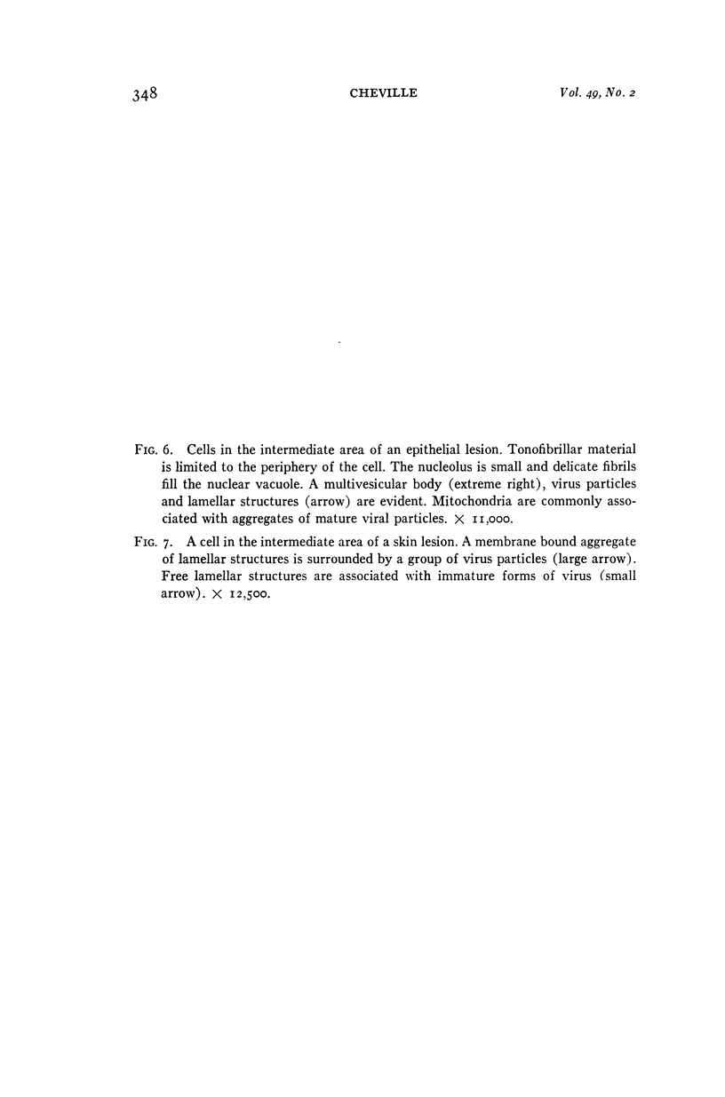
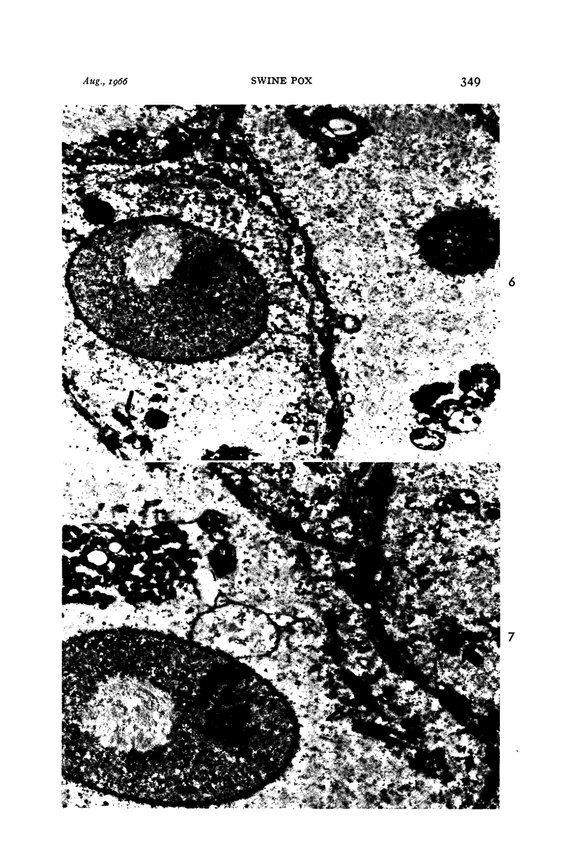
- BANFIELD W. G. Dense granule in the elementary body of molluscum contagiosum. J Biophys Biochem Cytol. 1959 May 25;5(3):513–514. doi: 10.1083/jcb.5.3.513. [DOI] [PMC free article] [PubMed] [Google Scholar]
- CONSTANTIN T., FEBVRE H. Evolution des acides nucléiques dans les corps d'inclusions provoqués par le virus du fibrome de shope. C R Hebd Seances Acad Sci. 1958 Jan 13;246(2):332–334. [PubMed] [Google Scholar]
- CROISSANT O., LEPINE P., WYCKOFF R. W. Sur le développement du virus vaccinal. Ann Inst Pasteur (Paris) 1958 Mar;94(3):294–297. [PubMed] [Google Scholar]
- DOURMASHKIN R., BERNHARD W. A study with the electron microscope of the skin tumour of molluscum contagiosum. J Ultrastruct Res. 1959 Oct;3:11–38. doi: 10.1016/s0022-5320(59)80011-3. [DOI] [PubMed] [Google Scholar]
- KARNOVSKY M. J. Simple methods for "staining with lead" at high pH in electron microscopy. J Biophys Biochem Cytol. 1961 Dec;11:729–732. doi: 10.1083/jcb.11.3.729. [DOI] [PMC free article] [PubMed] [Google Scholar]
- LEDUC E. H., BERNHARD W. Electron microscope study of mouse liver infected by ectromelia virus. J Ultrastruct Res. 1962 Jun;6:466–488. doi: 10.1016/s0022-5320(62)80003-3. [DOI] [PubMed] [Google Scholar]
- PLOWRIGHT W., FERRIS R. D. The growth and cytopathogenicity of sheep-pox virus in tissue cultures. Br J Exp Pathol. 1958 Aug;39(4):424–435. [PMC free article] [PubMed] [Google Scholar]
- RAKE G., BLANK H. The relationship of host and virus in molluscum contagiosum. J Invest Dermatol. 1950 Aug;15(2):81–93. doi: 10.1038/jid.1950.77. [DOI] [PubMed] [Google Scholar]
- RECZKO E. [Electron microscopic study of the abdominal skin of the young pig infected with original swine pox]. Arch Gesamte Virusforsch. 1959;9:193–213. [PubMed] [Google Scholar]