Abstract
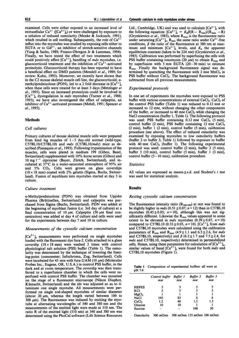
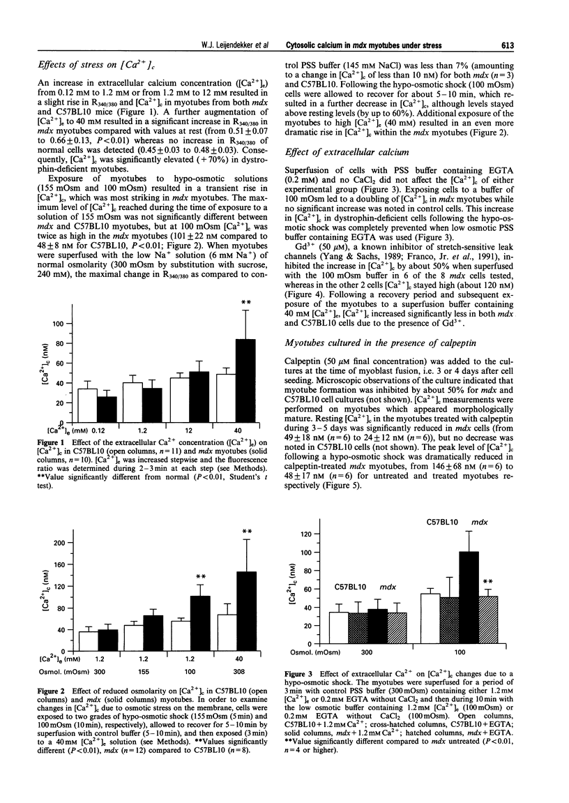
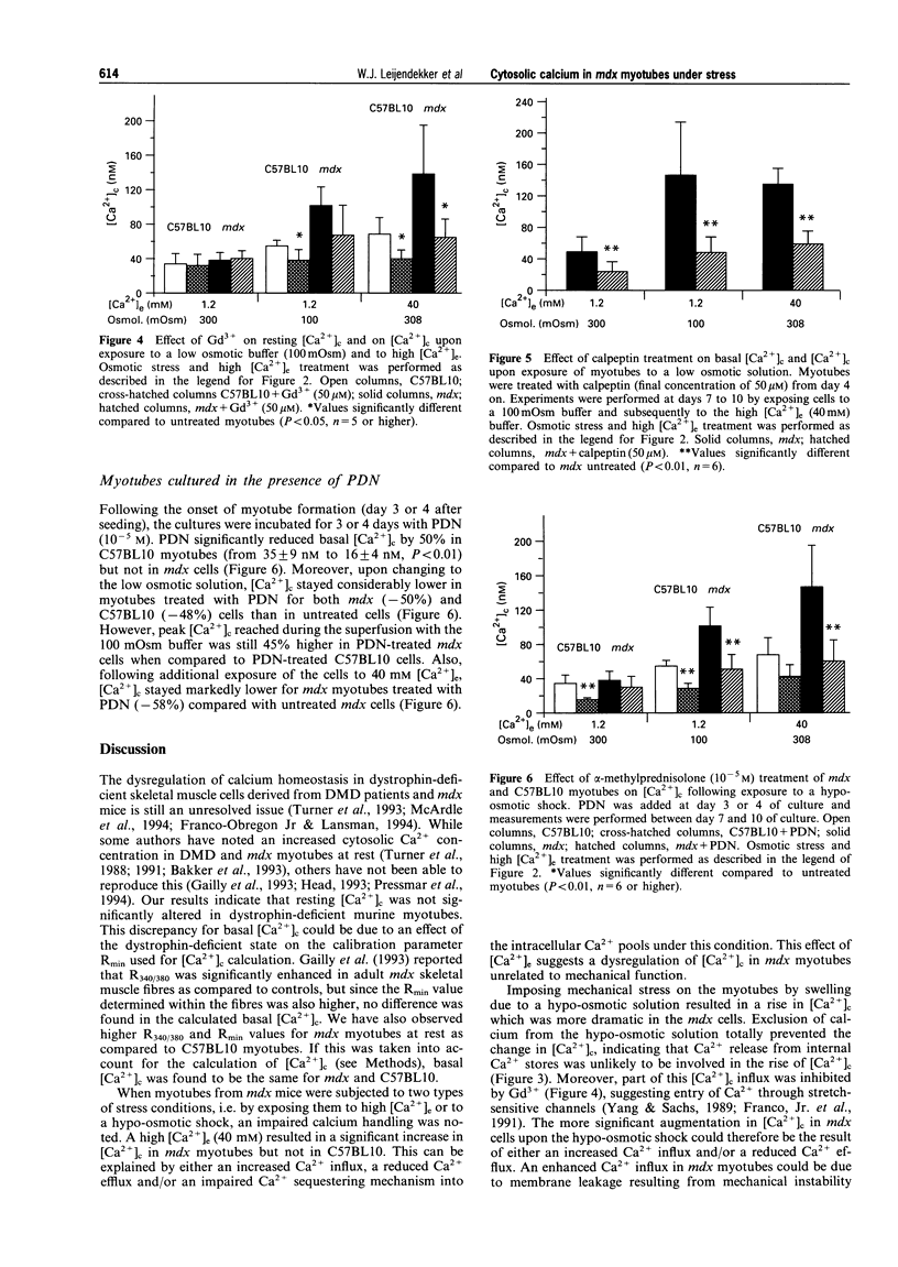
1. In Duchenne muscular dystrophy (DMD) dysregulation of cytosolic calcium appears to be involved in the degeneration of skeletal muscle fibres. Therefore, we have studied the regulation of the free cytosolic calcium concentration ([Ca2+]c) under specific stress conditions in cultured myotubes isolated from the hind limbs of wild-type (C57BL10) and dystrophin-deficient mutant mdx mice. [Ca2+]c in the myotubes was estimated by the use of the Ca(2+)-sensitive fluorescent dye, fura-2. 2. Resting [Ca2+]c was similar in mdx and normal myotubes (35 +/- 9 nM and 38 +/- 11 nM, respectively). However, when mdx myotubes were exposed to a high extracellular calcium concentration ([Ca2+]c) of 40 mM, the [Ca2+]c was elevated to 84 +/- 29 nM, compared to 49 +/- 7 nM in normal myotubes. 3. Lowering the osmolarity of the superfusion solution from 300 mOsm to 100 mOsm resulted also in a rise in [Ca2+]c which was about two times higher for mdx (243 +/- 65 nM) than for C57BL10 (135 +/- 37 nM). Replacing extracellular Ca2+ by EGTA (0.2 mM) prevented the rise in [Ca2+]c in both mdx and normal myotubes when exposed to the low osmolarity solution. 4. Gadolinium ion (50 microM), an inhibitor of Ca2+ entry, antagonized the rise in [Ca2+]c of myotubes superfused with 40 mM [Ca2+]c by 20-40% for both mdx and C57BL10 cells, but did not significantly reduce the rise in [Ca2+]c when the cells were exposed to the hypo-osmotic buffer (100 mOsm). 5. Incubation of the cell culture for 3-5 days from the onset of induction of myotube formation with the membrane permeable protease inhibitor, calpeptin (50 microM) abolished the rise in [Ca2+]c in mdx myotubes upon exposure to hypo-osmotic shock. 6. Treatment of the cell culture for 3-5 days with alpha-methylprednisolone (PDN, 10 microM) attenuated the rise in [Ca2+]c following hypo-osmotic stress for both normal and mdx myotubes by about 50%. 7. The results described here suggest an increased permeability of mdx myotubes to Ca2+ under specific stress conditions. The ameliorating effect of PDN on [Ca2+]c could explain, at least partly, the beneficial effect of this drug on DMD patients.
Full text
PDF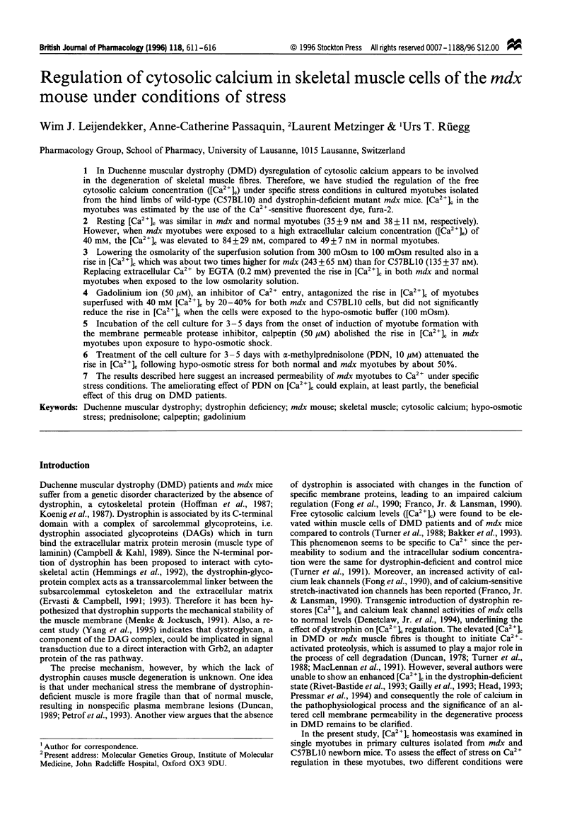
Selected References
These references are in PubMed. This may not be the complete list of references from this article.
- Bakker A. J., Head S. I., Williams D. A., Stephenson D. G. Ca2+ levels in myotubes grown from the skeletal muscle of dystrophic (mdx) and normal mice. J Physiol. 1993 Jan;460:1–13. doi: 10.1113/jphysiol.1993.sp019455. [DOI] [PMC free article] [PubMed] [Google Scholar]
- Campbell K. P., Kahl S. D. Association of dystrophin and an integral membrane glycoprotein. Nature. 1989 Mar 16;338(6212):259–262. doi: 10.1038/338259a0. [DOI] [PubMed] [Google Scholar]
- Denetclaw W. F., Jr, Hopf F. W., Cox G. A., Chamberlain J. S., Steinhardt R. A. Myotubes from transgenic mdx mice expressing full-length dystrophin show normal calcium regulation. Mol Biol Cell. 1994 Oct;5(10):1159–1167. doi: 10.1091/mbc.5.10.1159. [DOI] [PMC free article] [PubMed] [Google Scholar]
- Duncan C. J. Dystrophin and the integrity of the sarcolemma in Duchenne muscular dystrophy. Experientia. 1989 Feb 15;45(2):175–177. doi: 10.1007/BF01954866. [DOI] [PubMed] [Google Scholar]
- Duncan C. J. Role of intracellular calcium in promoting muscle damage: a strategy for controlling the dystrophic condition. Experientia. 1978 Dec 15;34(12):1531–1535. doi: 10.1007/BF02034655. [DOI] [PubMed] [Google Scholar]
- Ervasti J. M., Campbell K. P. A role for the dystrophin-glycoprotein complex as a transmembrane linker between laminin and actin. J Cell Biol. 1993 Aug;122(4):809–823. doi: 10.1083/jcb.122.4.809. [DOI] [PMC free article] [PubMed] [Google Scholar]
- Ervasti J. M., Campbell K. P. Membrane organization of the dystrophin-glycoprotein complex. Cell. 1991 Sep 20;66(6):1121–1131. doi: 10.1016/0092-8674(91)90035-w. [DOI] [PubMed] [Google Scholar]
- Fong P. Y., Turner P. R., Denetclaw W. F., Steinhardt R. A. Increased activity of calcium leak channels in myotubes of Duchenne human and mdx mouse origin. Science. 1990 Nov 2;250(4981):673–676. doi: 10.1126/science.2173137. [DOI] [PubMed] [Google Scholar]
- Franco-Obregón A., Jr, Lansman J. B. Mechanosensitive ion channels in skeletal muscle from normal and dystrophic mice. J Physiol. 1994 Dec 1;481(Pt 2):299–309. doi: 10.1113/jphysiol.1994.sp020440. [DOI] [PMC free article] [PubMed] [Google Scholar]
- Franco A., Jr, Lansman J. B. Calcium entry through stretch-inactivated ion channels in mdx myotubes. Nature. 1990 Apr 12;344(6267):670–673. doi: 10.1038/344670a0. [DOI] [PubMed] [Google Scholar]
- Franco A., Jr, Winegar B. D., Lansman J. B. Open channel block by gadolinium ion of the stretch-inactivated ion channel in mdx myotubes. Biophys J. 1991 Jun;59(6):1164–1170. doi: 10.1016/S0006-3495(91)82332-3. [DOI] [PMC free article] [PubMed] [Google Scholar]
- Gailly P., Boland B., Himpens B., Casteels R., Gillis J. M. Critical evaluation of cytosolic calcium determination in resting muscle fibres from normal and dystrophic (mdx) mice. Cell Calcium. 1993 Jun;14(6):473–483. doi: 10.1016/0143-4160(93)90006-r. [DOI] [PubMed] [Google Scholar]
- Grynkiewicz G., Poenie M., Tsien R. Y. A new generation of Ca2+ indicators with greatly improved fluorescence properties. J Biol Chem. 1985 Mar 25;260(6):3440–3450. [PubMed] [Google Scholar]
- Head S. I. Membrane potential, resting calcium and calcium transients in isolated muscle fibres from normal and dystrophic mice. J Physiol. 1993 Sep;469:11–19. doi: 10.1113/jphysiol.1993.sp019801. [DOI] [PMC free article] [PubMed] [Google Scholar]
- Hemmings L., Kuhlman P. A., Critchley D. R. Analysis of the actin-binding domain of alpha-actinin by mutagenesis and demonstration that dystrophin contains a functionally homologous domain. J Cell Biol. 1992 Mar;116(6):1369–1380. doi: 10.1083/jcb.116.6.1369. [DOI] [PMC free article] [PubMed] [Google Scholar]
- Hoffman E. P., Brown R. H., Jr, Kunkel L. M. Dystrophin: the protein product of the Duchenne muscular dystrophy locus. Cell. 1987 Dec 24;51(6):919–928. doi: 10.1016/0092-8674(87)90579-4. [DOI] [PubMed] [Google Scholar]
- Khan M. A. Corticosteroid therapy in Duchenne muscular dystrophy. J Neurol Sci. 1993 Dec 1;120(1):8–14. doi: 10.1016/0022-510x(93)90017-s. [DOI] [PubMed] [Google Scholar]
- Koenig M., Hoffman E. P., Bertelson C. J., Monaco A. P., Feener C., Kunkel L. M. Complete cloning of the Duchenne muscular dystrophy (DMD) cDNA and preliminary genomic organization of the DMD gene in normal and affected individuals. Cell. 1987 Jul 31;50(3):509–517. doi: 10.1016/0092-8674(87)90504-6. [DOI] [PubMed] [Google Scholar]
- Kwak K. B., Kambayashi J., Kang M. S., Ha D. B., Chung C. H. Cell-penetrating inhibitors of calpain block both membrane fusion and filamin cleavage in chick embryonic myoblasts. FEBS Lett. 1993 May 24;323(1-2):151–154. doi: 10.1016/0014-5793(93)81468-f. [DOI] [PubMed] [Google Scholar]
- MacLennan P. A., McArdle A., Edwards R. H. Effects of calcium on protein turnover of incubated muscles from mdx mice. Am J Physiol. 1991 Apr;260(4 Pt 1):E594–E598. doi: 10.1152/ajpendo.1991.260.4.E594. [DOI] [PubMed] [Google Scholar]
- Massa E. M., Morero R. D., Bloj B., Farías R. N. Hormone action and membrane fluidity: effect of insulin and cortisol on the Hill coefficients of rat erythrocyte membrane-bound acetylcholinesterase and (Na+ + K+)-ATPase. Biochem Biophys Res Commun. 1975 Sep 2;66(1):115–122. doi: 10.1016/s0006-291x(75)80302-0. [DOI] [PubMed] [Google Scholar]
- McArdle A., Edwards R. H., Jackson M. J. Time course of changes in plasma membrane permeability in the dystrophin-deficient mdx mouse. Muscle Nerve. 1994 Dec;17(12):1378–1384. doi: 10.1002/mus.880171206. [DOI] [PubMed] [Google Scholar]
- Mehdi S. Cell-penetrating inhibitors of calpain. Trends Biochem Sci. 1991 Apr;16(4):150–153. doi: 10.1016/0968-0004(91)90058-4. [DOI] [PubMed] [Google Scholar]
- Mendell J. R., Moxley R. T., Griggs R. C., Brooke M. H., Fenichel G. M., Miller J. P., King W., Signore L., Pandya S., Florence J. Randomized, double-blind six-month trial of prednisone in Duchenne's muscular dystrophy. N Engl J Med. 1989 Jun 15;320(24):1592–1597. doi: 10.1056/NEJM198906153202405. [DOI] [PubMed] [Google Scholar]
- Menke A., Jockusch H. Decreased osmotic stability of dystrophin-less muscle cells from the mdx mouse. Nature. 1991 Jan 3;349(6304):69–71. doi: 10.1038/349069a0. [DOI] [PubMed] [Google Scholar]
- Metzinger L., Passaquin A. C., Leijendekker W. J., Poindron P., Rüegg U. T. Modulation by prednisolone of calcium handling in skeletal muscle cells. Br J Pharmacol. 1995 Dec;116(7):2811–2816. doi: 10.1111/j.1476-5381.1995.tb15930.x. [DOI] [PMC free article] [PubMed] [Google Scholar]
- Metzinger L., Passaquin A. C., Vernier A., Thiriet N., Warter J. M., Poindron P. Lazaroids enhance skeletal myogenesis in primary cultures of dystrophin-deficient mdx mice. J Neurol Sci. 1994 Nov;126(2):138–145. doi: 10.1016/0022-510x(94)90263-1. [DOI] [PubMed] [Google Scholar]
- Metzinger L., Passaquin A. C., Warter J. M., Poindron P. alpha-Methylprednisolone promotes skeletal myogenesis in dystrophin-deficient and control mouse cultures. Neurosci Lett. 1993 Jun 11;155(2):171–174. doi: 10.1016/0304-3940(93)90700-u. [DOI] [PubMed] [Google Scholar]
- Passaquin A. C., Metzinger L., Léger J. J., Warter J. M., Poindron P. Prednisolone enhances myogenesis and dystrophin-related protein in skeletal muscle cell cultures from mdx mouse. J Neurosci Res. 1993 Jul 1;35(4):363–372. doi: 10.1002/jnr.490350403. [DOI] [PubMed] [Google Scholar]
- Petrof B. J., Shrager J. B., Stedman H. H., Kelly A. M., Sweeney H. L. Dystrophin protects the sarcolemma from stresses developed during muscle contraction. Proc Natl Acad Sci U S A. 1993 Apr 15;90(8):3710–3714. doi: 10.1073/pnas.90.8.3710. [DOI] [PMC free article] [PubMed] [Google Scholar]
- Pressmar J., Brinkmeier H., Seewald M. J., Naumann T., Rüdel R. Intracellular Ca2+ concentrations are not elevated in resting cultured muscle from Duchenne (DMD) patients and in MDX mouse muscle fibres. Pflugers Arch. 1994 Apr;426(6):499–505. doi: 10.1007/BF00378527. [DOI] [PubMed] [Google Scholar]
- Rivet-Bastide M., Imbert N., Cognard C., Duport G., Rideau Y., Raymond G. Changes in cytosolic resting ionized calcium level and in calcium transients during in vitro development of normal and Duchenne muscular dystrophy cultured skeletal muscle measured by laser cytofluorimetry using indo-1. Cell Calcium. 1993 Jul;14(7):563–571. doi: 10.1016/0143-4160(93)90077-j. [DOI] [PubMed] [Google Scholar]
- Salamino F., Sparatore B., Melloni E., Michetti M., Viotti P. L., Pontremoli S., Carafoli E. The plasma membrane calcium pump is the preferred calpain substrate within the erythrocyte. Cell Calcium. 1994 Jan;15(1):28–35. doi: 10.1016/0143-4160(94)90101-5. [DOI] [PubMed] [Google Scholar]
- Spencer M. J., Croall D. E., Tidball J. G. Calpains are activated in necrotic fibers from mdx dystrophic mice. J Biol Chem. 1995 May 5;270(18):10909–10914. doi: 10.1074/jbc.270.18.10909. [DOI] [PubMed] [Google Scholar]
- Turner P. R., Fong P. Y., Denetclaw W. F., Steinhardt R. A. Increased calcium influx in dystrophic muscle. J Cell Biol. 1991 Dec;115(6):1701–1712. doi: 10.1083/jcb.115.6.1701. [DOI] [PMC free article] [PubMed] [Google Scholar]
- Turner P. R., Schultz R., Ganguly B., Steinhardt R. A. Proteolysis results in altered leak channel kinetics and elevated free calcium in mdx muscle. J Membr Biol. 1993 May;133(3):243–251. doi: 10.1007/BF00232023. [DOI] [PubMed] [Google Scholar]
- Turner P. R., Westwood T., Regen C. M., Steinhardt R. A. Increased protein degradation results from elevated free calcium levels found in muscle from mdx mice. Nature. 1988 Oct 20;335(6192):735–738. doi: 10.1038/335735a0. [DOI] [PubMed] [Google Scholar]
- Vernier A., Metzinger L., Warter J. M., Poindron P., Passaquin A. C. Antioxidant lazaroids enhance differentiation of C2 skeletal muscle cells. Neurosci Lett. 1995 Feb 17;186(2-3):177–180. doi: 10.1016/0304-3940(95)11283-3. [DOI] [PubMed] [Google Scholar]
- Vilquin J. T., Braun S., Labouret P., Zuber G., Tranchant C., Poindron P., Warter J. M. Specific effect of corticoids on acetylcholine receptor expression in rat skeletal muscle cell cultures. J Neurosci Res. 1992 Feb;31(2):285–293. doi: 10.1002/jnr.490310209. [DOI] [PubMed] [Google Scholar]
- Wang K. K., Yuen P. W. Calpain inhibition: an overview of its therapeutic potential. Trends Pharmacol Sci. 1994 Nov;15(11):412–419. doi: 10.1016/0165-6147(94)90090-6. [DOI] [PubMed] [Google Scholar]
- Yang B., Jung D., Motto D., Meyer J., Koretzky G., Campbell K. P. SH3 domain-mediated interaction of dystroglycan and Grb2. J Biol Chem. 1995 May 19;270(20):11711–11714. doi: 10.1074/jbc.270.20.11711. [DOI] [PubMed] [Google Scholar]
- Yang X. C., Sachs F. Block of stretch-activated ion channels in Xenopus oocytes by gadolinium and calcium ions. Science. 1989 Feb 24;243(4894 Pt 1):1068–1071. doi: 10.1126/science.2466333. [DOI] [PubMed] [Google Scholar]


