Abstract
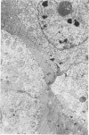
Mongolian gerbils fed diets containing lead acetate maintained body weight comparable to gerbils fed the same diet without added lead. Intranuclear lead inclusion bodies in epithelial cells of the proximal convoluted tubules of the kidney were first observed at 4 weeks, and increased in number to about 50 per high power field at 12 weeks. At this time, a corticomedullary area of empty-appearing tubules was prominent. Transmission electron microscopy confirmed the increase in number and size of nuclear lead inclusion over the 12-week period. Cytoplasmic changes observed in proximal tubule cells containing lead inclusions were considered indicative of acute lethal injury. Distinct cytoplasmic fibrillar structures, first apparent at 8 weeks, were present in some proximal tubular lining cells and strongly resembled newly formed intranuclear lead inclusions. After 12 weeks, the total amount of lead present in the gerbil kidney was four to six times greater than that in rat kidney as determined by atomic absorption spectrophotometry. A hypothesis has been formulated that relates the more efficient nephron of the gerbil kidney to the rapid and extensive development of intranuclear inclusion bodies and the greater accumulation of total lead.
Full text
PDF


Images in this article
Selected References
These references are in PubMed. This may not be the complete list of references from this article.
- BEAVER D. L. The ultrastructure of the kidney in lead intoxication with particular reference to intranuclear inclusions. Am J Pathol. 1961 Aug;39:195–208. [PMC free article] [PubMed] [Google Scholar]
- BOYLAND E., DUKES C. E., GROVER P. L., MITCHLEY B. C. The induction of renal tumors by feeding lead acetate to rats. Br J Cancer. 1962 Jun;16:283–288. doi: 10.1038/bjc.1962.33. [DOI] [PMC free article] [PubMed] [Google Scholar]
- Castellino N., Aloj S. Intracellular distribution of lead in the liver and kidney of the rat. Br J Ind Med. 1969 Apr;26(2):139–143. doi: 10.1136/oem.26.2.139. [DOI] [PMC free article] [PubMed] [Google Scholar]
- Castellino N., Lamanna P., Grieco B. Biliary excretion of lead in the rat. Br J Ind Med. 1966 Jul;23(3):237–239. doi: 10.1136/oem.23.3.237. [DOI] [PMC free article] [PubMed] [Google Scholar]
- Choie D. D., Richter G. W. Lead poisoning: rapid formation of intranuclear inclusions. Science. 1972 Sep 29;177(4055):1194–1195. doi: 10.1126/science.177.4055.1194. [DOI] [PubMed] [Google Scholar]
- DINISCHIOTU G. T., NESTORESCU B., RADULESCU I. C., IONESCU C., PREDA N., ILUTZA G. Studies on the chemical forms of urinary lead. Br J Ind Med. 1960 Apr;17:141–145. doi: 10.1136/oem.17.2.141. [DOI] [PMC free article] [PubMed] [Google Scholar]
- GOTTSCHALK C. W. OSMOTIC CONCENTRATION AND DILUTION OF THE URINE. Am J Med. 1964 May;36:670–685. doi: 10.1016/0002-9343(64)90179-2. [DOI] [PubMed] [Google Scholar]
- Goyer R. A., Krall A., Kimball J. P. The renal tubule in lead poisoning. II. In vitro studies of mitochondrial structure and function. Lab Invest. 1968 Jul;19(1):78–83. [PubMed] [Google Scholar]
- Goyer R. A. Lead toxicity: a problem in environmental pathology. Am J Pathol. 1971 Jul;64(1):167–182. [PMC free article] [PubMed] [Google Scholar]
- Goyer R. A., May P., Cates M. M., Krigman M. R. Lead and protein content of isolated intranuclear inclusion bodies from kidneys of lead-poisoned rats. Lab Invest. 1970 Mar;22(3):245–251. [PubMed] [Google Scholar]
- Goyer R. A. The renal tubule in lead poisoning. I. mMitochondrial swelling and aminoacidura. Lab Invest. 1968 Jul;19(1):71–77. [PubMed] [Google Scholar]
- PASSOW H., ROTHSTEIN A., CLARKSON T. W. The general pharmacology of the heavy metals. Pharmacol Rev. 1961 Jun;13:185–224. [PubMed] [Google Scholar]
- REYNOLDS E. S. The use of lead citrate at high pH as an electron-opaque stain in electron microscopy. J Cell Biol. 1963 Apr;17:208–212. doi: 10.1083/jcb.17.1.208. [DOI] [PMC free article] [PubMed] [Google Scholar]
- Richter G. W., Kress Y., Cornwall C. C. Another look at lead inclusion bodies. Am J Pathol. 1968 Aug;53(2):189–217. [PMC free article] [PubMed] [Google Scholar]
- SCHMIDT-NIELSEN K., SCHMIDT-NIELSEN B. Water metabolism of desert mammals 1. Physiol Rev. 1952 Apr;32(2):135–166. doi: 10.1152/physrev.1952.32.2.135. [DOI] [PubMed] [Google Scholar]
- Saltman P. The role of chelation in iron metabolism. J Chem Educ. 1965 Dec;42(12):682–687. doi: 10.1021/ed042p682. [DOI] [PubMed] [Google Scholar]
- Sun C. N., Mellies M., Yin M. W., Goyer R. A. The renal tubule in experimental lead intoxication. Arch Pathol. 1966 Aug;82(2):156–163. [PubMed] [Google Scholar]
- TANGE J. D., HAYWARD N. J., BREMNER D. A. RENAL LESIONS IN EXPERIMENTAL PLUMBISM AND THEIR CLINICAL IMPLICATIONS. Australas Ann Med. 1965 Feb;14:49–56. doi: 10.1111/imj.1965.14.1.49. [DOI] [PubMed] [Google Scholar]
- Vostál J., Heller J. Renal excretory mechanisms of heavy metals. I. Transtubular transport of heavy metal ions in the avian kidney. Environ Res. 1968 Sep;2(1):1–10. doi: 10.1016/0013-9351(68)90001-7. [DOI] [PubMed] [Google Scholar]
- van ESCH G., van GENDEREN, VINK H. H. The induction of renal tumours by feeding of basic lead acetate to rats. Br J Cancer. 1962 Jun;16:289–297. doi: 10.1038/bjc.1962.34. [DOI] [PMC free article] [PubMed] [Google Scholar]


