

Abstract
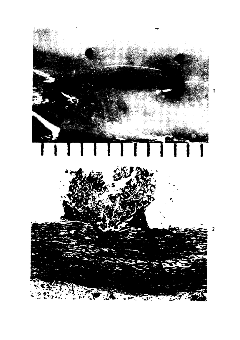
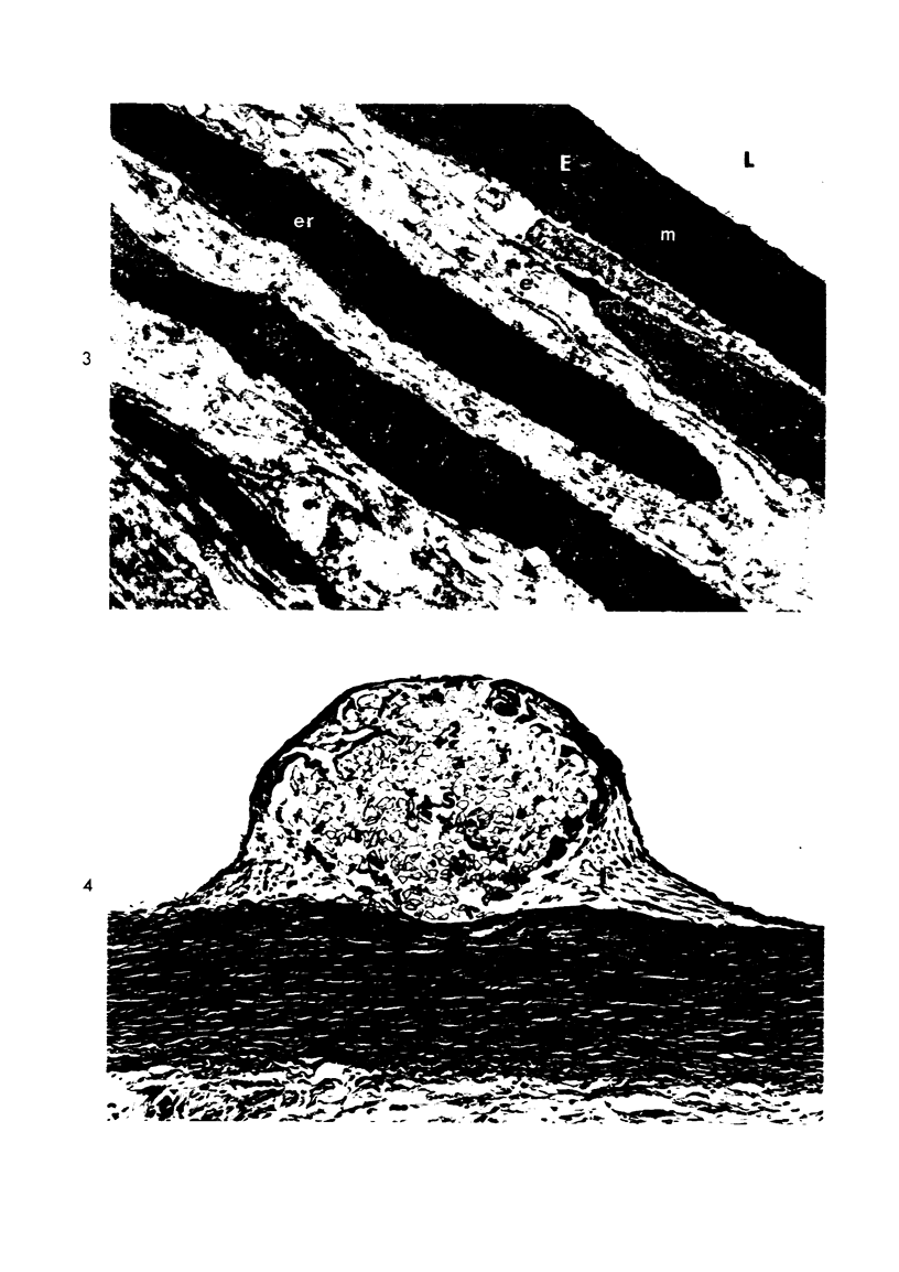
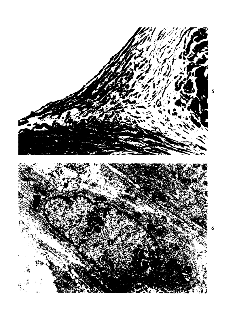
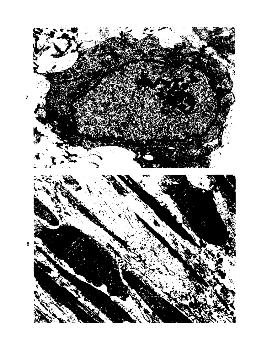
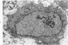
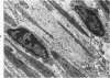
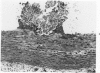
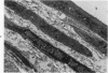
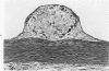
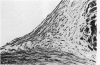
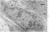
Repair to injury of the rabbit aorta was studied with special attention to the source of new intimal cells. Two types of injury, suture placement and electrocautery, were used to ascertain whether the type of injury significantly modifies the reparative response of the artery. Cell morphology and DNA synthesis, as determined by autoradiographic localization of tritiated thymidine uptake, were studied sequentially during the course of the repair reaction. Suture injury was the more reliable model for these studies because the magnitude of injury was quite comparable between animals, while with cauterization there was considerable variation in the magnitude of injury. The overall reparative response with the two types of injury was, however, similar. Cell division was first seen in the media adjacent to the injured focus and was followed by migration of medial cells into the intima. In the intima, medial cells continued to divide for a few days and then gradually matured from undifferentiated ovoid cells resembling monocytes to morphologically differentiated smooth muscle cells. Medial cells exhibiting DNA synthesis were both differentiated and undifferentiated smooth muscle cells, supporting previous reports that mature as well as undifferentiated smooth muscle cells are capable of mitosis. However, with injury and repair, the migrating cells appeared to be mainly undifferentiated since there was a predominance of these cells early in the repair reaction in the intima.
Full text
PDF
Images in this article
Selected References
These references are in PubMed. This may not be the complete list of references from this article.
- DAOUD A., JARMOLYCH J., ZUMBO A., FANI, FLORENTIN R. PREATHEROMA PHASE OF CORONARY ATHEROSCLEROSIS IN MAN. Exp Mol Pathol. 1964 Oct;90:475–484. doi: 10.1016/0014-4800(64)90028-0. [DOI] [PubMed] [Google Scholar]
- Geer J. C. Fine structure of human aortic intimal thickening and fatty streaks. Lab Invest. 1965 Oct;14(10):1764–1783. [PubMed] [Google Scholar]
- Gross L., Epstein E. Z., Kugel M. A. Histology of the Coronary Arteries and their Branches in the Human Heart. Am J Pathol. 1934 Mar;10(2):253–274.7. [PMC free article] [PubMed] [Google Scholar]
- Hassler O. Cell renewal in aortic necrosis following orthostatic collapse. An experimental autoradiographic study in the rabbit using 3 H-thymidine. Virchows Arch A Pathol Pathol Anat. 1973 Feb 19;358(4):295–299. doi: 10.1007/BF00543270. [DOI] [PubMed] [Google Scholar]
- Hassler O. The origin of the cells constituting arterial intima thickening. An experimental autoradiographic study with the use of H3-thymidine. Lab Invest. 1970 Apr;22(4):286–293. [PubMed] [Google Scholar]
- Haust M. D., More R. H., Bencosme S. A., Balis J. U. Elastogenesis in human aorta: an electron microscopic study. Exp Mol Pathol. 1965 Oct;4(5):508–524. doi: 10.1016/0014-4800(65)90015-8. [DOI] [PubMed] [Google Scholar]
- Jorgensen L., Rowsell H. C., Hovig T., Mustard J. F. Resolution and organization of platelet-rich mural thrombi in carotid arteries of swine. Am J Pathol. 1967 Nov;51(5):681–719. [PMC free article] [PubMed] [Google Scholar]
- Knieriem H. J., Bondjers G., Björkerud S. Electron microscopy of intimal plaques following induction of large superficial mechanical injury (transverse injury) in the rabbit aorta. Virchows Arch A Pathol Pathol Anat. 1973 Jun 29;359(4):267–282. doi: 10.1007/BF00548598. [DOI] [PubMed] [Google Scholar]
- Kádár A., Veress B., Jellinek H. Ultrastructural elements in experimental intimal thickening. II. Study of the development of elastic elements in intimal proliferation. Exp Mol Pathol. 1969 Oct;11(2):212–223. doi: 10.1016/0014-4800(69)90009-4. [DOI] [PubMed] [Google Scholar]
- MOVAT H. Z., MORE R. H., HAUST M. D. The diffuse intimal thickening of the human aorta with aging. Am J Pathol. 1958 Nov-Dec;34(6):1023–1031. [PMC free article] [PubMed] [Google Scholar]
- Moss N. S., Benditt E. P. The ultrastructure of spontaneous and experimentally induced arterial lesions. 3. The cholesterol-induced lesions and the effect of a cholesterol and oil diet on the preexisting spontaneous plaque in the chicken aorta. Lab Invest. 1970 Nov;23(5):521–535. [PubMed] [Google Scholar]
- Moss N. S., Benditt E. P. The ultrastructure of spontaneous and experimentally induced arterial lesions. II. The spontaneous plaque in the chicken. Lab Invest. 1970 Sep;23(3):231–245. [PubMed] [Google Scholar]
- Murray M., Schrodt G. R., Berg H. G. Role of smooth muscle cells in healing of injured arteries. Arch Pathol. 1966 Aug;82(2):138–146. [PubMed] [Google Scholar]
- Parker F. An Electron Microscopic Study of Experimental Atherosclerosis. Am J Pathol. 1960 Jan;36(1):19–53. [PMC free article] [PubMed] [Google Scholar]
- Poole J. C., Cromwell S. B., Benditt E. P. Behavior of smooth muscle cells and formation of extracellular structures in the reaction of arterial walls to injury. Am J Pathol. 1971 Mar;62(3):391–414. [PMC free article] [PubMed] [Google Scholar]
- Ross R., Klebanoff S. J. The smooth muscle cell. I. In vivo synthesis of connective tissue proteins. J Cell Biol. 1971 Jul;50(1):159–171. doi: 10.1083/jcb.50.1.159. [DOI] [PMC free article] [PubMed] [Google Scholar]
- Ross R. The smooth muscle cell. II. Growth of smooth muscle in culture and formation of elastic fibers. J Cell Biol. 1971 Jul;50(1):172–186. doi: 10.1083/jcb.50.1.172. [DOI] [PMC free article] [PubMed] [Google Scholar]
- Stemerman M. B., Ross R. Experimental arteriosclerosis. I. Fibrous plaque formation in primates, an electron microscope study. J Exp Med. 1972 Oct 1;136(4):769–789. doi: 10.1084/jem.136.4.769. [DOI] [PMC free article] [PubMed] [Google Scholar]
- THOMAS W. A., JONES R., SCOTT R. F., MORRISON E., GOODALE M. F., IMAI H. PRODUCTION OF EARLY ATHEROSCLEROTIC LESIONS IN RATS CHARACTERIZED BY PROLIFERATION OF "MODIFIED SMOOTH MUSCLE CELLS". Exp Mol Pathol. 1963 Aug;52:SUPPL1–SUPPL1:61. [PubMed] [Google Scholar]