

Abstract
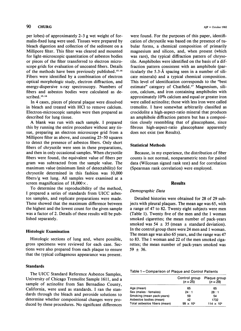
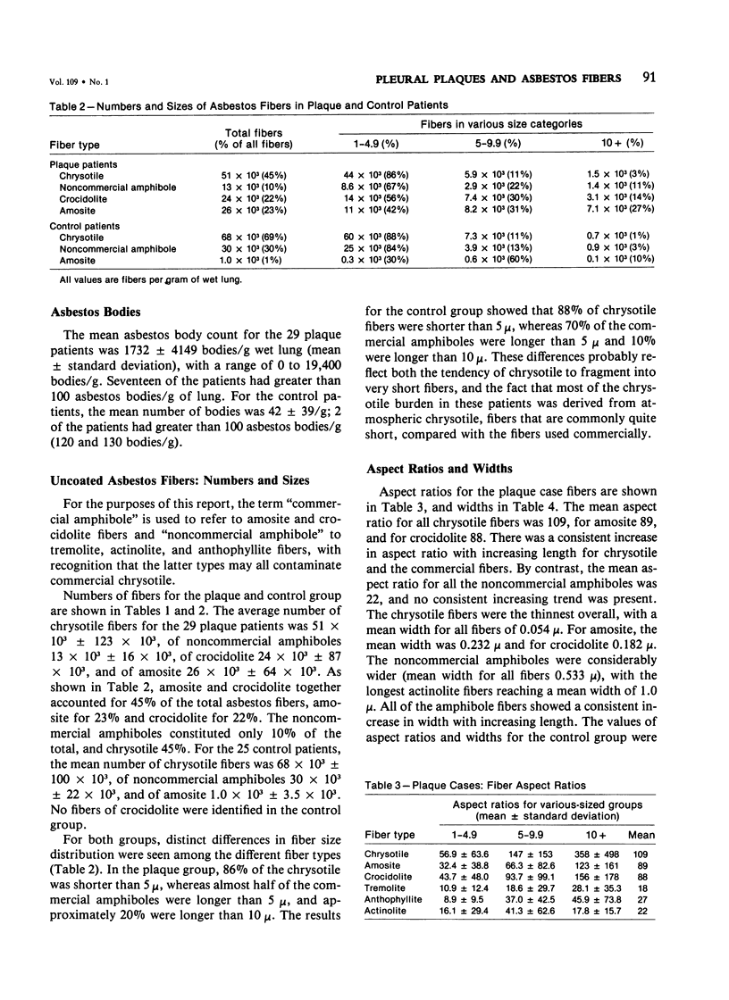
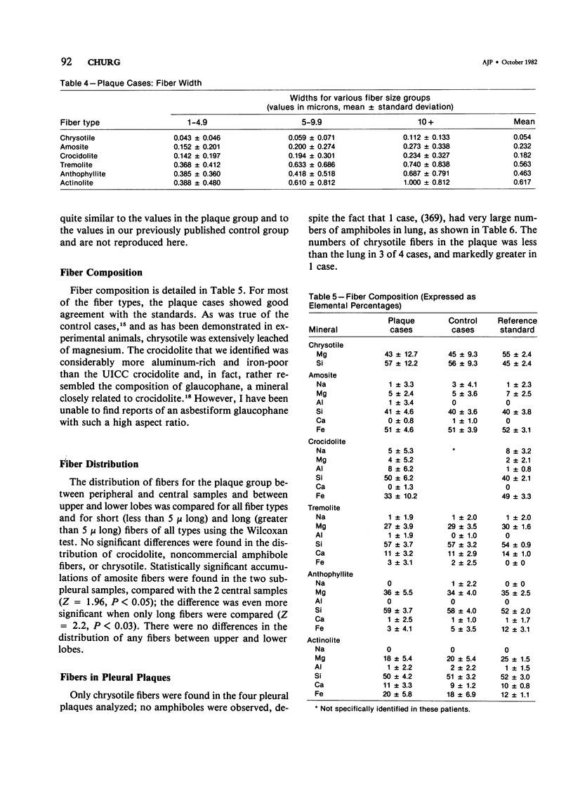
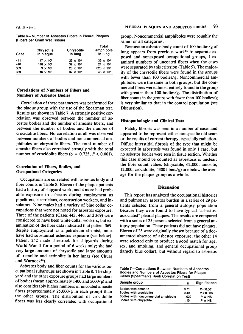
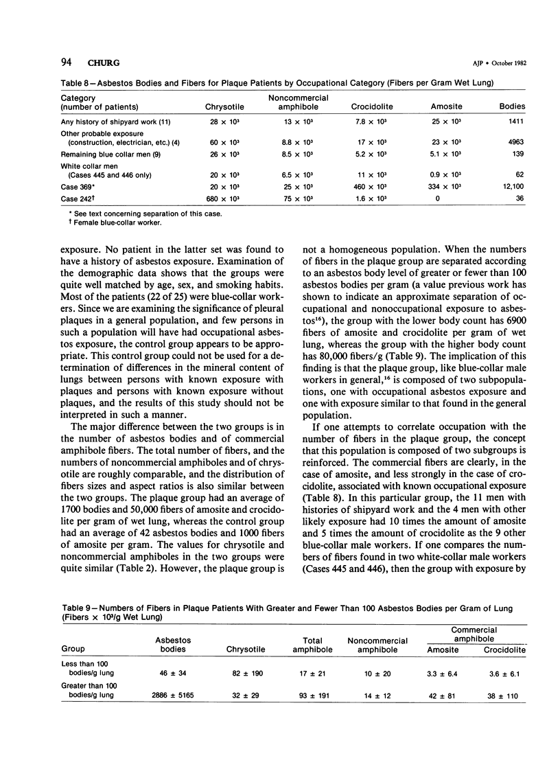
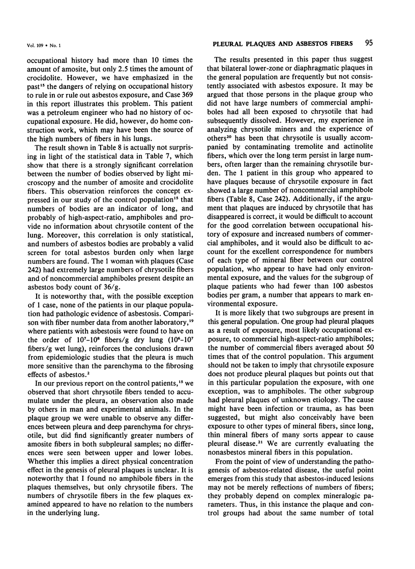
It has been claimed that symmetric lower zone pleural or diaphargmatic plaques are markers of asbestos exposure both in asbestos workers and the general population. In this study, total pulmonary asbestos burden was analyzed for 29 patients selected because pleural plaques were found at autopsy, and the results were compared with values obtained for 25 patients who had no occupational asbestos exposure. The average number of asbestos bodies in the plaque groups was 1732/g wet lung, and in the control group, 42/g wet lung. Uncoated asbestos fibers were extracted from lung and counted, measured, and identified by morphologic examination, electron diffraction, and energy-dispersive x-ray spectroscopy. The total number of fibers/per gram wet lung in the plaque group (114 x 10(3)) was similar to that in the control group (99 x 10(3), as was the number of chrysotile fibers (51 x 10(3) versus 29 x 10(3)). However, the plaque patients had a marked increase in the number of the commercially used high aspect ratio amphiboles, amosite and crocidolite (50 x 10(3) versus 1 x 10(3). A retrospective history of fairly certain asbestos exposure was obtained for 16 of the plaque patients, and such a history correlated strongly with increased numbers of commercial amphiboles in lung. It is concluded that 1) in this general autopsy population, two subgroups of patients are present. About one half of the patients appear to have developed pleural plaques as a result of asbestos exposure, while the etiology of the plaques in the other half is unclear; 2) the presence of pleural plaques correlates with a modest (50-fold) increase in numbers of long high-aspect ratio commercial amphiboles in lung tissue but does not correlate with numbers of chrysotile fibers, noncommercial amphiboles, or the total number of asbestos fibers; 3) asbestos-induced lesions are related to a complex set of mineralogic parameters and not to mere numbers of fibers in lung.
Full text
PDF

Selected References
These references are in PubMed. This may not be the complete list of references from this article.
- Ashcroft T., Heppleston A. G. The optical and electron microscopic determination of pulmonary asbestos fibre concentration and its relation to the human pathological reaction. J Clin Pathol. 1973 Mar;26(3):224–234. doi: 10.1136/jcp.26.3.224. [DOI] [PMC free article] [PubMed] [Google Scholar]
- Bariş Y. I., Artvinli M., Sahin A. A. Environmental mesothelioma in Turkey. Ann N Y Acad Sci. 1979;330:423–432. doi: 10.1111/j.1749-6632.1979.tb18744.x. [DOI] [PubMed] [Google Scholar]
- Becklake M. R. Asbestos-related diseases of the lung and other organs: their epidemiology and implications for clinical practice. Am Rev Respir Dis. 1976 Jul;114(1):187–227. doi: 10.1164/arrd.1976.114.1.187. [DOI] [PubMed] [Google Scholar]
- Churg A., Warnock M. L. Asbestos fibers in the general population. Am Rev Respir Dis. 1980 Nov;122(5):669–678. doi: 10.1164/arrd.1980.122.5.669. [DOI] [PubMed] [Google Scholar]
- Churg A., Warnock M. L. Correlation of quantitative asbestos body counts and occupation in urban patients. Arch Pathol Lab Med. 1977 Dec;101(12):629–634. [PubMed] [Google Scholar]
- Gibbs G. W. Etiology of pleural calcification: a study of Quebec chrysotile asbestos miners and millers. Arch Environ Health. 1979 Mar-Apr;34(2):76–83. doi: 10.1080/00039896.1979.10667372. [DOI] [PubMed] [Google Scholar]
- Hourihane D. O., Lessof L., Richardson P. C. Hyaline and calcified pleural plaques as an index of exposure to asbestos. Br Med J. 1966 Apr 30;1(5495):1069–1074. doi: 10.1136/bmj.1.5495.1069. [DOI] [PMC free article] [PubMed] [Google Scholar]
- Le Bouffant L. Investigation and analysis of asbestos fibers and accompanying minerals in biological materials. Environ Health Perspect. 1974 Dec;9:149–153. doi: 10.1289/ehp.749149. [DOI] [PMC free article] [PubMed] [Google Scholar]
- Meurman L. Asbestos bodies and pleural plaques in a Finnish series of autopsy cases. Acta Pathol Microbiol Scand. 1966;(Suppl):1+–1+. [PubMed] [Google Scholar]
- Navrátil M., Trippé F. Prevalence of pleural calcification in persons exposed to asbestos dust, and in the general population in the same district. Environ Res. 1972 Jun;5(2):210–216. doi: 10.1016/0013-9351(72)90035-7. [DOI] [PubMed] [Google Scholar]
- Roberts G. H. The pathology of parietal pleural plaques. J Clin Pathol. 1971 May;24(4):348–353. doi: 10.1136/jcp.24.4.348. [DOI] [PMC free article] [PubMed] [Google Scholar]
- Rous V., Studený J. Aetiology of pleural plaques. Thorax. 1970 May;25(3):270–284. doi: 10.1136/thx.25.3.270. [DOI] [PMC free article] [PubMed] [Google Scholar]
- Stanton M. F., Laynard M., Tegeris A., Miller E., May M., Kent E. Carcinogenicity of fibrous glass: pleural response in the rat in relation to fiber dimension. J Natl Cancer Inst. 1977 Mar;58(3):587–603. doi: 10.1093/jnci/58.3.587. [DOI] [PubMed] [Google Scholar]
- Whitwell F., Scott J., Grimshaw M. Relationship between occupations and asbestos-fibre content of the lungs in patients with pleural mesothelioma, lung cancer, and other diseases. Thorax. 1977 Aug;32(4):377–386. doi: 10.1136/thx.32.4.377. [DOI] [PMC free article] [PubMed] [Google Scholar]