

Abstract
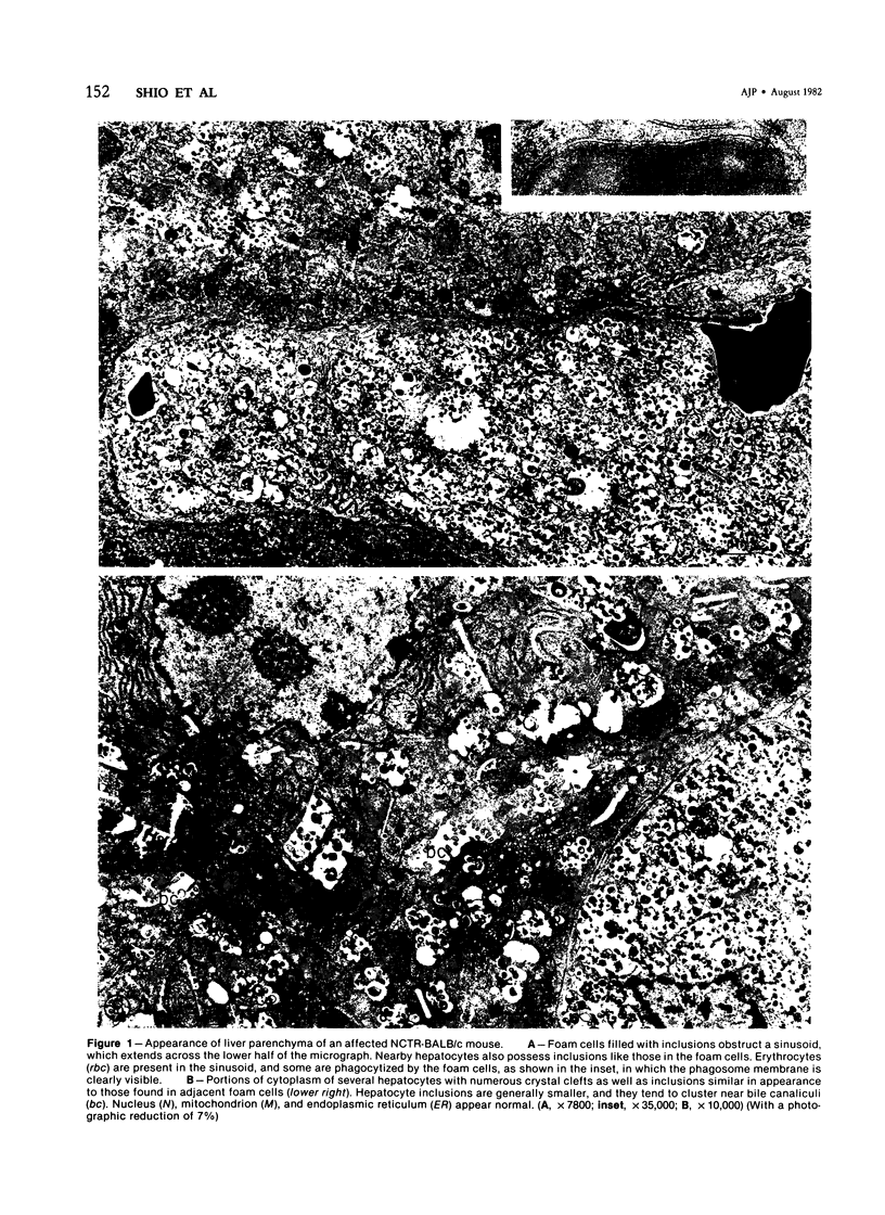
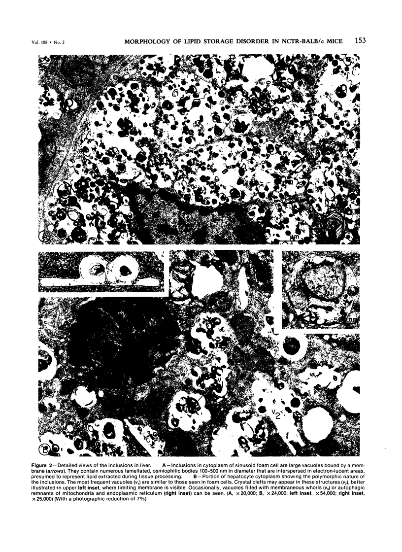
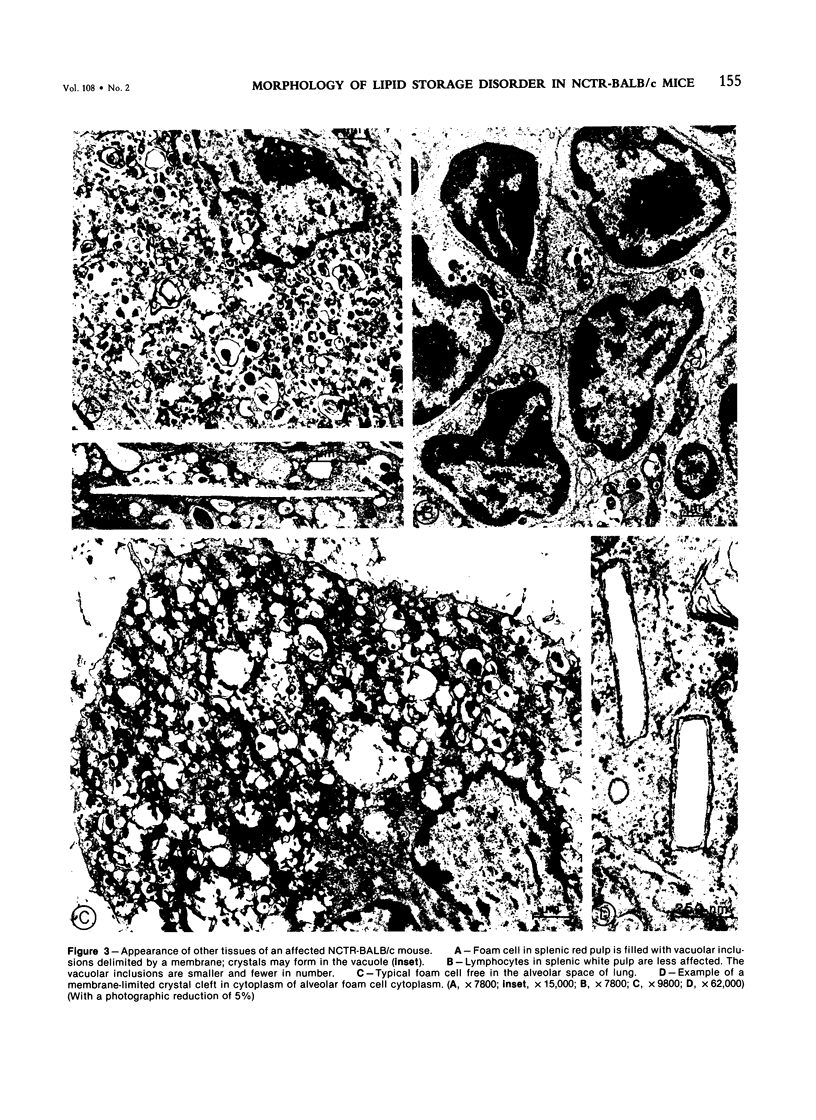
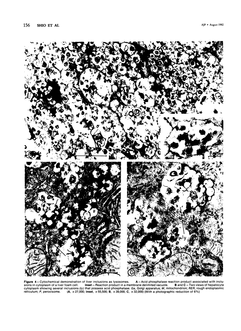
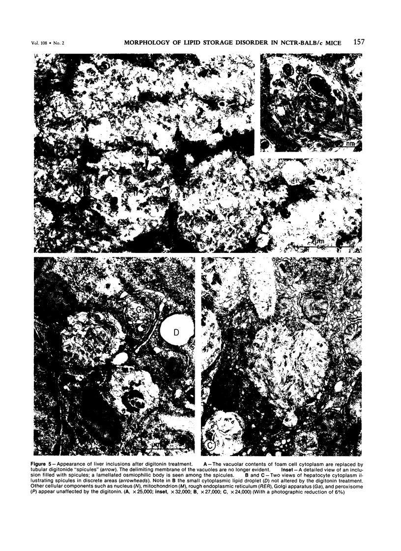
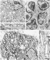
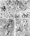
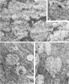
Electron-microscopic and cytochemical studies were carried out on tissues of NCTR-BALB/c mice. These mice are affected with a neurovisceral genetic disorder involving excessive tissue accumulation of lipid. Distinctive polymorphic intracellular inclusions, bounded by a membrane and containing lamellated bodies, were found in many cells of liver, spleen, lung, kidney, intestine, lymph nodes, and brain. The inclusions transformed reticuloendothelial cells into massive foam cells. Acid phosphatase cytochemical studies performed on sections of liver demonstrated that the inclusions were lysosomes. Fixation of liver in the presence of digitonin produced "spicules" in the inclusions characteristic of digitonin-cholesterol complexes. Clefts of cholesterol crystals were seen in the inclusions in liver, spleen, and lung. We conclude that the NCTR-BALB/c mice are affected by a lysosome lipid storage disease and that cholesterol is a major storage product.
Full text
PDF
Images in this article
Selected References
These references are in PubMed. This may not be the complete list of references from this article.
- Adachi M., Tsai C. Y., Hoffman L. M., Schneck L., Volk B. W. The central nervous system, liver, and spleen of FM mice. Ultrastructural, histochemical, and biochemical studies. Arch Pathol. 1974 Apr;97(4):232–238. [PubMed] [Google Scholar]
- Farquhar M. G., Palade G. E. Cell junctions in amphibian skin. J Cell Biol. 1965 Jul;26(1):263–291. doi: 10.1083/jcb.26.1.263. [DOI] [PMC free article] [PubMed] [Google Scholar]
- Karnovsky M. J. The ultrastructural basis of capillary permeability studied with peroxidase as a tracer. J Cell Biol. 1967 Oct;35(1):213–236. doi: 10.1083/jcb.35.1.213. [DOI] [PMC free article] [PubMed] [Google Scholar]
- LYNN R., TERRY R. D. LIPID HISTOCHEMISTRY AND ELECTRON MICROSCOPY IN ADULT NIEMANN-PICK DISEASE. Am J Med. 1964 Dec;37:987–994. doi: 10.1016/0002-9343(64)90139-1. [DOI] [PubMed] [Google Scholar]
- Lazarus S. S., Vethamany V. G., Schneck L., Volk B. W. Fine structure and histochemistry of peripheral blood cells in Niemann-Pick disease. Lab Invest. 1967 Aug;17(2):155–170. [PubMed] [Google Scholar]
- Morris M. D., Bhuvaneswaran C., Shio H., Fowler S. Lysosome lipid storage disorder in NCTR-BALB/c mice. I. Description of the disease and genetics. Am J Pathol. 1982 Aug;108(2):140–149. [PMC free article] [PubMed] [Google Scholar]
- Novikoff P. M., Novikoff A. B., Quintana N., Hauw J. J. Golgi apparatus, GERL, and lysosomes of neurons in rat dorsal root ganglia, studied by thick section and thin section cytochemistry. J Cell Biol. 1971 Sep;50(3):859–886. doi: 10.1083/jcb.50.3.859. [DOI] [PMC free article] [PubMed] [Google Scholar]
- Okrös I. Digitonin reaction in electron microscopy. Histochemie. 1968;13(1):91–96. doi: 10.1007/BF00303878. [DOI] [PubMed] [Google Scholar]
- REYNOLDS E. S. The use of lead citrate at high pH as an electron-opaque stain in electron microscopy. J Cell Biol. 1963 Apr;17:208–212. doi: 10.1083/jcb.17.1.208. [DOI] [PMC free article] [PubMed] [Google Scholar]
- Scallen T. J., Dietert S. E. The quantitative retention of cholesterol in mouse liver prepared for electron microscopy by fixation in a digitonin-containing aldehyde solution. J Cell Biol. 1969 Mar;40(3):802–813. doi: 10.1083/jcb.40.3.802. [DOI] [PMC free article] [PubMed] [Google Scholar]
- Shio H., Haley N. J., Fowler S. Characterization of lipid-laden aortic cells from cholesterol-fed rabbits. III. Intracellular localization of cholesterol and cholesteryl ester. Lab Invest. 1979 Aug;41(2):160–167. [PubMed] [Google Scholar]
- Vethamany V. G., Welch J. P., Vethamany S. K. Type D Niemann-Pick disease (Nova Scotia variant). Ultrastructure of blood, skin fibroblasts, and bone marrow. Arch Pathol. 1972 Jun;93(6):537–543. [PubMed] [Google Scholar]
- Volk B. W., Wallace B. J. The liver in lipidosis. An electron miscroscopic and histochemical study. Am J Pathol. 1966 Aug;49(2):203–225. [PMC free article] [PubMed] [Google Scholar]
- WATSON M. L. Staining of tissue sections for electron microscopy with heavy metals. J Biophys Biochem Cytol. 1958 Jul 25;4(4):475–478. doi: 10.1083/jcb.4.4.475. [DOI] [PMC free article] [PubMed] [Google Scholar]
- Wallace B. J., Schneck L., Kaplan H., Volk B. W. Fine structure of the cerebellum of children with lipidoses. Arch Pathol. 1965 Nov;80(5):466–486. [PubMed] [Google Scholar]
- Williamson J. R. Ultrastructural localization and distribution of free cholesterol (3 beta-hydroxysterols) in tissues. J Ultrastruct Res. 1969 Apr;27(1):118–133. [PubMed] [Google Scholar]