

Abstract
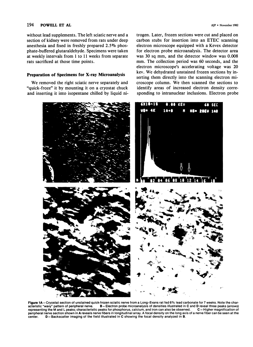
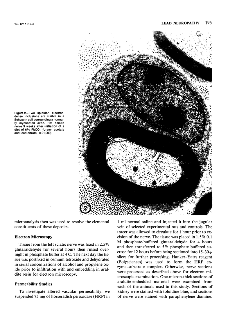
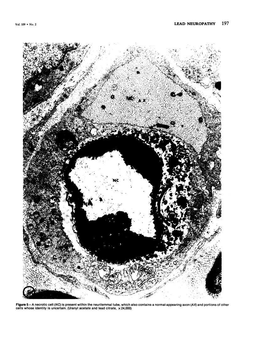
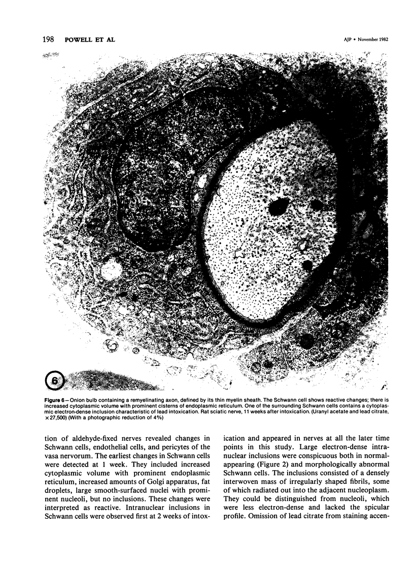
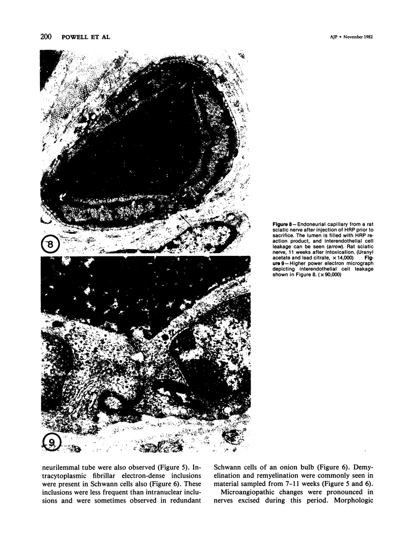
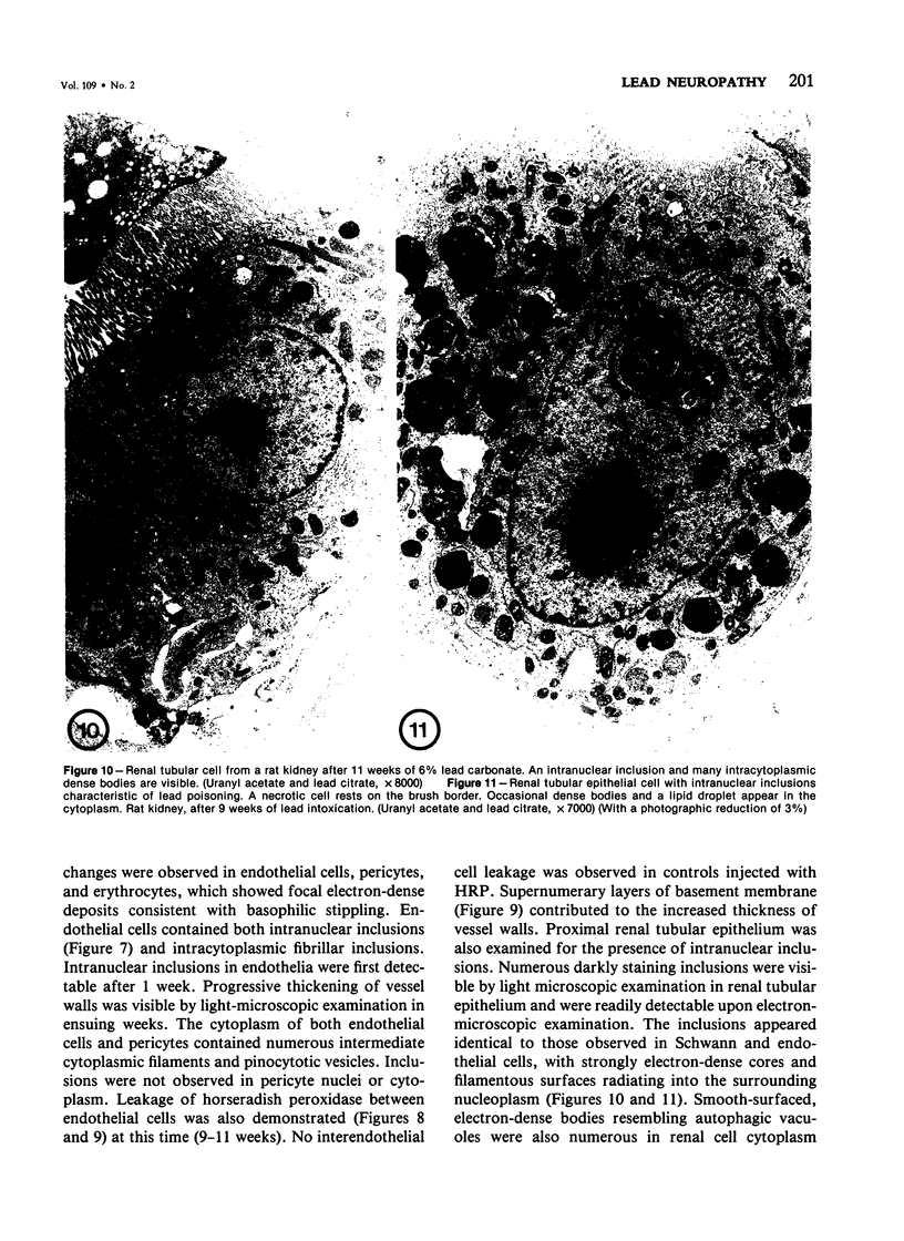
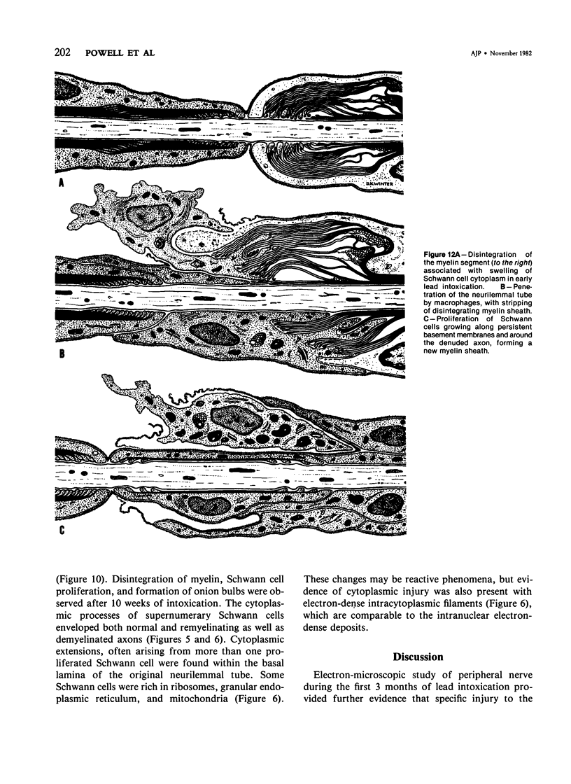
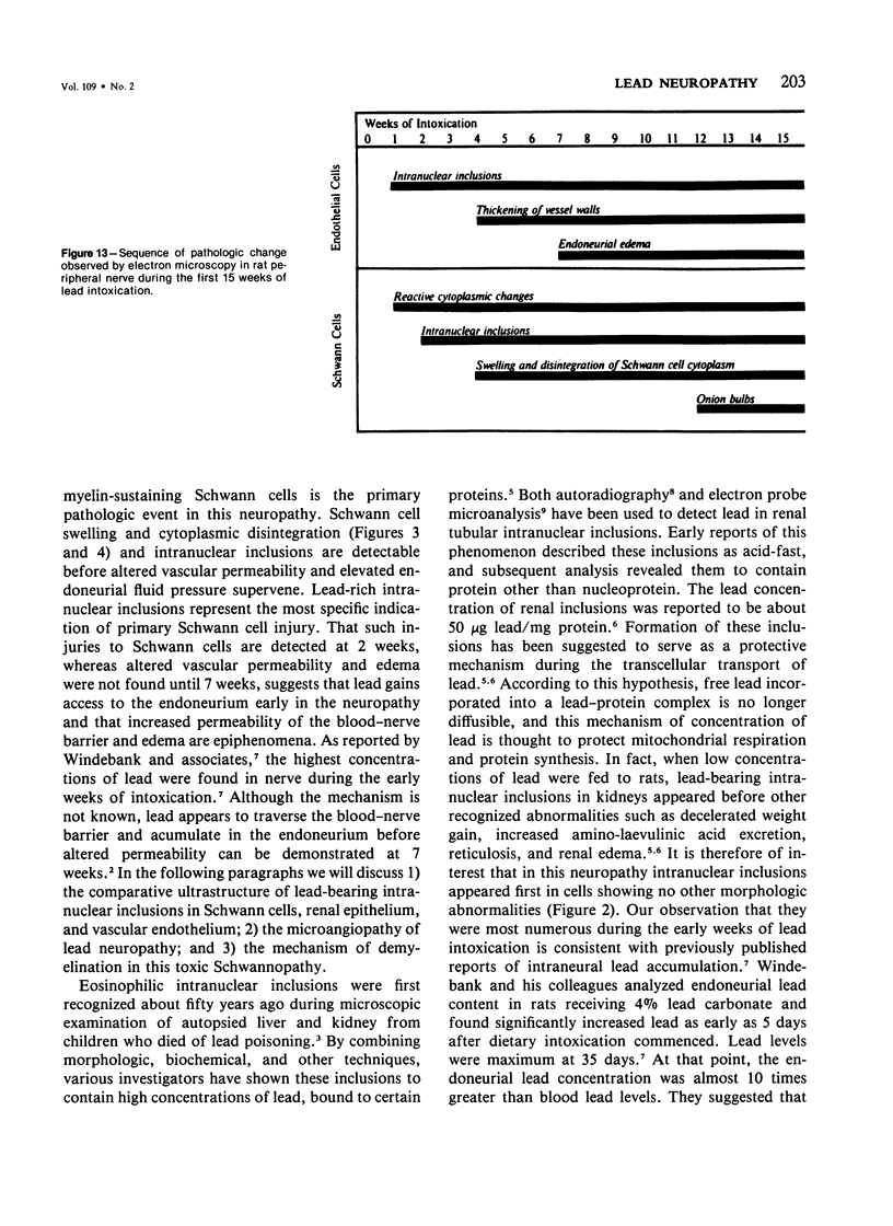
Transmission electron microscopy (TEM) of peripheral nerve in rats receiving 6% lead carbonate for 4-10 weeks provided evidence of a specific Schwann cell injury, associated with demyelination. Intranuclear inclusions in Schwann cells appeared within 2 weeks of administration of a lead-containing diet. Swelling of Schwann cells and disintegration of their cytoplasm was evident at 4 weeks. Distinctive electron-dense inclusions appeared in both Schwann and endothelial cells during the period of intoxication and were ultrastructurally identical to pathognomonic inclusions of lead poisoning seen in renal tubular epithelial cells. Scanning microscopy (SEM) with electron-probe microanalysis was used to identify the lead-containing deposits. In addition to Schwann cell changes, vessels revealed endothelial cell injury and alteread permeability to macromolecules. Since morphologic changes of Schwann cells precede the development of altered vascular permeability and endoneurial edema, it appears that lead gains access to the endoneurium prior to the development of altered vascular permeability, suggesting that edema and altered endoneurial fluid pressure are epiphenomena that supervene after demyelination occurs. Remyelination, Schwann cell proliferation and formation of onion bulbs are manifestations of persistent toxic injury to myelin-sustaining cells, resulting in chronic demyelination.
Full text
PDF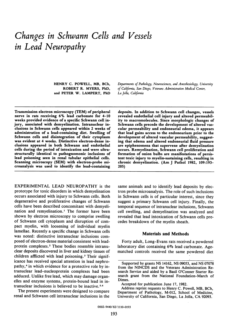

Images in this article

Selected References
These references are in PubMed. This may not be the complete list of references from this article.
- Carroll K. G., Spinelli F. R., Goyer R. A. Electron probe microanalyser localization of lead in kidney tissue of poisoned rats. Nature. 1970 Sep 5;227(5262):1056–1056. doi: 10.1038/2271056a0. [DOI] [PubMed] [Google Scholar]
- Choie D. D., Richter G. W. Lead poisoning: rapid formation of intranuclear inclusions. Science. 1972 Sep 29;177(4055):1194–1195. doi: 10.1126/science.177.4055.1194. [DOI] [PubMed] [Google Scholar]
- Clasen R. A., Hartmann J. F., Starr A. J., Coogan P. S., Pandolfi S., Laing I., Becker R., Hass G. M. Electron microscopic and chemical studies of the vascular changes and edema of lead encephalopathy. A comparative study of the human and experimental disease. Am J Pathol. 1974 Feb;74(2):215–240. [PMC free article] [PubMed] [Google Scholar]
- Goyer R. A., Leonard D. L., Moore J. F., Rhyne B., Krigman M. R. Lead dosage and the role of the intranuclear inclusion body. An experimental study. Arch Environ Health. 1970 Jun;20(6):705–711. doi: 10.1080/00039896.1970.10665647. [DOI] [PubMed] [Google Scholar]
- Goyer R. A., May P., Cates M. M., Krigman M. R. Lead and protein content of isolated intranuclear inclusion bodies from kidneys of lead-poisoned rats. Lab Invest. 1970 Mar;22(3):245–251. [PubMed] [Google Scholar]
- Hsu J. S., Herman M. M., Hsu H. J., Mortell P. The pathogenesis of lead encephalopathy. Effects of lead carbonate feedings on morphology, lead content, and mitochondrial respiration in brains of immature and adult rats. Virchows Arch A Pathol Anat Histol. 1980;387(2):147–164. doi: 10.1007/BF00430696. [DOI] [PubMed] [Google Scholar]
- Lampert P. W., Schochet S. S., Jr Demyelination and remyelination in lead neuropathy. Electron microscopic studies. J Neuropathol Exp Neurol. 1968 Oct;27(4):527–545. [PubMed] [Google Scholar]
- Low P. A., Dyck P. J. Increased endoneurial fluid pressure in experimental lead neuropathy. Nature. 1977 Sep 29;269(5627):427–428. doi: 10.1038/269427a0. [DOI] [PubMed] [Google Scholar]
- Myers R. R., Powell H. C., Shapiro H. M., Costello M. L., Lampert P. W. Changes in endoneurial fluid pressure, permeability, and peripheral nerve ultrastructure in experimental lead neuropathy. Ann Neurol. 1980 Oct;8(4):392–401. doi: 10.1002/ana.410080410. [DOI] [PubMed] [Google Scholar]
- Richter G. W. Evolution of cytoplasmic fibrillar bodies induced by lead in rat and mouse kidneys. Am J Pathol. 1976 Apr;83(1):135–148. [PMC free article] [PubMed] [Google Scholar]
- Silbergeld E. K., Wolinsky J. S., Goldstein G. W. Electron probe microanalysis of isolated brain capillaries poisoned with lead. Brain Res. 1980 May 12;189(2):369–376. doi: 10.1016/0006-8993(80)90097-9. [DOI] [PubMed] [Google Scholar]
- Windebank A. J., McCall J. T., Hunder H. G., Dyck P. J. The endoneurial content of lead related to the onset and severity of segmental demyelination. J Neuropathol Exp Neurol. 1980 Nov;39(6):692–699. doi: 10.1097/00005072-198011000-00008. [DOI] [PubMed] [Google Scholar]