Abstract
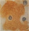
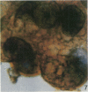
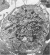
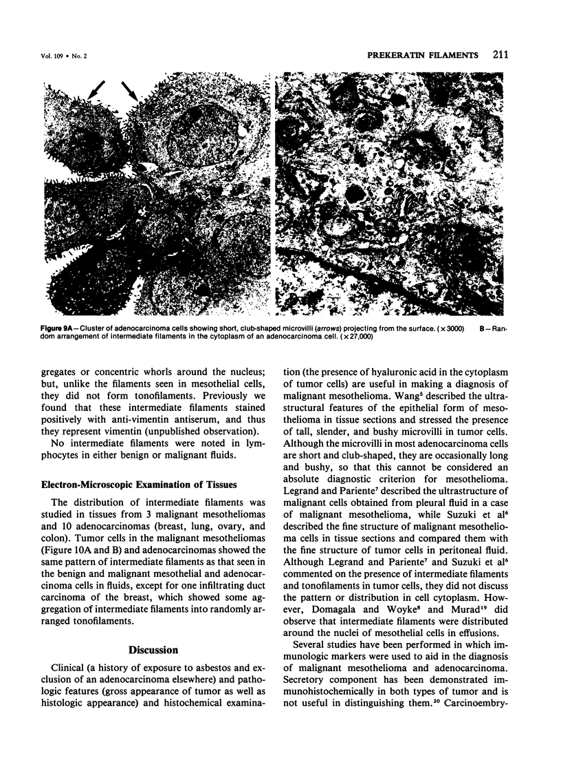
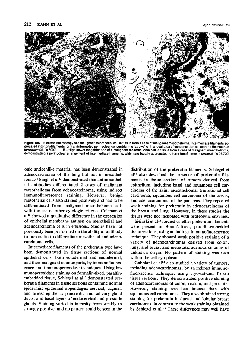
An immunoperoxidase technique employing antibody to prekeratin was used to study distribution and pattern of staining of prekeratin filaments in cytological smears obtained from 42 specimens of pleural and peritoneal effusions (27 benign, 15 malignant). The smears were either air-dried or ethanol-fixed. Both benign and malignant mesothelial cells showed distinctive peripheral or perinuclear staining patterns which differed from the characteristic arborizing pattern in adenocarcinoma cells. The ultrastructure of these 2 cell types studied in 27 body fluids (12 benign, 15 malignant) and in 13 malignant tumors (3 mesotheliomas, 10 adenocarcinomas) showed a distinctive localizaton of intermediate filaments which corresponded to and could explain the pattern of staining obtained using the immunoperoxidase technique. The immunohistochemical and ultrastructural findings appeared characteristic for benign and malignant mesothelial cells as well as for adenocarcinoma cells, and could be used as markers to differentiate mesothelial tumors and reactive mesothelial cells from adenocarcinomas.
Full text
PDF
Images in this article
Selected References
These references are in PubMed. This may not be the complete list of references from this article.
- Coleman D. V., To A., Ormerod M. G., Dearnaley D. P. Immunoperoxidase staining in tumor marker distribution studies in cytologic specimen. Acta Cytol. 1981 May-Jun;25(3):205–206. [PubMed] [Google Scholar]
- Domagala W., Woyke S. Transmission and scanning electron microscopic studies of cells in effusions. Acta Cytol. 1975 May-Jun;19(3):214–224. [PubMed] [Google Scholar]
- Ernst C. S., Brooks J. J. Immunoperoxidase localization of secretory component in reactive mesothelium and mesotheliomas. J Histochem Cytochem. 1981 Sep;29(9):1102–1104. doi: 10.1177/29.9.7026669. [DOI] [PubMed] [Google Scholar]
- Franke W. W., Appelhans B., Schmid E., Freudenstein C., Osborn M., Weber K. Identification and characterization of epithelial cells in mammalian tissues by immunofluorescence microscopy using antibodies to prekeratin. Differentiation. 1979;15(1):7–25. doi: 10.1111/j.1432-0436.1979.tb01030.x. [DOI] [PubMed] [Google Scholar]
- Franke W. W., Schmid E., Osborn M., Weber K. Different intermediate-sized filaments distinguished by immunofluorescence microscopy. Proc Natl Acad Sci U S A. 1978 Oct;75(10):5034–5038. doi: 10.1073/pnas.75.10.5034. [DOI] [PMC free article] [PubMed] [Google Scholar]
- Franke W. W., Weber K., Osborn M., Schmid E., Freudenstein C. Antibody to prekeratin. Decoration of tonofilament like arrays in various cells of epithelial character. Exp Cell Res. 1978 Oct 15;116(2):429–445. doi: 10.1016/0014-4827(78)90466-4. [DOI] [PubMed] [Google Scholar]
- Gabbiani G., Kapanci Y., Barazzone P., Franke W. W. Immunochemical identification of intermediate-sized filaments in human neoplastic cells. A diagnostic aid for the surgical pathologist. Am J Pathol. 1981 Sep;104(3):206–216. [PMC free article] [PubMed] [Google Scholar]
- Hameed K., Morgan D. A. Papillary adenocarcinoma of endometrium with psammoma bodies. Histology and fine structure. Cancer. 1972 May;29(5):1326–1335. doi: 10.1002/1097-0142(197205)29:5<1326::aid-cncr2820290530>3.0.co;2-x. [DOI] [PubMed] [Google Scholar]
- Kannerstein M., Churg J. Peritoneal mesothelioma. Hum Pathol. 1977 Jan;8(1):83–94. doi: 10.1016/s0046-8177(77)80067-1. [DOI] [PubMed] [Google Scholar]
- Legrand M., Pariente R. Ultrastructural study of pleural fluid in mesothelioma. Thorax. 1974 Mar;29(2):164–171. doi: 10.1136/thx.29.2.164. [DOI] [PMC free article] [PubMed] [Google Scholar]
- McCaughey W. T. Asbestos and neoplasia: diffuse mesothelial tumors. Criteria for diagnosis of diffuse mesothelial tumors. Ann N Y Acad Sci. 1965 Dec 31;132(1):603–613. doi: 10.1111/j.1749-6632.1965.tb41140.x. [DOI] [PubMed] [Google Scholar]
- Murad T. M. Electron microscopic studies of cells in pleural and peritoneal effusions. Acta Cytol. 1973 Sep-Oct;17(5):401–409. [PubMed] [Google Scholar]
- NAYLOR B. THE EXFOLIATIVE CYTOLOGY OF DIFFUSE MALIGNANT MESOTHELIOMA. J Pathol Bacteriol. 1963 Oct;86:293–298. doi: 10.1002/path.1700860204. [DOI] [PubMed] [Google Scholar]
- Roberts G. H., Campbell G. M. Exfoliative cytology of diffuse mesothelioma. J Clin Pathol. 1972 Jul;25(7):577–582. doi: 10.1136/jcp.25.7.577. [DOI] [PMC free article] [PubMed] [Google Scholar]
- Schlegel R., Banks-Schlegel S., McLeod J. A., Pinkus G. S. Immunoperoxidase localization of keratin in human neoplasms: a preliminary survey. Am J Pathol. 1980 Oct;101(1):41–49. [PMC free article] [PubMed] [Google Scholar]
- Schlegel R., Banks-Schlegel S., Pinkus G. S. Immunohistochemical localization of keratin in normal human tissues. Lab Invest. 1980 Jan;42(1):91–96. [PubMed] [Google Scholar]
- Sieinski W., Dorsett B., Ioachim H. L. Identification of prekeratin by immunofluorescence staining in the differential diagnosis of tumors. Hum Pathol. 1981 May;12(5):452–458. doi: 10.1016/s0046-8177(81)80026-3. [DOI] [PubMed] [Google Scholar]
- Singh G., Whiteside T. L., Dekker A. Immunodiagnosis of mesothelioma: use of antimesothelial cell serum in an indirect immunofluorescence assay. Cancer. 1979 Jun;43(6):2288–2296. doi: 10.1002/1097-0142(197906)43:6<2288::aid-cncr2820430619>3.0.co;2-n. [DOI] [PubMed] [Google Scholar]
- Sternberger L. A., Hardy P. H., Jr, Cuculis J. J., Meyer H. G. The unlabeled antibody enzyme method of immunohistochemistry: preparation and properties of soluble antigen-antibody complex (horseradish peroxidase-antihorseradish peroxidase) and its use in identification of spirochetes. J Histochem Cytochem. 1970 May;18(5):315–333. doi: 10.1177/18.5.315. [DOI] [PubMed] [Google Scholar]
- Sun T. T., Green H. Immunofluorescent staining of keratin fibers in cultured cells. Cell. 1978 Jul;14(3):469–476. doi: 10.1016/0092-8674(78)90233-7. [DOI] [PubMed] [Google Scholar]
- Suzuki Y., Kannerstein M. Ultrastructure of human malignant diffuse mesothelioma. Am J Pathol. 1976 Nov;85(2):241–262. [PMC free article] [PubMed] [Google Scholar]
- Wang N. S. Electron microscopy in the diagnosis of pleural mesotheliomas. Cancer. 1973 May;31(5):1046–1054. doi: 10.1002/1097-0142(197305)31:5<1046::aid-cncr2820310502>3.0.co;2-p. [DOI] [PubMed] [Google Scholar]
- Wang N. S., Huang S. N., Gold P. Absence of carcinoembryonic antigen-like material in mesothelioma: an immunohistochemical differentiation from other lung cancers. Cancer. 1979 Sep;44(3):937–943. doi: 10.1002/1097-0142(197909)44:3<937::aid-cncr2820440322>3.0.co;2-k. [DOI] [PubMed] [Google Scholar]


