Abstract
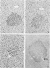
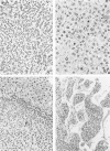
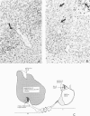
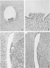
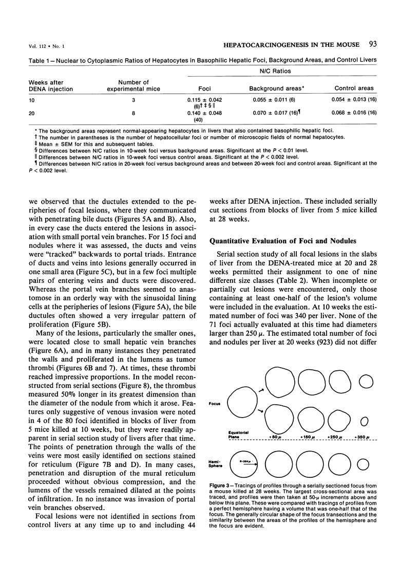
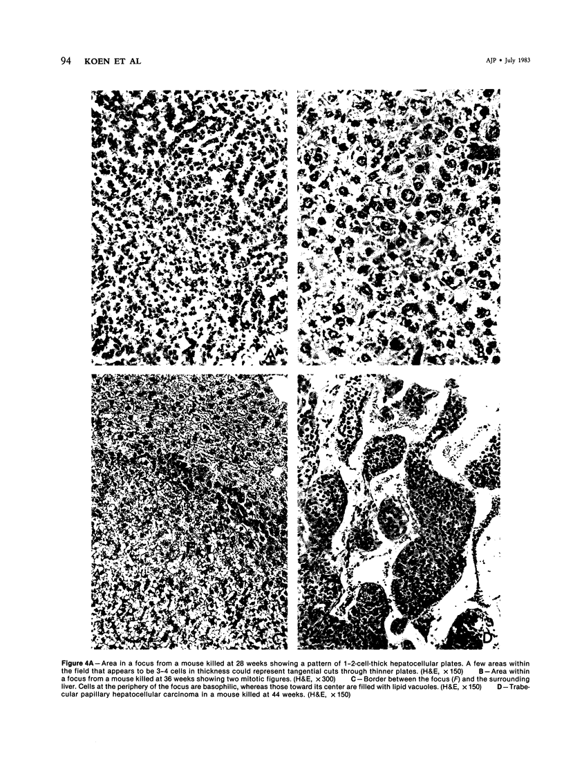
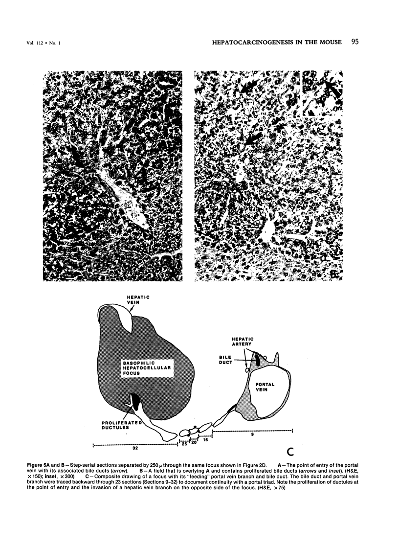
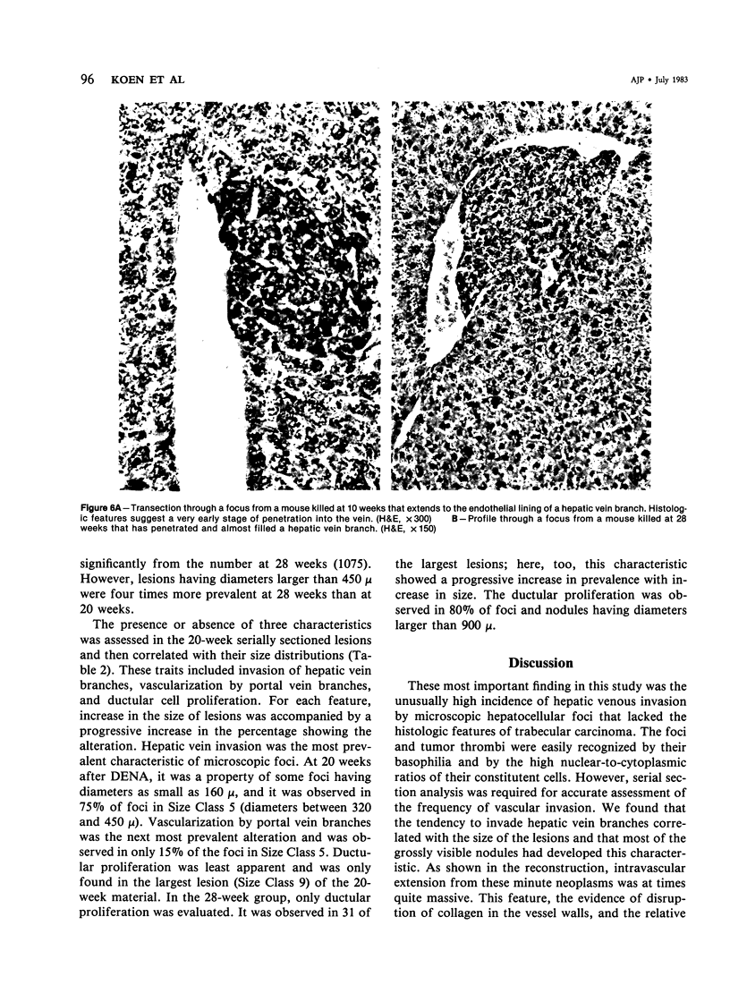
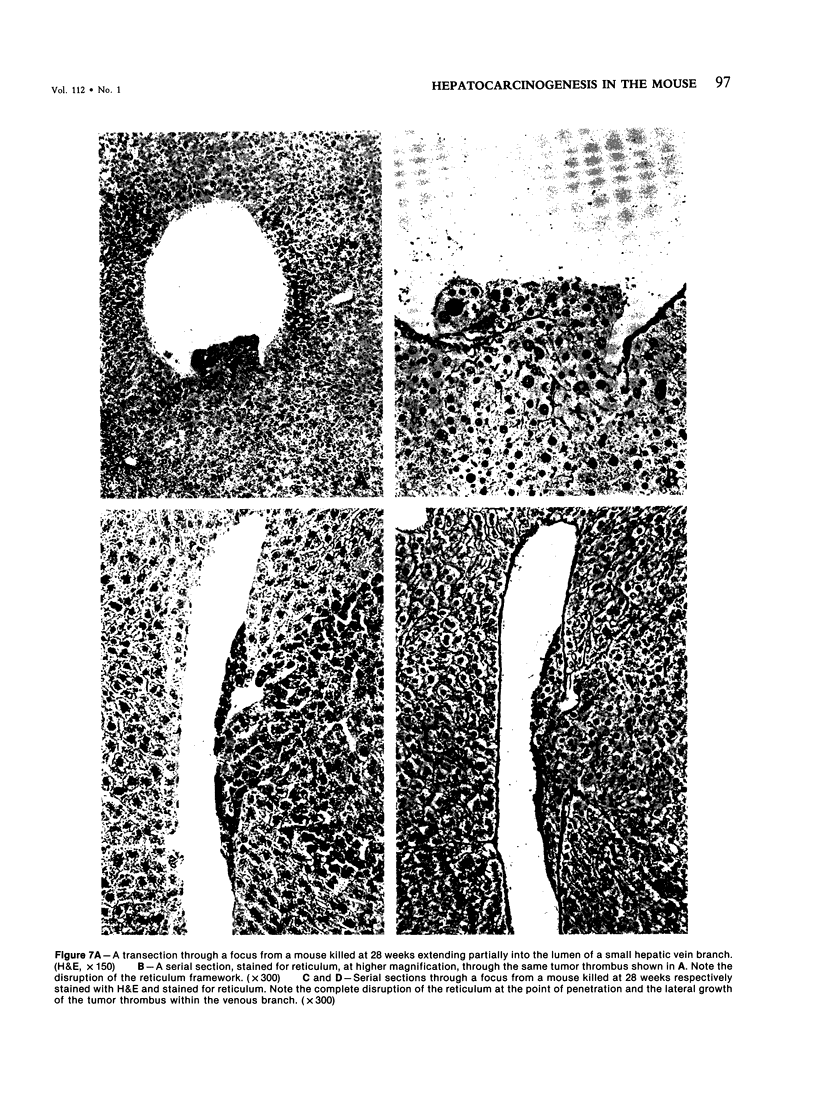
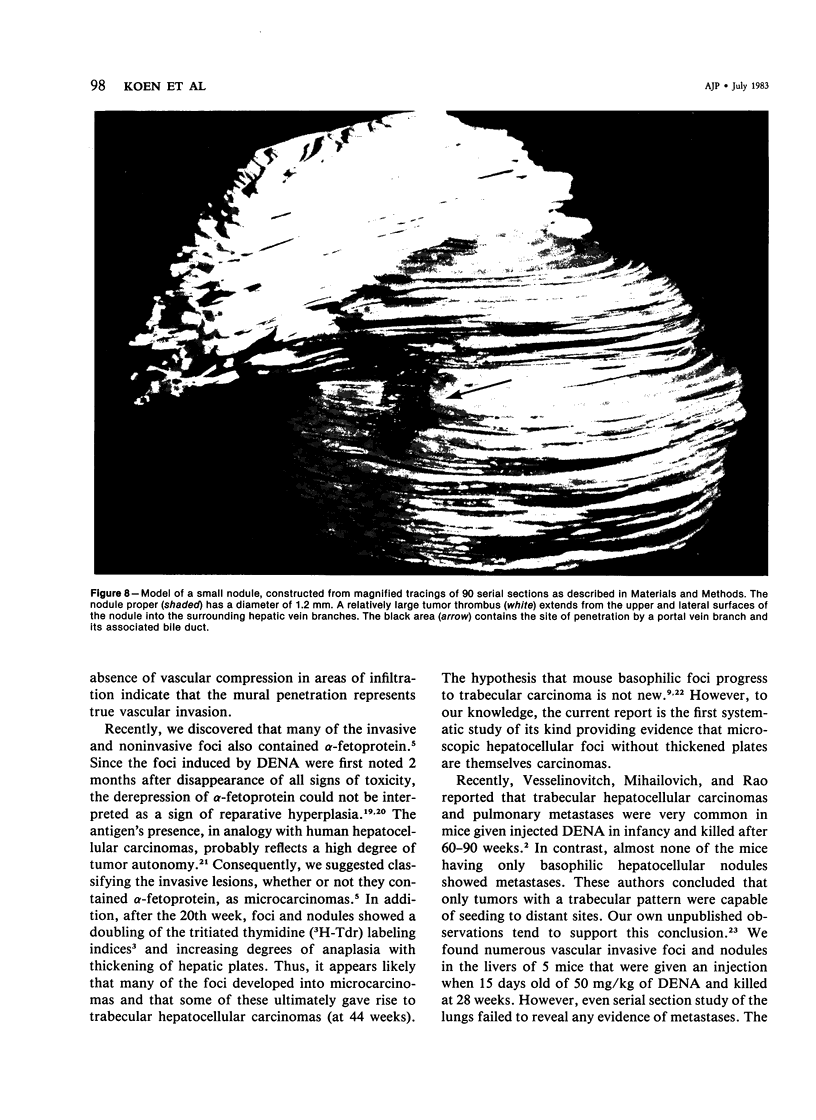
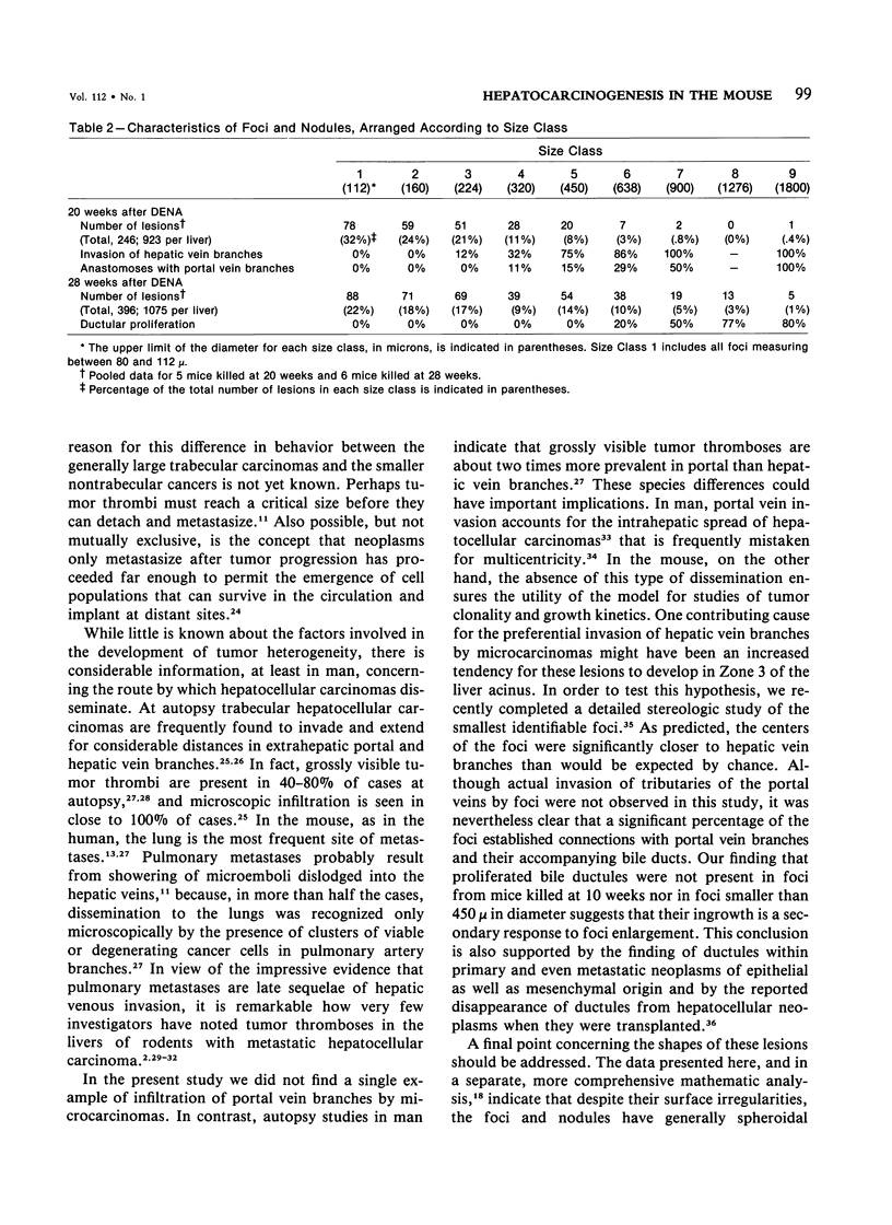
The histogenesis of trabecular hepatocellular carcinomas was studied in male B6C3 F1 mice that were given injections of 5 micrograms diethylnitrosamine (DENA)/g body wt when they were 15 days old. Fully developed trabecular carcinomas with characteristically thickened hepatic plates were not seen until 44 weeks after DENA injection. However, focal microscopic collections of RNA-rich hepatocytes, referred to as basophilic hepatic foci, were first noted at 10 weeks after DENA injection. Hepatocytes in the foci were characterized by a twofold increase in the nuclear to cytoplasmic ratios, which imparted an easily recognized crowded appearance to the lesions, and by a marked tendency to invade hepatic vein branches. Blocks of liver from 16 mice, killed at 10, 20, and 28 weeks, were serially sectioned; and all foci and nodules with a diameter greater than 80 mu were identified. At 20 weeks, only 6 of 51 foci (12%) with diameters between 160 and 224 mu showed venous invasion; whereas 15 of 20 (75%) in the range 320-450 mu showed this feature. Thus, the predisposition to invade hepatic vein branches correlated with an increase in the size of foci and preceded their development of thickened hepatic plates. Since other studies have documented the presence of alpha-fetoprotein, an oncofetal marker, in some invasive foci, and since the late-appearing trabecular carcinomas metastasize to the lungs, we have suggested that the tiny infiltrating lesions be classified as microcarcinomas that are predisposed to develop into trabecular hepatocellular carcinomas. Proliferated bile ductules were found in some of the larger foci and nodules. This feature also became more prevalent with an increase in the size of the lesions. The ductules were derived from bile ducts, which, in association with portal vein branches, entered the lesions at localized areas at their peripheries. Thus, the presence of ductules within foci appeared to result from an encroachment on bile ducts of the enlarging foci. Since the foci were generally spherical and did not disseminate within the liver, this single-dose carcinogen model should be particularly useful for further studies of tumor growth kinetics.
Full text
PDF
Images in this article
Selected References
These references are in PubMed. This may not be the complete list of references from this article.
- Abelev G. I. Alpha-fetoprotein in ontogenesis and its association with malignant tumors. Adv Cancer Res. 1971;14:295–358. doi: 10.1016/s0065-230x(08)60523-0. [DOI] [PubMed] [Google Scholar]
- Abelev G. I., Assecritova I. V., Kraevsky N. A., Perova S. D., Perevodchikova N. I. Embryonal serum alpha-globulin in cancer patients: diagnostic value. Int J Cancer. 1967 Sep 15;2(5):551–558. doi: 10.1002/ijc.2910020517. [DOI] [PubMed] [Google Scholar]
- Dempo K., Chisaka N., Yoshida Y., Kaneko A., Onoé T. Immunofluorescent study on alpha-fetoprotein-producing cells in the early stage of 3'-methyl-4-dimethylaminoazobenzene carcinogenesis. Cancer Res. 1975 May;35(5):1282–1287. [PubMed] [Google Scholar]
- Fidler I. J., Hart I. R. Biological diversity in metastatic neoplasms: origins and implications. Science. 1982 Sep 10;217(4564):998–1003. doi: 10.1126/science.7112116. [DOI] [PubMed] [Google Scholar]
- Frith C. H., Baetcke K. P., Nelson C. J., Schieferstein G. Sequential morphogenesis of liver tumors in mice given benzidine dihydrochloride. Eur J Cancer. 1980 Sep;16(9):1205–1216. doi: 10.1016/0014-2964(80)90180-2. [DOI] [PubMed] [Google Scholar]
- Koen H., Pugh T. D., Nychka D., Goldfarb S. Presence of alpha-fetoprotein-positive cells in hepatocellular foci and microcarcinomas induced by single injections of diethylnitrosamine in infant mice. Cancer Res. 1983 Feb;43(2):702–708. [PubMed] [Google Scholar]
- Moore M. R., Drinkwater N. R., Miller E. C., Miller J. A., Pitot H. C. Quantitative analysis of the time-dependent development of glucose-6-phosphatase-deficient foci in the livers of mice treated neonatally with diethylnitrosamine. Cancer Res. 1981 May;41(5):1585–1593. [PubMed] [Google Scholar]
- PREHN R. T. A CLONAL SELECTION THEORY OF CHEMICAL CARCINOGENESIS. J Natl Cancer Inst. 1964 Jan;32:1–17. [PubMed] [Google Scholar]
- Pugh T. D., King J. H., Koen H., Nychka D., Chover J., Wahba G., He Y. H., Goldfarb S. Reliable stereological method for estimating the number of microscopic hepatocellular foci from their transections. Cancer Res. 1983 Mar;43(3):1261–1268. [PubMed] [Google Scholar]
- Reuber M. D. Histopathology of carcinomas of the liver in mice ingesting heptachlor or heptachlor epoxide. Exp Cell Biol. 1977;45(3-4):147–157. doi: 10.1159/000162866. [DOI] [PubMed] [Google Scholar]
- SHANMUGARATNAM K. Primary carcinomas of the liver and biliary tract. Br J Cancer. 1956 Jun;10(2):232–246. doi: 10.1038/bjc.1956.27. [DOI] [PMC free article] [PubMed] [Google Scholar]
- Vesselinovitch S. D., Mihailovich N., Rao K. V. Morphology and metastatic nature of induced hepatic nodular lesions in C57BL x C3H F1 mice. Cancer Res. 1978 Jul;38(7):2003–2010. [PubMed] [Google Scholar]
- Vesselinovitch S. D., Rao K. V., Mihailovich N. Neoplastic response of mouse tissues during perinatal age periods and its significance in chemical carcinogenesis. Natl Cancer Inst Monogr. 1979 May;(51):239–250. [PubMed] [Google Scholar]
- Ward J. M., Vlahakis G. Evaluation of hepatocellular neoplasms in mice. J Natl Cancer Inst. 1978 Sep;61(3):807–811. [PubMed] [Google Scholar]
- Ward J. M., Vlahakis G. Evaluation of hepatocellular neoplasms in mice. J Natl Cancer Inst. 1978 Sep;61(3):807–811. [PubMed] [Google Scholar]
- Williams G. M., Hirota N., Rice J. M. The resistance of spontaneous mouse hepatocellular neoplasms to iron accumulation during rapid iron loading by parenteral administration and their transplantability. Am J Pathol. 1979 Jan;94(1):65–74. [PMC free article] [PubMed] [Google Scholar]


