

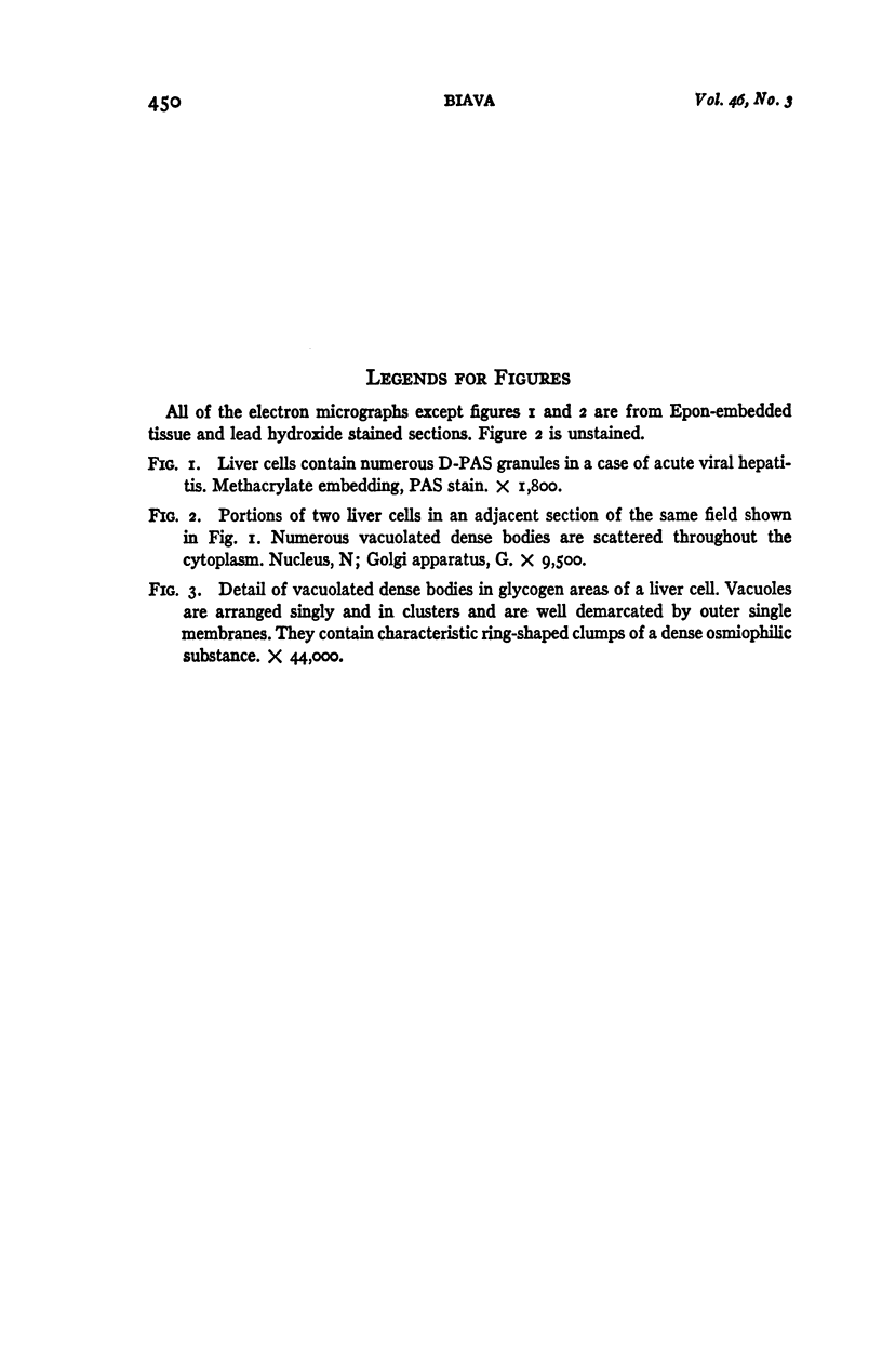
Full text
PDF
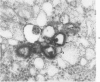
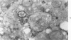
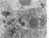
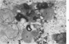
Images in this article
Selected References
These references are in PubMed. This may not be the complete list of references from this article.
- ALBOT G., JEZEQUEL A. M., LUNEL J. [Cholostatic episodes during the course of jaundice caused by viral hepatitis. Their biological manifestations and their aspect in optical and electron microscopy]. Acta Hepatosplenol. 1962 May-Jun;9:140–154. [PubMed] [Google Scholar]
- ASHFORD T. P., PORTER K. R. Cytoplasmic components in hepatic cell lysosomes. J Cell Biol. 1962 Jan;12:198–202. doi: 10.1083/jcb.12.1.198. [DOI] [PMC free article] [PubMed] [Google Scholar]
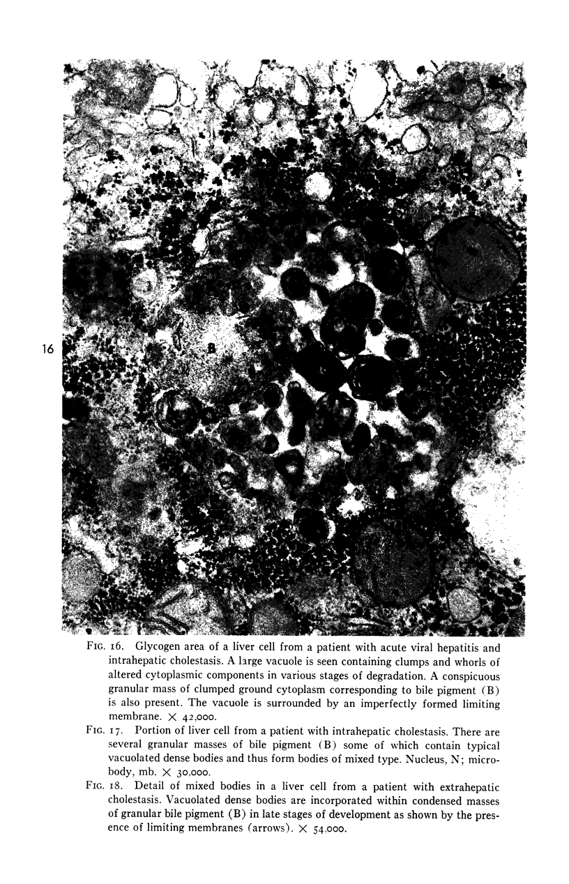
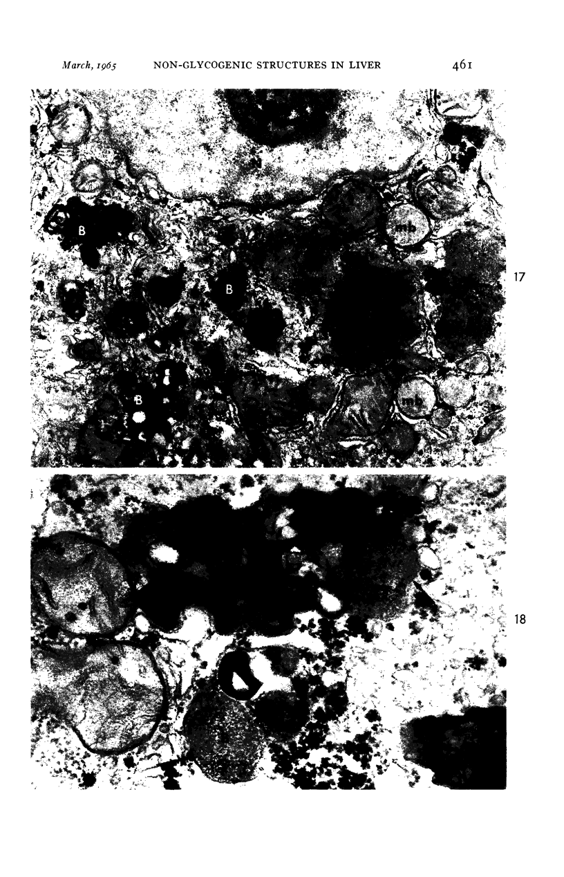
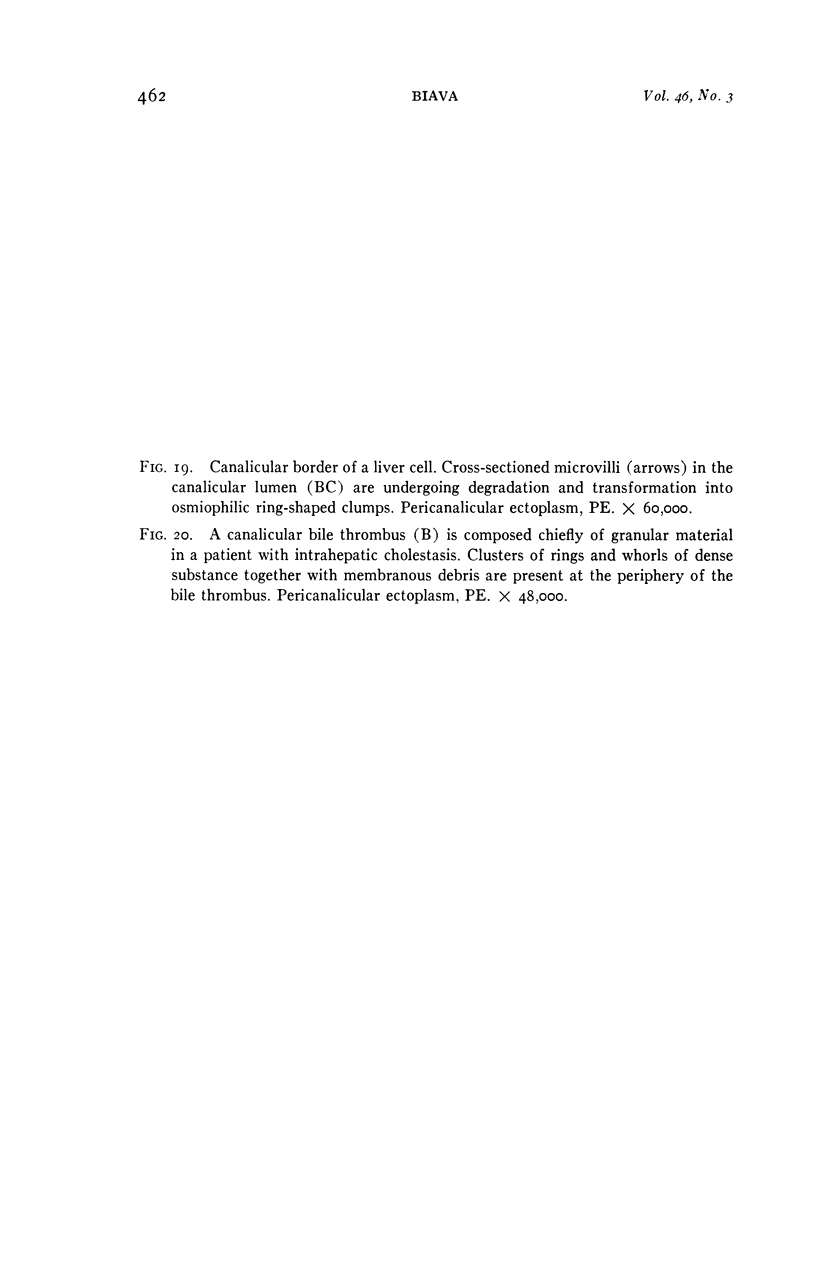
- BIAVA C. STUDIES ON CHOLESTASIS. THE FINE STRUCTURE AND MORPHOGENESIS OF HEPATOCELLULAR AND CANALICULAR BILE PIGMENT. Lab Invest. 1964 Sep;13:1099–1123. [PubMed] [Google Scholar]
- CECIO A. ELECTRON MICROSCOPIC OBSERVATIONS OF YOUNG RAT LIVER. I. DISTRIBUTION AND STRUCTURE OF THE MYELIN FIGURES (LAMELLAR BODIES). Z Zellforsch Mikrosk Anat. 1964 May 29;62:717–742. doi: 10.1007/BF00342180. [DOI] [PubMed] [Google Scholar]
- COSSEL L. [Electron microscope studies in chronic viral hepatitis and liver cirrhosis]. Virchows Arch Pathol Anat Physiol Klin Med. 1963;336:354–367. [PubMed] [Google Scholar]
- GUEFT B. Viral hepatitis under the electron microscope. Arch Pathol. 1961 Jul;72:61–69. [PubMed] [Google Scholar]
- HALL M. J. A staining reaction for bilirubin in sections of tissue. Am J Clin Pathol. 1960 Oct;34:313–316. doi: 10.1093/ajcp/34.4.313. [DOI] [PubMed] [Google Scholar]
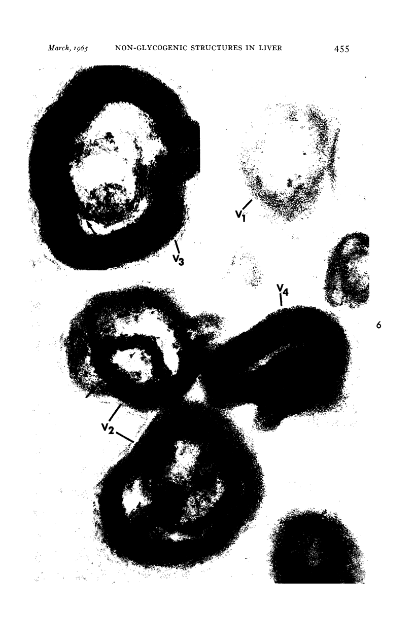
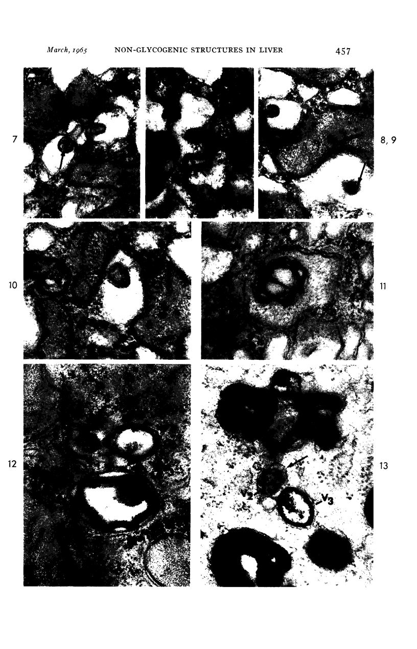
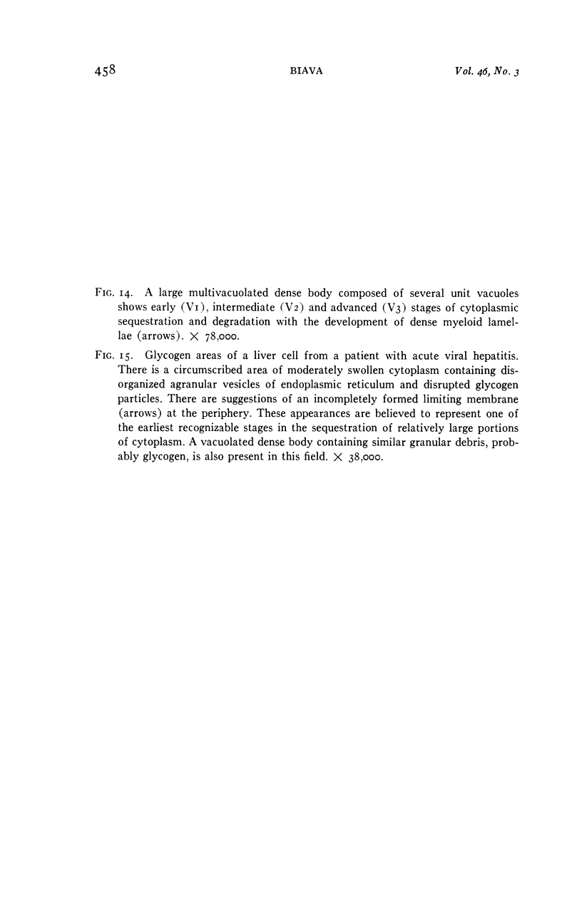
- HRUBAN Z., SPARGO B., SWIFT H., WISSLER R. W., KLEINFELD R. G. Focal cytoplasmic degradation. Am J Pathol. 1963 Jun;42:657–683. [PMC free article] [PubMed] [Google Scholar]
- HRUBAN Z., SWIFT H., WISSLER R. W. Analog-induced inclusions in pancreatic acinar cells. J Ultrastruct Res. 1962 Oct;7:273–285. doi: 10.1016/s0022-5320(62)90023-0. [DOI] [PubMed] [Google Scholar]
- JENNINGS B. M., FARQUHAR M. G., MOON H. D. Staining methods for osmium-methacrylate sections. Am J Pathol. 1959 Sep-Oct;35:991–997. [PMC free article] [PubMed] [Google Scholar]
- JORDAN S. W. ELECTRON MICROSCOPY OF HEPATIC REGENERATION. Exp Mol Pathol. 1964 Jun;86:183–200. doi: 10.1016/0014-4800(64)90052-8. [DOI] [PubMed] [Google Scholar]
- KIKKAWA Y., GUEFT B. Virus-like hepatic cell particles in patients with chronic alcoholism. Gastroenterology. 1963 Mar;44:243–250. [PubMed] [Google Scholar]
- LUFT J. H. Improvements in epoxy resin embedding methods. J Biophys Biochem Cytol. 1961 Feb;9:409–414. doi: 10.1083/jcb.9.2.409. [DOI] [PMC free article] [PubMed] [Google Scholar]
- MENEFEE M. G., EVANS V. J. Structural differences produced in mammalian cells by changes in their environment. J Natl Cancer Inst. 1960 Dec;25:1303–1310. [PubMed] [Google Scholar]
- NAPOLITANO L. CYTOLYSOMES IN METABOLICALLY ACTIVE CELLS. J Cell Biol. 1963 Aug;18:478–481. doi: 10.1083/jcb.18.2.478. [DOI] [PMC free article] [PubMed] [Google Scholar]
- NOVIKOFF A. B., BEAUFAY H., DE DUVE C. Electron microscopy of lysosomerich fractions from rat liver. J Biophys Biochem Cytol. 1956 Jul 25;2(4 Suppl):179–184. [PMC free article] [PubMed] [Google Scholar]
- NOVIKOFF A. B., ESSNER E. Cytolysomes and mitochondrial degeneration. J Cell Biol. 1962 Oct;15:140–146. doi: 10.1083/jcb.15.1.140. [DOI] [PMC free article] [PubMed] [Google Scholar]
- POLICARD A., BESSIS M., BRETONGORIUS J. Structures myéliniques observées au microscope électronique sur des coupes de globules rouges en voie de lyse. Exp Cell Res. 1957 Aug;13(1):184–186. doi: 10.1016/0014-4827(57)90066-6. [DOI] [PubMed] [Google Scholar]
- POPPER H., PARONETTO F., BARKA T. PAS-positive structures of nonglycogenic character in normal and abnormal liver. Arch Pathol. 1960 Sep;70:300–313. [PubMed] [Google Scholar]
- ROBBINS E., MARCUS P. I., GONATAS N. K. DYNAMICS OF ACRIDINE ORANGE-CELL INTERACTION. II. DYE-INDUCED ULTRASTRUCTURAL CHANGES IN MULTIVESICULAR BODIES (ACRIDINE ORANGE PARTICLES). J Cell Biol. 1964 Apr;21:49–62. doi: 10.1083/jcb.21.1.49. [DOI] [PMC free article] [PubMed] [Google Scholar]
- RUEBNER B. H., MIYAI K. THE PATHOLOGY OF NEONATAL HEPATITIS AND BILIARY ATRESIA WITH PARTICULAR REFERENCE TO HEMOPOIESIS AND HEMOSIDERIN DEPOSITION. Ann N Y Acad Sci. 1963 Dec 30;111:375–391. doi: 10.1111/j.1749-6632.1963.tb36978.x. [DOI] [PubMed] [Google Scholar]
- STOECKENIUS W. Some electron microscopical observations on liquid-crystalline phases in lipid-water systems. J Cell Biol. 1962 Feb;12:221–229. doi: 10.1083/jcb.12.2.221. [DOI] [PMC free article] [PubMed] [Google Scholar]
- Steiner J. W., Carruthers J. S. Studies on the Fine Structure of the Terminal Branches of the Biliary Tree: II. Observations of Pathologically Altered Bile Canaliculi. Am J Pathol. 1961 Jul;39(1):41–63. [PMC free article] [PubMed] [Google Scholar]
- TAKAHASHI N., MOTTET N. K. Further observations on the cytoplasmic spherical bodies in HEp-3 carcinoma cells. Lab Invest. 1962 Sep;11:743–752. [PubMed] [Google Scholar]
- TRUMP B. F., GOLDBLATT P. J., STOWELL R. E. An electron microscopic study of early cytoplasmic alterations in hepatic parenchymal cells of mouse liver during necrosis in vitro (autolysis). Lab Invest. 1962 Nov;11:986–1015. [PubMed] [Google Scholar]
- TRUMP B. F., JANIGAN D. T. The pathogenesis of cytologic vacuolization in sucrose nephrosis. An electron microscopic and histochemical study. Lab Invest. 1962 May;11:395–411. [PubMed] [Google Scholar]