

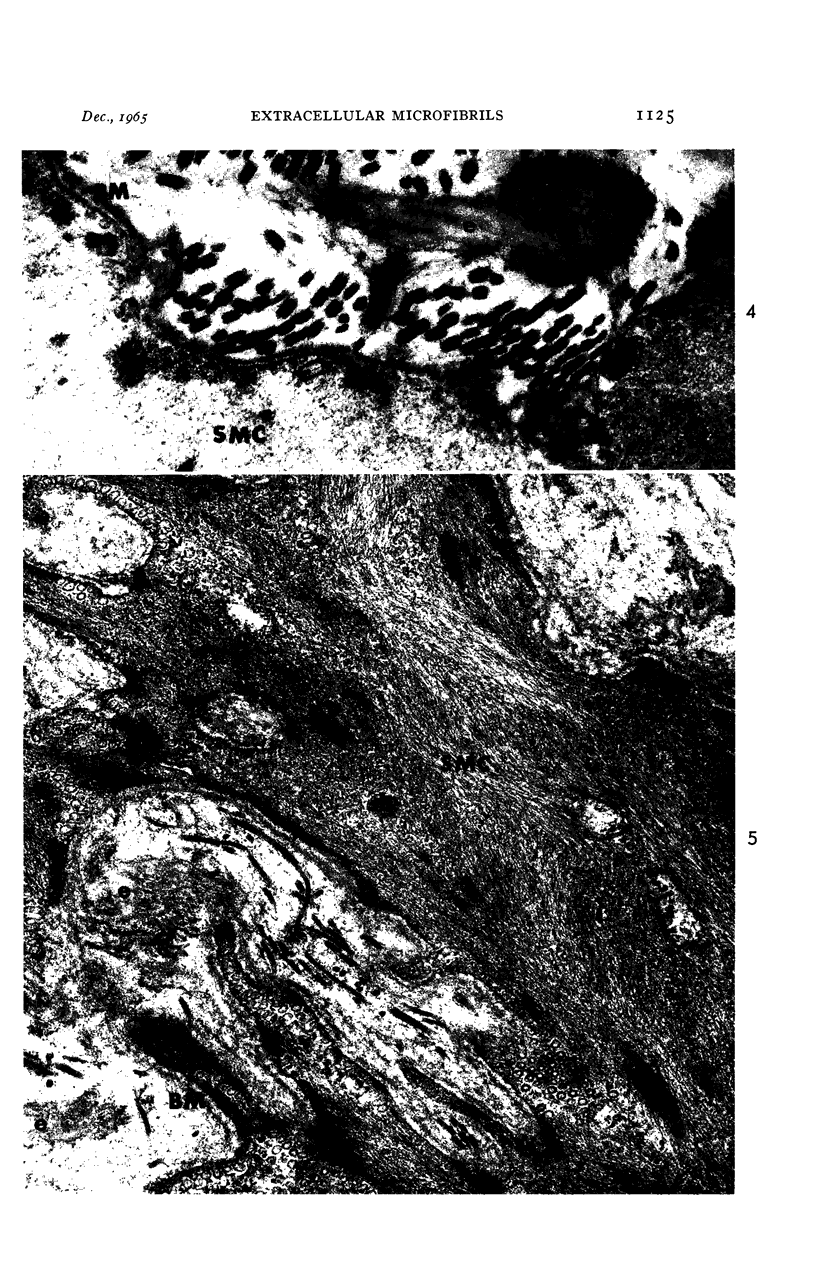
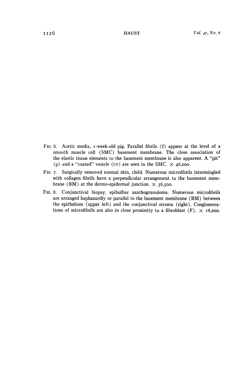
Full text
PDF


Images in this article


Selected References
These references are in PubMed. This may not be the complete list of references from this article.
- BIERRING F., KOBAYASI T. Electron microscopy of the normal rabbit aorta. Acta Pathol Microbiol Scand. 1963;57:154–168. doi: 10.1111/j.1699-0463.1963.tb03440.x. [DOI] [PubMed] [Google Scholar]
- CAULFIELD J. B. Effects of varying the vehicle for OsO4 in tissue fixation. J Biophys Biochem Cytol. 1957 Sep 25;3(5):827–830. doi: 10.1083/jcb.3.5.827. [DOI] [PMC free article] [PubMed] [Google Scholar]
- CHAPMAN J. A. Morphological and chemical studies of collagen formation. I. The fine structure of guinea pig granulomata. J Biophys Biochem Cytol. 1961 Mar;9:639–651. doi: 10.1083/jcb.9.3.639. [DOI] [PMC free article] [PubMed] [Google Scholar]
- CHASE W. H. Distribution and fine structure of elastic fibers in mouse lung. Exp Cell Res. 1959 Apr;17(1):121–130. doi: 10.1016/0014-4827(59)90158-2. [DOI] [PubMed] [Google Scholar]
- DETTMER N. Elektronenmikroskopisch Untersuchungen am elastischen Fasersystem des Ligamentum nuchae. Z Zellforsch Mikrosk Anat. 1956;45(3):265–279. [PubMed] [Google Scholar]
- FERNANDO N. V., MOVAT H. Z. Fibrillogenesis in regenerating tendon. Lab Invest. 1963 Feb;12:214–229. [PubMed] [Google Scholar]
- GOLDBERG B., GREEN H. AN ANALYSIS OF COLLAGEN SECRETION BY ESTABLISHED MOUSE FIBROBLAST LINES. J Cell Biol. 1964 Jul;22:227–258. doi: 10.1083/jcb.22.1.227. [DOI] [PMC free article] [PubMed] [Google Scholar]
- GROSS J., SOKAL Z., ROUGVIE M. Structural and chemical studies on the connective tissue of marine sponges. J Histochem Cytochem. 1956 May;4(3):227–246. doi: 10.1177/4.3.227. [DOI] [PubMed] [Google Scholar]
- HALL D. A., REED R., TUNBRIDGE R. E. Electron miscroscope studies of elastic tissue. Exp Cell Res. 1955 Feb;8(1):35–48. doi: 10.1016/0014-4827(55)90041-0. [DOI] [PubMed] [Google Scholar]
- IWAMOTO T. LIGHT AND ELECTRON MICROSCOPY OF THE PRESUMED ELASTIC COMPONENTS OF THE TRABECULAE AND SCLERAL SPUR OF THE HUMAN EYE. Invest Ophthalmol. 1964 Apr;3:144–156. [PubMed] [Google Scholar]
- JAKUS M. A. Further observations on the fine structure of the cornea. Invest Ophthalmol. 1962 Apr;1:202–225. [PubMed] [Google Scholar]
- JAKUS M. A. Studies on the cornea. I. The fine structure of the rat cornea. Am J Ophthalmol. 1954 Jul;38(12):40–53. [PubMed] [Google Scholar]
- KARRER H. E. An electron microscope study of the aorta in young and in aging mice. J Ultrastruct Res. 1961 Mar;5:1–27. doi: 10.1016/s0022-5320(61)80002-6. [DOI] [PubMed] [Google Scholar]
- KARRER H. E. Electron microscope study of developing chick embryo aorta. J Ultrastruct Res. 1960 Dec;4:420–454. doi: 10.1016/s0022-5320(60)80032-9. [DOI] [PubMed] [Google Scholar]
- KARRER H. E. The striated musculature of blood vessels. II. Cell interconnections and cell surface. J Biophys Biochem Cytol. 1960 Sep;8:135–150. doi: 10.1083/jcb.8.1.135. [DOI] [PMC free article] [PubMed] [Google Scholar]
- KEECH M. K. Electron microscope study of the normal rat aorta. J Biophys Biochem Cytol. 1960 Jun;7:533–538. doi: 10.1083/jcb.7.3.533. [DOI] [PMC free article] [PubMed] [Google Scholar]
- LOW F. N. Microfibrils: fine filamentous components of the tissue space. Anat Rec. 1962 Feb;142:131–137. doi: 10.1002/ar.1091420205. [DOI] [PubMed] [Google Scholar]
- LUFT J. H. Improvements in epoxy resin embedding methods. J Biophys Biochem Cytol. 1961 Feb;9:409–414. doi: 10.1083/jcb.9.2.409. [DOI] [PMC free article] [PubMed] [Google Scholar]
- PARKER F. An electron microscope study of coronary arteries. Am J Anat. 1958 Sep;103(2):247–273. doi: 10.1002/aja.1001030206. [DOI] [PubMed] [Google Scholar]
- PARTRIDGE S. M., ELSDEN D. F., THOMAS J. Constitution of the cross-linkages in elastin. Nature. 1963 Mar 30;197:1297–1298. doi: 10.1038/1971297a0. [DOI] [PubMed] [Google Scholar]
- PAULE W. J. Electron microscopy of the newborn rat aorta. J Ultrastruct Res. 1963 Apr;8:219–235. doi: 10.1016/s0022-5320(63)90004-2. [DOI] [PubMed] [Google Scholar]
- PEACH R., WILLIAMS G., CHAPMAN J. A. Alight and electron optical study of regenerating tendon. Am J Pathol. 1961 Apr;38:495–513. [PMC free article] [PubMed] [Google Scholar]
- PEASE D. C., MOLINARI S. Electron microscopy of muscular arteries; pial vessels of43 the cat and monkey. J Ultrastruct Res. 1960 Jun;3:447–468. doi: 10.1016/s0022-5320(60)90022-8. [DOI] [PubMed] [Google Scholar]
- PEASE D. C., PAULE W. J. Electron microscopy of elastic arteries; the thoracic aorta of the rat. J Ultrastruct Res. 1960 Jun;3:469–483. doi: 10.1016/s0022-5320(60)90023-x. [DOI] [PubMed] [Google Scholar]
- REYNOLDS E. S. The use of lead citrate at high pH as an electron-opaque stain in electron microscopy. J Cell Biol. 1963 Apr;17:208–212. doi: 10.1083/jcb.17.1.208. [DOI] [PMC free article] [PubMed] [Google Scholar]
- RHODIN J., DALHAMN T. Electron microscopy of collagen and elastin in lamina propria of the tracheal muscosa of rat. Exp Cell Res. 1955 Oct;9(2):371–375. doi: 10.1016/0014-4827(55)90116-6. [DOI] [PubMed] [Google Scholar]
- ROBERTSON J. D. Some features of the ultrastructure of reptilian skeletal muscle. J Biophys Biochem Cytol. 1956 Jul 25;2(4):369–380. doi: 10.1083/jcb.2.4.369. [DOI] [PubMed] [Google Scholar]
- ROMHANYI G. Submicroscopic structure of elastic fibres as observed in the polarization microscope. Nature. 1958 Oct 4;182(4640):929–930. doi: 10.1038/182929a0. [DOI] [PubMed] [Google Scholar]
- ROTH T. F., PORTER K. R. YOLK PROTEIN UPTAKE IN THE OOCYTE OF THE MOSQUITO AEDES AEGYPTI. L. J Cell Biol. 1964 Feb;20:313–332. doi: 10.1083/jcb.20.2.313. [DOI] [PMC free article] [PubMed] [Google Scholar]
- TROMANS W. J., HORNE R. W., GRESHAM G. A., BAILEY A. J. Electron microscope studies on the structure of collagen fibrils by negative staining. Z Zellforsch Mikrosk Anat. 1963;58:798–802. doi: 10.1007/BF00410661. [DOI] [PubMed] [Google Scholar]
- WATSON M. L. Staining of tissue sections for electron microscopy with heavy metals. J Biophys Biochem Cytol. 1958 Jul 25;4(4):475–478. doi: 10.1083/jcb.4.4.475. [DOI] [PMC free article] [PubMed] [Google Scholar]