Abstract
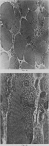
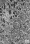
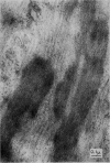
Examination by light and electron microscopy of more than 100 muscle biopsies revealed one very unusual case. A 4-year-old boy with non-progressive muscle weakness and hypotonia was found to have small particles, termed “myogranules”, in many muscle fibres from two gastrocnemius biopsies. Paraffin sections and thin sections of plastic-embedded muscle showed that the rod-shaped myogranules measured between 0.1 and 5 microns in length, and were usually orientated in the long axis of the fibre. Normal cross-striations could not be seen in areas occupied by myogranules, although adjacent parts of the same fibre were normal. Electron micrographs showed myofilaments running through the myogranules and a periodicity similar to sections of recrystallized muscle protein paramyosin. It is possible that this child has a disturbance of muscle proteins.
Full text
PDF


Images in this article
Selected References
These references are in PubMed. This may not be the complete list of references from this article.
- MULDAL S., OCKEY C. H. Deletion of Y chromosome in a family with muscular dystrophy and hypospadias. Br Med J. 1962 Feb 3;1(5274):291–294. doi: 10.1136/bmj.1.5274.291. [DOI] [PMC free article] [PubMed] [Google Scholar]
- PAINE R. S. The future of the 'floppy infant': a follow-up study of 133 patients. Dev Med Child Neurol. 1963 Apr;5:115–124. doi: 10.1111/j.1469-8749.1963.tb05010.x. [DOI] [PubMed] [Google Scholar]


