Abstract
Regulatory genes of the Antp class have been a major factor for the invention and radiation of animal bauplans. One of the most diverse animal phyla are the Cnidaria, which are close to the root of metazoan life and which often appear in two distinct generations and a remarkable variety of body forms. Hox-like genes have been known to be involved in axial patterning in the Cnidaria and have been suspected to play roles in the genetic control of many of the observed bauplan changes. Unfortunately RNAi mediated gene silencing studies have not been satisfactory for marine invertebrate organisms thus far. No direct evidence supporting Hox-like gene induced bauplan changes in cnidarians have been documented as of yet. Herein, we report a protocol for RNAi transfection of marine invertebrates and demonstrate that knock downs of Hox-like genes in Cnidaria create substantial bauplan alterations, including the formation of multiple oral poles (“heads”) by Cnox-2 and Cnox-3 inhibition, deformation of the main body axis by Cnox-5 inhibition and duplication of tentacles by Cnox-1 inhibition. All phenotypes observed in the course of the RNAi studies were identical to those obtained by morpholino antisense oligo experiments and are reminiscent of macroevolutionary bauplan changes. The reported protocol will allow routine RNAi studies in marine invertebrates to be established.
Introduction
RNAi and morpholino antisense studies on regulatory genes have offered crucial insights into the genetic mechanisms underlying bauplan changes in metazoan animals [e.g. 1]–[8]. It is highly unfortunate that only a very limited number of gene silencing studies in basal metazoan lineages have yet been possible due to methodological limitations resulting from working with multicellular marine animals [5], [7]–[10]. Functional studies on regulatory genes in the Cnidaria are particularly desirable as the diversity of bauplans within the phyla is one of the richest. Cnidaria harbor a minimal set of 5 (e.g. the hydrozoan Eleutheria) to 10 (e.g. the anthozoan Nematostella) Hox-like genes [11], [12] and expression studies have shown that these genes are regionally expressed along the aboral-oral axis [e.g. 13]–[17]. Some of these expression studies on homologous Hox-like genes revealed different expression patterns in different species and have led to incongruent results [11]. Functional studies may help to resolve these inconsistencies and learn about the function of Hox-like genes in early metazoan evolution. We here report a useful means for gene silencing by RNAi in marine invertebrates, validated with the use of several controls, including morpholino antisense oligos and information from expression data, demonstrate gene specific effects. This initial report will be followed by further in depth studies of the expression and function of all genes in each developmental stage of the complex 3-stage metagenic life-cycle of Eleutheria dichotoma.
Results and Discussion
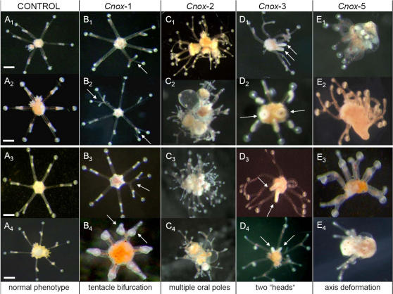
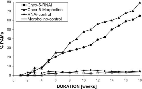
In addition to three previously cloned Hox-like genes, Cnox-3 to Cnox-5 [18], we also isolated full-length cDNA sequences of Cnox-1 and Cnox-2 from the hydrozoan Eleutheria dichotoma. We have developed a protocol enabling RNAi studies on marine invertebrates and performed different gene inhibition studies using RNAi and morpholino antisense oligos for all five genes. Based on partial and full length homeobox sequences, all five Cnox genes show a clear relationship to Antp class genes [18]. The full length cDNA sequences support the previous suggestion, as well as the recent view that the Cnidaria possess Hox related genes (Hox-like) [12]. All Cnox genes are relatively short, harboring coding sequences of between 684bp (Cnox-3-Ed) and 924bp (Cnox-1-Ed; see suppl. data, Fig. S1). Outside the homeobox, no similarity to Hox or other Hox related genes is seen, except for the presence of a heptapeptide (RELENRR) in Cnox-2, also found in an orthologous gene from Cassiopeia xamanchana [19] (for a possible derived amino terminal HEP-motif see [20], [21]). No other conserved motifs were found outside the homeobox, making the design of specific antisense oligos and dsRNA unproblematic (see suppl. data Fig. S1). In several repetitions vegetative medusae of E. dichotoma were subjected to gene silencing experiments (see Fig. S2). Transfected medusae of the hydrozoan Eleutheria dichotoma showed remarkable alterations in morphology if Cnox-1-Ed, Cnox-2-Ed, Cnox-3-Ed or Cnox-5-Ed were inhibited (see Fig. 1 and Tab. 1; [e.g. 5, 13, 22]). The most striking knock down phenotype is medusae developing multiple oral poles or “heads”. This phenotype regularly developed as a result of inhibition of the Cnox-2 or Cnox-3 gene, or inhibition of both together. If medusae developed two manubriae (“heads”), both were fully functional and capable of independent feeding. More than two manubriae (i.e. multiple heads) were regularly observed when Cnox-2 was inhibited. Another striking phenotype from the same inhibition experiment relates to a massive deformation of the primary body axis, the oral-aboral axis. Budding medusae did not develop defined aboral and oral body poles; the tentacles, however, were well developed. Identical phenotypes were regularly observed also when Cnox-5 was inhibited (up to 80% PAMs [phenotypic abnormal medusae] after 18 weeks). A third type of phenotype resulted from the inhibition of Cnox-1, which predominantly created additional tentacle bifurcations. Remarkably all of the above described, RNAi driven phenotypes were also observed when the same genes were inhibited by morpholino antisense oligos (Fig. 1). The latter were more effective inhibiting single or combinations of Cnox genes, while qualitatively no differences were detected between RNAi and morpholino oligo inhibition experiments. In morpholino oligo studies the percentage of PAMs was significantly higher (Fig. 2). This observation is consistent with former observations [23], [24]. The findings that both, morpholino and double stranded RNA inhibition, resulted in the same phenotypes, suggests specific and successful inhibition of the target genes, and indicates that the observed phenotypic changes are not due to morpholaxis or regeneration processes. In more than five decades of experimental research on Eleutheria medusae, none of the observed phenotypes–with one exception-has ever been seen in medusae under a variety of favorable and unfavorable physiological conditions [25]–[28]. Only tentacle deformations were occasionally seen (but never exceeding more than 1% in a population; Carl Hauenschild, pers. comm.). The presence of supplementary mouths or bifurcated tentacles can be normal in some other hydrozoans (Ferdinando Boero, pers. comm.). Interestingly Bouillon et al. (1997; [29]) and Boero et al. (2006; [30]), suggested that multiple manubriae in the senescent medusa of Codonorchis octaedrus might be due to the activation or repression of genes leading to clonal morphs. Since medusa buds develop from a very small number of epithelia and undifferentiated I-cells, it seems likely that transfection manifests here and subsequently enters the majority of cells in the daughter medusae. Here transfection of neighboring cells would be very efficient [31]. In future studies, it will be interesting to examine the potential for overlapping functions of these Hox-like genes. It would also be desirable to perform silencing studies of Cnox-1 to Cnox-5 genes on earlier developmental, i.e. larval stages. However, this would clearly exceed the scope of this paper.
Figure 1. Observed phenotypic changes in knock down experiments of Hox-like genes in the hydrozoan Eleutheria dichotoma.
Four of the five Hox-like genes produced phenotypically abnormal medusae (PAMs) in gene knock down studies. The main features of bauplan change relate to additional tentacle bifurcation (Cnox-1; see arrows), multiple oral poles or heads (Cnox-2), head duplication (Cnox-3; see arrows), and oral-aboral body axis deformation (Cnox-5). The two upper panels show life pictures of medusae transfected with double stranded RNA (B1,2, C1,2, D1,2, E1,2), and the lower panel medusae transfected with morpholino oligos (B3,4, C3,4, D3,4, E3,4). A1,2 and A3,4 are controls, scale bar is 100 µm. While we here unambiguously show that Hox-like genes can be silenced we think it would be premature to derive final conclusions on their functions yet.
Table 1. Cnox gene expression and knock down phenotypes in the hydrozoan Eleutheria dichotoma.
| Cnox-gene | In situ expression | Dominant knock down phenotype |
| Cnox-1 | Medusa: + ; ectodermal, oral ring in the “cnidoblast channel” | >71% of medusae with abnormal tentacle structures |
| Planula: − ; | ||
| Polyp: − ; | ||
| Cnox-2 | Medusa: + ; in early buds, entodermal | >66% of medusae with multiple oral poles |
| Planula: + ; ectodermal, oral | ||
| Polyp: + ; in polyps and primary polyps | ||
| ectodermal, oral | ||
| Cnox-3 | Medusa: + ; ectodermal, oral ring around the manubrium | >60% of medusae with double oral pole (two heads) |
| Planula: − ; | ||
| Polyp: − ; | ||
| Cnox-4 | Medusa: − ; | No visible phenotypic effect |
| Planula: − ; | ||
| Polyp: + ; in primary polyps ectodermal, aboral | ||
| Cnox-5 | Medusa: − ; | >79% of medusae with deformed aboral-oral body axis |
| Planula: + ; ectodermal, aboral. | ||
| Polyp: + ; ecto- and entodermal, oral and aboral |
Explanation is given in the text.
Figure 2. Time course for the development of phenotypic effects resulting from Cnox-5 gene knock down.
Both morpholino oligo and RNAi experiments result in an increase of phenotypically abnormal medusae (PAMs) over time. Shown is the time course of increasing numbers of PAMs as a result of inhibition by double stranded RNA and inhibition by morpholino-antisense oligonucleotides. In each case control 1 is untreated animals cultured in normal seawater, and control 2 is a sense morpholino oligonucleotide or dsRNA targeted to the Trox-2 gene from the placozoan Trichoplax adhaerens respectively. Independent controls were run for each single experiment.
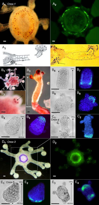
With respect to gene expression, all five Cnox genes revealed cum grano salis unambiguous patterns (Fig. 3). Cnox-1 and Cnox-3 were expressed in the medusa generation only, while Cnox-4 and Cnox-5 were detectable in the polyp generation (including planula larva) only. Cnox-2 was the only gene which was expressed in all life cycle stages; planula, polyp, and medusa (see table 1). Relating the observed knock down phenotypes to expression profiles leads to some intriguing speculations regarding possible gene function. Oral ectodermal expression in the cnidoblast channel directly suggests a role for Cnox-1 in tentacle formation and regeneration. The cnidoblast channel harbors differentiating cnidocytes and undifferentiated totipotent cells, both of which are essential for tentacle development [25]. A possible function of Cnox-2 in axis formation has previously been suggested before, and is supported by both the multiple oral head phenotypes (resulting from gene knock down) and the observed entodermal expression pattern in early medusa buds, where oral-aboral axis formation initiates. In the absence of Cnox-2 expression, either regulation of the aboral pole or a head inhibitor is missing [14], [15], [32]–[34]. As a consequence several oral poles (heads) develop, and orient themselves to the single aboral pole. A complementary function seems suggestive for the Cnox-3 gene, and it may play a crucial role in oral-aboral pole formation later in ontogeny. In adult medusae it is expressed orally in the ectoderm and possibly inhibits the formation of additional oral poles. When inhibited, a second oral pole can develop in adult medusae (although note that more than two oral poles have not been observed in Cnox-3 studies). For Cnox-5 we suggest a more upstream regulatory function. Although this gene is expressed at a very low level (detectable by semi-quantitative PCR but not by in situ hybridization; see suppl. data, Fig. S3), inhibition of this gene results in the highest number of PAMs; more than 80% of medusae show a dramatically deformed oral-aboral axis structures, except for the tentacles which develop normally. The observation that Cnox-5 produces a robust phenotype but not a visible in situ signal seems puzzling. We may conclude that expression is very low and possibly limited to a small number of undifferentiated I-cells. Alternatively, we can not exclude that Cnox-5 may be expressed in only a very short time window in the medusa stage. The expression of the Cnox-5-Ed gene of Eleutheria, an orthologue of both Anthox-6-Nv (Nematostella) and Cnox-1-Pc (Podocoryne; see table 2), may provide a clue. Indeed, the expression of this gene is dynamic during the development of Eleutheria, moving from the aboral (like Podocoryne) to the oral pole (like Nematostella). Hence a heterochronic change in the regulation of this gene might explain the evolution of its expression in the Cnidaria. It is an interesting speculation that Cnox-5 might be involved in the regulation of Cnox-2 and Cnox-3 in the medusa generation. Any further conclusions would be premature and clearly exceed the scope of this paper.
Figure 3. In situ expression of Hox-like genes in the hydrozoan Eleutheria dichotoma.
The five Hox-like genes, Cnox-1 to Cnox-5, display differential spatio-temporal expression patterns along the oral-aboral body axis. Cnox-1 (A1–A4) is expressed ectodermally in the so-called “Nesselring”, an area of undifferentiated cells lining the ring canal of medusae (cross section: A3, A4). Cnox-2 is expressed in the entoderm of developing medusa buds (B1, B2), in 2-day-old planula larvae ectodermally and orally (B5), in 5-day-old planula larvae along the body column ectodermally (B7) and at the oral pole of primary (B9) and adult polyps (B3; single polyp with 4 tentacles). Cnox-4 is exclusively expressed at the aboral pole of primary polyps (C2). Cnox-3 expression perfectly marks the most ectodermal oral part of the manubrium (D1, D2). Cnox-5 shows a remarkable pattern of expression moving from aboral only in the planula larva (E2) to both oral and aboral simultaneously in the metamorphosing polyp (E4). NBT/X-phosphate (A1, A4, B1–B3, D1) and fluorescein-labeled probes (A2, B5, B7, B9, C2, D2, E2, E4). Signals in B5, B7, B9, C2, E2 and E4 are overlaid with DAPI staining. Morphologies are shown in light microscopy (B4, B6, B8, C1, E1, E3). Scale bar is 50µm. A2, D1, E1–E4 are reprinted with permission from Elsevier Publishers [11].
Table 2. Homology nomenclature and expression of Hox/ParaHox-like genes in Cnidaria.
| GENE | ORGANISM | EXPRESSION | REFERENCE |
| Cnox -1-Ed Hox-like | Nematostella vectensis; Anthox-1 | polyp: ectodermal&aboral | Finnerty, 1998, 2003; [44], [45] |
| Hydra magnipapillata; Cnox-1 | ??? | Naito et al., 1993; Gauchat et al., 2000; [46], [40] | |
| Cnox -2-Ed ParaHox-like | Hydractinia symbiolongicarpus; Cnox-2 | polyp: ectodermal, oral&aboral, throuout the length of the body column | Murtha et al., 1991; Cartwright&Buss, 1999; Cartwright et al., 1999, 2006; [47], [48], [32] [33] |
| Hydra vulgaris; Cnox-2 | polyp: suppressed oral1; body column1,2; ectodermal & oral2, | Endl et al., 19991; Gauchat et al., 20002; [34], [43] | |
| Chlorohydra viridissima; Cnox-2 | polyp: ectodermal&oral (high level) | Schummer et al., 1992; [49] | |
| Nematostella vectensis; Anthox-2 | polyp: ectodermal&oral | Finnerty et al., 2003; [45] | |
| Acropora millipora; Cnox-2-Am | polyp: ectodermal&oral | Hayward et al., 2001; [50] | |
| planula: ectodermal&vegetal (oral) | |||
| Hydra magnipapillata; Cnox-2 | polyp: ectodermal&oral (low level), aboral (high level); body column | Senk et al., 1993a, b; [13], [14] | |
| Podocoryne carnea; gsx | polyp: ectodermal&oral | Yanze et al., 2001; [16] | |
| early medusa: ectodermal&oral; adult: entodermal; planula: entodermal&animal (aboral) | |||
| Cnox -3-Ed Hox-like | Hydra vulgaris; Cnox-3 | polyp: ectodermal&oral | Gauchat et al., 2000; [43] |
| Cnox -4-Ed ParaHox-like | Metridium senile; Anthox-4 | ??? | Finnerty&Martindale, 1997; [51] |
| Cnox -5-Ed Hox-like | Podocoryne carnea; Cnox-1-Pc | (primary-) polyp: moves from aboral to oral (same in Eleutheria); medusa: striated muscle tissue cells; planula: ecto-&entodermal animal (aboral) | Aerne et al., 1995; Yanze et al., 2001; Galliot&Schmid, 2002; [52], [16], [53] |
| Hydra vulgaris; Cnox-11,2; Cnox-33,4,5 | polyp: ectodermal&oral1,3; around the hypostom; tentacle zone2,3 | Schummer et al., 19921; Gauchat et al., 20002; Smith et al., 20003; Bode, 20014; Senk et al., 1993a5; [49], [43], [54], [55] | |
| Chlorohydra viridissima; Cnox-1 | polyp: oral | Schummer et al., 1992; [49] | |
| Hydra magnipapillata; Cnox-1 | ??? | Naito et al., 1993; [46] | |
| Nematostella vectensis; Anthox-6 | Polyp&planula: entodermal, oral. | Finnerty&Martindale, 1999; Finnerty, 2003; Finnerty et al., 2004 [56], [45], [17] |
Notably, some of the observed gene inhibition phenotypes look like direct links to the bauplan patterns found in other hydrozoan taxa. The multiple oral poles (or multiple heads) phenotype is strikingly suggestive of colonial hydrozoans, or colonial cnidarians in general. Many colonial cnidarians form colonies by adding oral poles to a common stalk (stolon or hydrocaulus). Each head is functional in feeding and connects to a shared gastrovascular cavity, just as in some Cnox-2, Cnox-3 and Cnox-5 inhibited Eleutheria medusae. Other cnidarian bauplans derive from multiplication of tentacle structures or whole tentacles. The already bifurcated tentacles in Eleutheria duplicate further if Cnox-1 or Cnox-2 is inhibited, and the resulting phenotypes are similar to those found in several hydrozoan groups such as Cladonema radiatum [35]. As a working hypothesis, it seems to us an intriguing idea that Cnox genes in Cnidaria may provide a very efficient means for macroevolutionary bauplan alteration, for example by multiplication of body parts. The RNAi transfection protocol developed here opens useful and efficient avenues to functional evolutionary genomics not only in Cnidaria, but to basal marine invertebrates in general and probably also to vertebrate larvae.
Methods
Animal material
Eleutheria dichotoma medusae were collected in Southern France (Banyuls-sur-mer) and have been maintained in cultures for several years under constant laboratory conditions as described earlier [28]. Medusae were fed twice a week with 3–4 days old brine shrimp larvae, Artemia salina. 6 hours after feeding and again on the following day, the water was changed. Under laboratory conditions, E. dichotoma reproduces alternatively by vegetative budding or bisexual reproduction. For in situ hybridization, growing, budding, and sexual medusae, planula larvae, primary polyps, as well as growing and budding polyps were analyzed.
Cloning and sequencing of Eleutheria dichotoma Antp genes
Total RNA was isolated from whole tissue of medusae and polyps using a total RNA-Isolation kit (Promega). First-strand cDNA was synthesized with an oligo (dT)-adapter-primer, reverse transcriptase (Superscript, Invitrogen), and total RNA as template, following the manufacturer's protocol (for detail see [19]). Using sequence information from the homeobox fragments, 3′RACE and 5′RACE [36] were performed to amplify the cDNA ends of the homeobox genes in order to obtain full-length sequences [37].
In situ hybridization
Whole mount in situ hybridization followed a slightly modified protocol developed for Placozoa [38]. Deviations from the original protocol include: (i) proteinase K treatment was done for 10 min at 37°C; (ii) no post-fixation in 4% paraformaldehyde/0.2% glutaraldehyde was allowed; (iii) Digoxigenin and fluorescein labeled RNA sense and antisense probes of variable length were synthesized from the PCR amplified 5′ regions of the different genes (Cnox-1: 401 bp; Cnox-2: 426 bp; Cnox-3: 241 bp; Cnox-4: 199 bp; Cnox-5: 219 bp; see suppl. data).
Gene inhibition studies
For the gene inhibition experiments replicates each of ten vegetative medusae per experiment were maintained in single culture dishes in a volume of 5 ml artificial seawater.
a) Cnox gene inhibition by RNAi
Double stranded RNA was synthesized from cDNA templates (cloned into pGEM-T vectors) by using Sp6/T7-RNA polymerase. Single RNA strands of the 5′ ends of the genes (Cnox-1: 401 bp; Cnox-2: 426 bp; Cnox-3: 241 bp; Cnox-4: 199 bp; Cnox-5: 219 bp) were synthesized at 37°C for 2 h in 20 µl transcription buffer (Roche) in the presence of 250 ng template, 40 U RNase-inhibitor, 20 U T7 or SP6 RNA polymerase, and 10 mM each NTP, including fluorescein UTP. After digestion of the DNA template, ssRNA was precipitated and resuspended in ddH2O, containing 1 μl RNase inhibitor, before complementary strands were annealed. Before transfection with dsRNA, vegetative medusae were gently acclimated from artificial seawater conditions in a stepwise manner, from 35 ppt to 10 ppt salinity. First we added 10 μl FuGENE™-6 (HD)-Transfection Reagent to a total volume of 150 µl of sterile ddH2O containing 5 ppt artificial seawater salt (the undiluted FuGENE ™-6 (HD)-reagent may not come into contact with any surface other than the pipette tip). After adding 5 µl of dsRNA (∼5μg; FuGENE-Reagent: dsRNA 2:1, µl and µg respectively), contents were very gently mixed and incubated at 20°C for 45 min. Transfection of animals with dsRNA was done in a total volume of 500 µl for 3 hours. Animals were reacclimatized stepwise back to normal seawater conditions (35 ppt salinity). The first phenotypes were observed 12 days after transfection. By the end of the experiments, i.e. after 18 weeks, 60-80% of all animals showed abnormal phenotypes (Cnox-1: 71,3%; Cnox-2: 66,4%; Cnox-3: 60,3%; Cnox-5: 79,7%; N = 90–140 medusae per experiment). In all control populations, the percentage of PAMs was fewer than 3%.
b) Cnox gene inhibition by morpholino antisense oligonucleotides
For each Cnox gene, a 25 nucleotide long chemically modified morpholino antisense oligonucleotide [Gene Tools, LLC; e.g. 23, 24, 39, 40] was designed, complementary to the 5′ region close to the start methionine codon (suppl. data, Fig. S1). Transfection with morpholino antisense oligonucleotides was done with the aid of EPEI solution (ethoxilated polyethylenimine; Gene Tools, LLC) in a total volume of 10 ml [cf. 24]. Before transfection, animals were acclimated as described above. Modified oligonucleotides were added to the seawater to a final concentration of 1 µM. The first phenotypes were observed two weeks after transfection. By the end of the experiments, i.e. after 18 weeks, 65–88% of all animals showed abnormal phenotypes (Cnox-1: 76,8%; Cnox-2: 69,4%; Cnox-3: 65,1%; Cnox-5: 87,7%; N = 95–160 medusae per experiment). In all control populations, the percentage of PAMs was fewer than 2%.
c) Controls
We used several means to obtain controls for the RNAi and morpholino antisense oligo experiments, including: (i) Transfection success with the described protocol was verified by means of RT-PCR of silenced target-genes. For this 25 Cnox-1 to Cnox-5 transfected medusae and 25 Trox-2 (Hox-like gene from T. adhaerens, Placozoa) transfected control medusae were collected two days after transfection. Total RNA was extracted and RT-PCR was carried out according to Li et al. (2000) [41] (see Fig. S3 and Fig. S4; e.g. [40], [42]). (ii) In vivo detection of fluorescein-labeled dsRNA and morpholino oligos after transfection using fluorescence microscopy (see Fig. S5; e.g. [38]). (iii) Target gene in situ hybridization of knock down animals (see Fig. S6; e.g. [8]). (iv) Usage of two independent inhibition methods (RNAi and morpholino antisense oligomers) giving qualitatively the same results (see Fig. 2). Controls for RNAi comprised (a) untreated animals in normal seawater, and (b) animals transfected with dsRNA of the Trox-2 gene of the placozoan Trichoplax adhaerens. Control morpholino oligos comprised (a) a random sequence of the same length and (b) the sense sequence of the experimental Cnox gene morpholino oligos (e.g. [40]).
RNAi studies were repeated ten times and morpholino oligo studies six times, according to the design in Fig. S2. All reported results were highly reproducible. The observed phenotypes were qualitatively identical in all experiments and variation in percentage of PAMs was statistically not significant (p≤0.01, t-test, two-sided; in all cases).
Supporting Information
cDNA sequences for in situ probes, and RNAi and morpholino antisense oligo binding sites of all five Hox-like genes from Eleutheria dichotoma. Cnox-1 to Cnox-5 cDNA sequences from Eleutheria dichotoma. Homeodomain sequences are underlined, double-stranded RNA used in RNAi experiments as well as in situ probe sequences are highlighted in yellow. The complement of the morpholino antisense oligonucleotides is shown in red.
(0.04 MB PDF)
Experimental design for gene silencing studies on vegetative medusae of the hydrozoan Eleutheria dichotoma. At day 0, each of the 10 vegetative i.e. budding medusae were placed in a Boveri dish and treated either with dsRNA (RNAi) or antisense morpholinos, or treated in different ways as controls (Control). After approximately one week, the first medusa buds are released from the parent medusa (circled and highlighted), which then start reproducing vegetatively. In this way, by the end of the experiment the total number of medusae increases to values between 90 and 160 medusae. The original 10 parent medusae at the beginning of the experiment thus represent some 6 to 11% of the final population. If one assumes that only medusa buds are transfected, the percentage of PAMs could be as high as 90% by the end of the experiment.
(0.15 MB PDF)
RT-PCR of all five Hox-like genes in Eleutheria dichotoma. Differential expression of Cnox-1 to Cnox-5 gene in Eleutheria dichotoma medusae. Shown are products of RT-PCR after 27 cycles of Cnox-1 (lane 1; 401bp), Cnox-2 (lane 2; 426bp), Cnox-3 (lane 3; 241bp), Cnox-4 (lane 4; 199bp), Cnox-5 (lane 5; 219bp) and Eleutheria-actin (lane 6; 152bp). Cnox-4 and Cnox-5 are only weakly expressed.
(0.10 MB PDF)
RT-PCR of Cnox-1-Ed after gene knock down. Transfection with dsRNA significantly reduces Cnox gene expression. RT-PCR products from untreated control animals are shown in lanes 1 and 2, from control dsRNA animals in lanes 3 and 4, and from Cnox-1 dsRNA-infected animals in lanes 5 and 6. Products in lanes 1, 3, and 5 are actin controls. Note the strong decline of Cnox product in lane 6 relative to lanes 2 and 4. Shown here is the example for Cnox-1 in Eleutheria dichotoma; RT-PCR controls look similar for all five Hox-like genes (data not shown). RT-PCR controls were taken as subsets from the transfected animals used in the gene silencing studies.
(0.10 MB PDF)
In vivo detection of fluorescein-labeled dsRNA (A) and morpholino oligomers (B) after transfection.
(0.10 MB PDF)
Target gene in situ hybridization of knock down animals. In situ hybridization of Cnox-1 gene in a Cnox-1 knock down animal (A'), in a RNAi-control animal (B), in a Morpholino-control animal (B') and Cnox-3 in situ hybridization in a Cnox-1 knock down animal. Medusa morphology is shown in light microscopy (A) and DAPI staining of the same medusa (A').
(0.29 MB PDF)
Acknowledgments
We thank Danielle De Jong and three anonymous referees for invaluable comments on the manuscript. Special thanks go to Max.
Footnotes
Competing Interests: The authors have declared that no competing interests exist.
Funding: DFG Schi-277/20-1.
References
- 1.Amali AA, Ji-Fan Lin C, Chen Y, Wang W, Gong H et al. Up-Regulation of Muscle-Specific Transcription Factors During Embryonic Somitogenesis of Zebrafish (Danio rerio) by Knock-Down of Myo-statin-1. Dev Dyn. 2004;229:847–856. doi: 10.1002/dvdy.10454. [DOI] [PubMed] [Google Scholar]
- 2.Deutsch J. Hox and wings. Bioessays. 2005;27:673–675. doi: 10.1002/bies.20260. [DOI] [PubMed] [Google Scholar]
- 3.Herke SW, Serio NV, Rogers BT. Functional analyses of tiptop and antennapedia in the embryonic development of Oncopeltus fasciatus suggests an evolutionary pathway from ground state to insect legs. Development. 2005;132:27–34. doi: 10.1242/dev.01561. [DOI] [PubMed] [Google Scholar]
- 4.Hughes CL, Kaufman TC. RNAi analysis of Deformed, proboscipedia and Sex combs reduced in the milkweed bug Oncopeltus fasciatus: novel roles for Hox genes in the hemipteran head. Development. 2000;127:3683–3694. doi: 10.1242/dev.127.17.3683. [DOI] [PubMed] [Google Scholar]
- 5.Lohmann JU, Endl I, Bosch TCG. Silencing of developmental genes in Hydra. Dev Biol. 1999;214:211–214. doi: 10.1006/dbio.1999.9407. [DOI] [PubMed] [Google Scholar]
- 6.Van Auken K, Weaver DC, Edgar LG, Wood WB. Caenorhabditis elegans embryonic axial patterning requires two recently discovered posterior-group Hox genes. Proc Natl Acad Sci U S A. 2000;97:4499–4503. doi: 10.1073/pnas.97.9.4499. [DOI] [PMC free article] [PubMed] [Google Scholar]
- 7.Schubert M, Holland ND, Laudet V, Holland LZ. A retinoic acid-Hox hierarchy controls both anterior/posterior patterning and neuronal specification in the developing central nervous system of the cephalochordate amphioxus. Dev Biol. 2006;296:192–202. doi: 10.1016/j.ydbio.2006.04.457. [DOI] [PubMed] [Google Scholar]
- 8.Momose T, Houliston E. Two oppositely localised frizzled RNAs as axis determinants in a cnidarian embryo. PloS ONE. 2007;5:0889–0899. doi: 10.1371/journal.pbio.0050070. [DOI] [PMC free article] [PubMed] [Google Scholar]
- 9.Wikramanayake AH, Peterson R, Chen J, Huang L, Bince JM, et al. Nuclear beta-catenin-dependent Wnt8 signaling in vegetal cells of the early sea urchin embryo regulates gastrulation and differentiation of endoderm and mesodermal cell lineages. Genesis. 2004;39:194–205. doi: 10.1002/gene.20045. [DOI] [PubMed] [Google Scholar]
- 10.Yamada L, Shoguchi E, Wada S, Kobayashi K, Mochizuki Y, et al. Morpholino-based gene knockdown screen of novel genes with developmental function in Ciona intestinalis. Development. 2003;130:6485–6495. doi: 10.1242/dev.00847. [DOI] [PubMed] [Google Scholar]
- 11.Kamm K, Schierwater B, Jakob W, Dellaporta SL, Miller DJ. Axial Patterning and Diversification in the Cnidaria Predate the Hox System. Curr Biol. 2006;16:1–7. doi: 10.1016/j.cub.2006.03.036. [DOI] [PubMed] [Google Scholar]
- 12.Ryan JF, Mazza ME, Pang K, Matus DQ, Baxevanis AD, et al. Pre-Bilaterian Origins of the Hox Cluster and the Hox Code: Evidence from the Sea Anemone, Nematostella vectensis. PLoS ONE. 2007;2:e153. doi: 10.1371/journal.pone.0000153. [DOI] [PMC free article] [PubMed] [Google Scholar]
- 13.Shenk MA, Bode HR, Steele RE. Expression of Cnox-2, a HOM/Hox homeobox gene in hydra, is correlated with axial pattern formation. Development. 1993a;117:657–667. doi: 10.1242/dev.117.2.657. [DOI] [PubMed] [Google Scholar]
- 14.Shenk MA, Gee L, Steele RE, Bode HR. Expression of Cnox-2, a HOM/Hox gene, is suppressed during head formation in hydra. Dev Biol. 1993b;160:108–118. doi: 10.1006/dbio.1993.1290. [DOI] [PubMed] [Google Scholar]
- 15.Mokady O, Dick MH, Lackschewitz D, Schierwater B, Buss LW. Over one-half billion years of head conservation? Expression of an ems class gene in Hydractinia symbiolongicarpus (Cnidaria: Hydrozoa). Proc Natl Acad Sci U S A. 1998;95:3673–3678. doi: 10.1073/pnas.95.7.3673. [DOI] [PMC free article] [PubMed] [Google Scholar]
- 16.Yanze N, Spring J, Schmidli C, Schmid V. Conservation of Hox/ParaHox-related genes in the early development of a cnidarian. Dev Biol. 2001;236:89–98. doi: 10.1006/dbio.2001.0299. [DOI] [PubMed] [Google Scholar]
- 17.Finnerty JR, Pang K, Burton P, Paulson D, Martindale MQ. Origins of Bilateral Symmetry: Hox and Dpp Expression in a Sea Anemone. Science. 2004;304:1335–1337. doi: 10.1126/science.1091946. [DOI] [PubMed] [Google Scholar]
- 18.Kuhn K, Streit B, Schierwater B. Homeobox genes in the cnidarian Eleutheria dichotoma: Evolutionary implications for the origin of Antennapedia-class (HOM/Hox) genes. Mol Phylogenet Evol. 1996;6:30–38. doi: 10.1006/mpev.1996.0055. [DOI] [PubMed] [Google Scholar]
- 19.Schierwater B, Kuhn K. Homology of Hox genes and the zootype concept in early metazoan evolution. Mol Phylogenet Evol. 1998;9:375–381. doi: 10.1006/mpev.1998.0489. [DOI] [PubMed] [Google Scholar]
- 20.Ferrier DEK, Holland PWH. Sipunculan ParaHox genes. Evol Dev. 2001;3:263–270. doi: 10.1046/j.1525-142x.2001.003004263.x. [DOI] [PubMed] [Google Scholar]
- 21.Finnerty JR, Paulson D, Pang K, Martindale MQ. Early evolution of a homeobox gene: the parahox gene Gsx in the Cnidaria and the bilateria. Evol Dev. 2003;5:331–345. doi: 10.1046/j.1525-142x.2003.03041.x. [DOI] [PubMed] [Google Scholar]
- 22.Blackstone NW, Jasker BD. Phylogenetic considerations of clonality, coloniality, and mode of germline development in animals. J Exp Zool. 2003;297B:35–47. doi: 10.1002/jez.b.16. [DOI] [PubMed] [Google Scholar]
- 23.Summerton J, Weller D. Morpholino antisense oligomers: design, preparation and properties. Antisense Nuc Acid Drug Dev. 1997;7:187. doi: 10.1089/oli.1.1997.7.187. [DOI] [PubMed] [Google Scholar]
- 24.Morcos PA. Achieving efficient delivery of morpholino oligos in cultured cells. Genesis. 2001;30:94–102. doi: 10.1002/gene.1039. [DOI] [PubMed] [Google Scholar]
- 25.Hauenschild C. Versuche über die Wanderung der Nesselzellen bei der Meduse von Eleutheria dichotoma. Z Naturforsch. 1957;12b, 7 [Google Scholar]
- 26.Hauenschild C. Experimentelle Untersuchungen über die Entstehung asexueller Klone bei der Hydromeduse Eleutheria dichotoma. Z Naturforsch. 1956;11b, 7 [Google Scholar]
- 27.Hauenschild C. Ergänzende Mitteilung über die asexuellen Medusenklone bei Eleutheria dichotoma. Z Naturforsch. 1957;12b, 6 [Google Scholar]
- 28.Schierwater B. Allometric changes during growth and reproduction in Eleutheria dichotoma (Hydrozoa, Athecata) and the problem of estimating body size in a microscopic animal. J Morphol. 1989;200:255–267. doi: 10.1002/jmor.1052000304. [DOI] [PubMed] [Google Scholar]
- 29.Boero F, Gravili C, Denitto F, Miglietta MP, Bouillon J. The rediscovery of Codonorchis octaedrus (Hydroidomedusae, Anthomedusae, Pandeidae), with an update of the Mediterranean hydroidomedusan biodiversity. It J Zool. 1997;64:359–365. [Google Scholar]
- 30.Bouillon J, Gravili C, Pagès F, Gili JM, Boero F. Paris: Publications Scientifiques du Muséum; 2006. An introduction to hydrozoa. p. 591. [Google Scholar]
- 31.Buchon N, Vaury C. RNAi: a defensive RNA-silencing against viruses and transposable elements. Heredity. 2006;96:195–202. doi: 10.1038/sj.hdy.6800789. [DOI] [PubMed] [Google Scholar]
- 32.Cartwright P, Bowsher J, Buss LW. Expression of a Hox gene, Cnox-2, and the division of labor in a colonial hydroid. Proc Natl Acad Sci U S A. 1999;96:2183–2186. doi: 10.1073/pnas.96.5.2183. [DOI] [PMC free article] [PubMed] [Google Scholar]
- 33.Cartwright P, Schierwater B, Buss LW. Expression of a Gsx Parahox Gene, Cnox-2, in Colony Ontogeny in Hydractinia (Cnidaria: Hydrozoa). J Exp Zool (Mol Dev Evol) 2006;306B:1–10. doi: 10.1002/jez.b.21106. [DOI] [PubMed] [Google Scholar]
- 34.Endl I, Lohmann JU, Bosch TCG. Head-specific gene expression in Hydra: Complexity of DNA-protein interactions at the promotor of ks1 is inversely correlated to the head activation potential. Proc Natl Acad Sci U S A. 1999;96:1445–1450. doi: 10.1073/pnas.96.4.1445. [DOI] [PMC free article] [PubMed] [Google Scholar]
- 35.Bouillon J, Boero F. Phylogeny and classification of Hydroidomedusae. Thal Sal. 2000:296. [Google Scholar]
- 36.Frohman M A, Dush MK, Martin GR. Rapid production of full-length cDNAs from rare transcripts: Amplification using a single gene-specific oligonucleotide primer. Proc Natl Acad Sci U S A. 1988;85:8998–9002. doi: 10.1073/pnas.85.23.8998. [DOI] [PMC free article] [PubMed] [Google Scholar]
- 37.Schierwater B, Murtha MT, Dick MH, Ruddle FH, Buss LW. Homeoboxes in cnidarians. J Exp Zool. 1991;260:413–416. doi: 10.1002/jez.1402600316. [DOI] [PubMed] [Google Scholar]
- 38.Jakob W, Sagasser S, Dellaporta S, Holland PWH, Kuhn K, et al. The Trox-2 Hox/ParaHox gene of Trichoplax (Placozoa) marks an epithelial boundary. Dev Genes Evol. 2004;214:170–175. doi: 10.1007/s00427-004-0390-8. [DOI] [PubMed] [Google Scholar]
- 39.Partridge M, Vincent A, Matthews P, Puma J, Stein D, et al. A simple method for delivering morpholino antisense oligos into the cytoplasm of cells. Antisense Nuc Acid Drug Dev. 1996;6:169. doi: 10.1089/oli.1.1996.6.169. [DOI] [PubMed] [Google Scholar]
- 40.Summerton J, Stein D, Huang SB, Matthews P, Weller D, et al. Morpholino and phosphorothioate antisense oligomers compared in cell-free and cell-system. Antisense Nuc Acid Drug Dev. 1997;7:63. doi: 10.1089/oli.1.1997.7.63. [DOI] [PubMed] [Google Scholar]
- 41.Li YX, Farrell MJ, Liu R, Mohanty N, Kirby ML. Doublestranded RNA injection produces null phenotypes in zebrafish. Dev Biol. 2000;217:394–405. doi: 10.1006/dbio.1999.9540. [DOI] [PubMed] [Google Scholar]
- 42.Li M, Rohrer B. Gene silencing in Xenopus laevis by DNA vector-based RNA interference and transgenesis. Cell Res. 2006;16:99–105. doi: 10.1038/sj.cr.7310013. [DOI] [PubMed] [Google Scholar]
- 43.Gauchat D, Mazet F, Berney C, Schummer M, Kreger S, et al. Evolution of Antp-class genes and differential expression of Hydra Hox/paraHox genes in anterior patterning. Proc Natl Acad Sci U S A. 2000;97:4493–4498. doi: 10.1073/pnas.97.9.4493. [DOI] [PMC free article] [PubMed] [Google Scholar]
- 44.Finnerty JR. Homeoboxes in sea anemones and other non-bilaterian animals: impli-cations for the evolution of the Hox cluster and the zootype. Curr Dev Biol. 1998;40:212–251. doi: 10.1016/s0070-2153(08)60368-3. [DOI] [PubMed] [Google Scholar]
- 45.Finnerty JR. The origins of axial patterning in the metazoa: how old is bilateral symmetry? Int J Dev Biol. 2003;47:523–529. [PubMed] [Google Scholar]
- 46.Naito M, Ishiguro H, Fujisawa T, Kurosawa Y. Presence of eight distinct homeobox-containing genes in cnidarians. FEBS Letters. 1993;333:271–274. doi: 10.1016/0014-5793(93)80668-k. [DOI] [PubMed] [Google Scholar]
- 47.Murtha MT, Leckmann JF, Ruddle FH. Detection of homeobox genes in development and evolution. Proc Natl Acad Sci U S A. 1991;88:10711–10715. doi: 10.1073/pnas.88.23.10711. [DOI] [PMC free article] [PubMed] [Google Scholar]
- 48.Cartwright P, Buss LW. Colony integration and the expression of the Hox gene, Cnox-2, in Hydractinia symbiolongicarpus (Cnidaria: Hydrozoa). J Exp Zool. 1999;285:57–62. doi: 10.1002/(sici)1097-010x(19990415)285:1<57::aid-jez7>3.0.co;2-p. [DOI] [PubMed] [Google Scholar]
- 49.Schummer M, Scheurlen I, Schaller C, Galliot B. HOM/Hox homeobox genes are present in hydra (Chlorohydra viridissima) and are differentially expressed during regeneration. EMBO J. 1992;11:1815–1823. doi: 10.1002/j.1460-2075.1992.tb05233.x. [DOI] [PMC free article] [PubMed] [Google Scholar]
- 50.Hayward DC, Catmull J, Reece-Hoyes JS, Berghammer H, Dodd H, et al. Gene structure and larval expression of cnox-2Am from the coral Acropora millepora. Dev Genes Evol. 2001;211:10–19. doi: 10.1007/s004270000112. [DOI] [PubMed] [Google Scholar]
- 51.Finnerty JR, Martindale MQ. Homeoboxes in sea anemones (Cnidaria: Anthozoa): a PCR-based survey of Nematostella vectensis and Metridium senile. Biol Bull. 1997;193:62–76. doi: 10.2307/1542736. [DOI] [PubMed] [Google Scholar]
- 52.Aerne BL, Baader CD, Schmid V. Life stage and tissue-specific expression of the homeobox gene Cnox-1-Pc of the hydrozoan Podocoryne carnea. Dev Biol. 1995;169:547–556. doi: 10.1006/dbio.1995.1168. [DOI] [PubMed] [Google Scholar]
- 53.Galliot B, Schmid V. Cnidarians as a model system for understanding evolution and regeneration. Int J Dev Biol. 2002;46:39–48. [PubMed] [Google Scholar]
- 54.Smith KM, Gee L, Bode HR. HyAlx, an aristaless-related gene, is involved in tentacle formation in hydra. Development. 2000;127:4743–4752. doi: 10.1242/dev.127.22.4743. [DOI] [PubMed] [Google Scholar]
- 55.Bode HR. The role of Hox genes in axial patterning in Hydra. Am Zool: 2001;41:621–628. [Google Scholar]
- 56.Finnerty JR, Martindale MQ. Ancient origins of axial patterning genes: Hox genes and ParaHox genes in the cnidaria. Evol Dev. 1999;1:16–23. doi: 10.1046/j.1525-142x.1999.99010.x. [DOI] [PubMed] [Google Scholar]
Associated Data
This section collects any data citations, data availability statements, or supplementary materials included in this article.
Supplementary Materials
cDNA sequences for in situ probes, and RNAi and morpholino antisense oligo binding sites of all five Hox-like genes from Eleutheria dichotoma. Cnox-1 to Cnox-5 cDNA sequences from Eleutheria dichotoma. Homeodomain sequences are underlined, double-stranded RNA used in RNAi experiments as well as in situ probe sequences are highlighted in yellow. The complement of the morpholino antisense oligonucleotides is shown in red.
(0.04 MB PDF)
Experimental design for gene silencing studies on vegetative medusae of the hydrozoan Eleutheria dichotoma. At day 0, each of the 10 vegetative i.e. budding medusae were placed in a Boveri dish and treated either with dsRNA (RNAi) or antisense morpholinos, or treated in different ways as controls (Control). After approximately one week, the first medusa buds are released from the parent medusa (circled and highlighted), which then start reproducing vegetatively. In this way, by the end of the experiment the total number of medusae increases to values between 90 and 160 medusae. The original 10 parent medusae at the beginning of the experiment thus represent some 6 to 11% of the final population. If one assumes that only medusa buds are transfected, the percentage of PAMs could be as high as 90% by the end of the experiment.
(0.15 MB PDF)
RT-PCR of all five Hox-like genes in Eleutheria dichotoma. Differential expression of Cnox-1 to Cnox-5 gene in Eleutheria dichotoma medusae. Shown are products of RT-PCR after 27 cycles of Cnox-1 (lane 1; 401bp), Cnox-2 (lane 2; 426bp), Cnox-3 (lane 3; 241bp), Cnox-4 (lane 4; 199bp), Cnox-5 (lane 5; 219bp) and Eleutheria-actin (lane 6; 152bp). Cnox-4 and Cnox-5 are only weakly expressed.
(0.10 MB PDF)
RT-PCR of Cnox-1-Ed after gene knock down. Transfection with dsRNA significantly reduces Cnox gene expression. RT-PCR products from untreated control animals are shown in lanes 1 and 2, from control dsRNA animals in lanes 3 and 4, and from Cnox-1 dsRNA-infected animals in lanes 5 and 6. Products in lanes 1, 3, and 5 are actin controls. Note the strong decline of Cnox product in lane 6 relative to lanes 2 and 4. Shown here is the example for Cnox-1 in Eleutheria dichotoma; RT-PCR controls look similar for all five Hox-like genes (data not shown). RT-PCR controls were taken as subsets from the transfected animals used in the gene silencing studies.
(0.10 MB PDF)
In vivo detection of fluorescein-labeled dsRNA (A) and morpholino oligomers (B) after transfection.
(0.10 MB PDF)
Target gene in situ hybridization of knock down animals. In situ hybridization of Cnox-1 gene in a Cnox-1 knock down animal (A'), in a RNAi-control animal (B), in a Morpholino-control animal (B') and Cnox-3 in situ hybridization in a Cnox-1 knock down animal. Medusa morphology is shown in light microscopy (A) and DAPI staining of the same medusa (A').
(0.29 MB PDF)


