Abstract
An association between swarming and hemolysin BL secretion was observed in a collection of 42 Bacillus cereus isolates (P = 0.029). The highest levels of toxin were detected in swarmers along with swarm cell differentiation (P = 0.021), suggesting that swarming B. cereus strains may have a higher virulence potential than nonswarming strains.
Bacterial swarming is a specialized form of surface translocation that enables flagellate bacteria to coordinately move atop solid surfaces (11). The ability to swarm depends on a complex differentiation process that leads short and oligoflagellate swimmer cells to produce long, multinucleate, and hyperflagellate swarm cells actively migrating over surfaces in organized groups of tightly bound cells (reviewed in reference 10). The widespread nature of swarming-proficient species suggests that this type of flagellum-aided motility is a successful strategy developed by flagellate bacteria to rapidly colonize environmental surfaces (10). Moreover, swarming can be influential in host-pathogen interactions, since it contributes to the virulence potential that certain pathogens may exert by facilitating host colonization (1, 7, 8, 14) and/or leads to an increase in the production of specific virulence factors (2, 15). We have previously described the swarming behavior exhibited by laboratory strains of Bacillus cereus and Bacillus thuringiensis (9, 19), two closely related species that produce common genome-encoded virulence factors (17); among these, the tripartite toxin (B, L1, and L2 components) hemolysin BL (HBL) exerts enterotoxic, hemolytic, cytotoxic, and dermonecrotic activity (3-5). In B. thuringiensis 407 Cry−, a mutation in flhA, a component of the flagellar export apparatus (12, 16), was found to coordinately abolish swarming and secretion of HBL (9). In B. cereus NCIB 8122, which produces only the L2 component of HBL, L2 secretion was detectable exclusively in differentiated swarm cells (19). These findings suggested that swarming and HBL secretion could be associated phenomena.
In this study, we assessed the motility behavior of and the secretion of HBL by B. cereus strains isolated from different sources to evaluate whether (i) HBL secretion requires intact flagella, (ii) swarming and HBL secretion are prevalent traits in natural isolates, and (iii) an increase in HBL secretion occurs along with swarm cell differentiation.
Swimming and swarming motility in B. cereus isolates.
B. cereus strains were collected from clinical, environmental, or food samples (Table 1) and identified by the API 50 CH assay (Bio-Merieux, France). Identification of B. cereus was confirmed, excluding the presence of parasporal crystals, which are discriminative for B. thuringiensis, in preparations of sporulating cultures stained with 0.5% basic fuchsin. Assays for swimming (on 1% tryptone-0.5% NaCl plates containing 0.25% agar [TrM]) and chemotaxis (on TrM supplemented with 2.0 mM mannitol or glutamine) were performed as described previously (9, 19). Swarming differentiation (on 1% tryptone-0.5% NaCl plates containing 0.7% agar [TrA]) was ascertained by visualizing the presence of hyperflagellate and elongated cells (at least 2.5 times longer than cells growing in liquid medium).
TABLE 1.
B. cereus strains used in this study: analysis of motility and HBL secretion
| Origin and strain | Liquid environment
|
Solid surface
|
Secreted HBL activityf | |||||
|---|---|---|---|---|---|---|---|---|
| FLAa | Max cell length (μm) | MOTb | CHEc | Hyper-FLAd | Max cell length (μm) | SWe | ||
| Patient | ||||||||
| F837/76 | + | 3.5 | + | + | + | 18 | + | + |
| Soc 67 | + | 4.0 | + | + | + | 18 | + | + |
| MGBC 145 | + | 3.0 | + | + | − | 9 | − | + |
| F3748/75 | + | 3.1 | + | + | − | 6 | − | − |
| PGb1 | + | 5.0 | + | + | + | 23 | + | + |
| PGd3 | + | 3.5 | + | + | − | 7 | − | + |
| 296 | + | 4.0 | + | + | + | 12 | + | − |
| SVe1 | + | 2.5 | + | + | + | 10 | + | + |
| SEe1 | + | 3.0 | + | + | + | 8 | + | + |
| SEe2 | + | 3.5 | + | + | − | 6 | − | + |
| Ve1 | + | 3.5 | + | + | + | 10 | + | + |
| Ve2 | + | 4.5 | + | + | + | 11 | + | + |
| Ve3 | + | 4.5 | + | + | + | 13 | + | + |
| Te1 | + | 3.5 | + | + | − | 8 | − | + |
| Pic1 | + | 3.0 | + | + | + | 8 | + | + |
| Sme1 | + | 4.5 | + | + | − | 12 | − | − |
| Environment | ||||||||
| S3-7 | + | 3.5 | + | + | + | 15 | + | + |
| S1C | + | 4.0 | + | − | − | 14 | − | + |
| S3-4 | + | 3.5 | − | − | − | 9 | − | + |
| S2-8 | + | 4.5 | + | + | + | 15 | + | − |
| Food | ||||||||
| HRm 44 | + | 3.5 | + | + | + | 20 | + | + |
| FP | + | 4.5 | + | + | + | 11 | + | + |
| F1589/77 | + | 3.5 | + | + | + | 17 | + | − |
| D5 | + | 3.5 | + | + | − | 12 | − | − |
| D7 | + | 2.5 | + | + | + | 7 | + | + |
| D21 | + | 4.5 | + | + | + | 15 | + | − |
| D26 | + | 3.5 | − | − | − | 8 | − | − |
| D31 | + | 3.0 | + | − | − | 9 | − | − |
| D33 | + | 3.0 | + | + | − | 6 | − | − |
| R-6 | + | 6.5 | + | + | − | 30 | − | − |
| R-12 | − | 2.5 | − | − | − | 7 | − | − |
| B-4ac | + | 3.5 | + | + | + | 12 | + | + |
| 1230-88 | + | 4.0 | − | − | − | 7 | − | + |
| FM-1 | + | 3.5 | + | − | − | 8 | − | + |
| F4433/73 | + | 3.5 | − | − | − | 9 | − | + |
| F4429/73 | + | 3.5 | + | + | − | 10 | − | − |
| F4431/73 | + | 3.5 | + | − | − | 10 | − | − |
| F3502/73 | +/− | 3.5 | − | − | − | 9 | − | − |
| F4810/72 | + | 3.5 | + | − | − | 10 | − | − |
| ATCC | ||||||||
| 10876 | + | 3.5 | + | − | − | 8 | − | + |
| 14579 | + | 3.5 | + | + | + | 12 | + | + |
| 33018 | + | 3.5 | − | − | − | 8 | − | − |
Cell flagellation analyzed by flagellar staining.
Swimming motility evaluated on tryptone-NaCl plates containing 0.25% agar (TrM).
Chemotactic activity determined by comparing migration halos obtained on TrM with those obtained on TrM supplemented with mannitol or glutamine.
Cell hyperflagellation was defined as a >3-fold increase in flagellation from liquid to solid medium.
Swarming proficiency was deduced from the presence of hyperflagellated and elongated cells.
Determined on sheep blood agar plates from the formation of a discontinuous pattern of hemolysis.
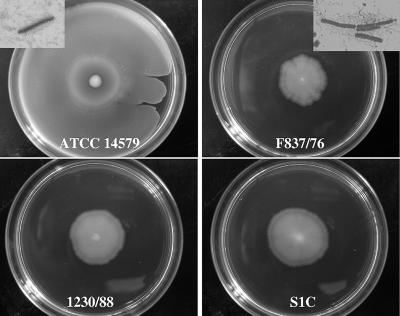
Among the 42 strains analyzed, seven failed to swim (16.7%) and six were able to swim but could not perform chemotaxis (14.3%) (Table 1). Hyperflagellate and elongated swarm cells, mainly localized at the colony rim, were evidenced for 45.2% of the strains. The possibility of predicting swarming proficiency by measuring colony diameter was proven to be inapplicable. Indeed, although the isolates produced differently sized colonies, the strains truly undergoing swarming, as demonstrated by production of differentiated swarm cells, did not always develop wider colonies than did strains unable to swarm (Fig. 1). As already demonstrated for B. cereus and B. thuringiensis laboratory strains (9, 19), swimming- and chemotaxis-deficient strains were unable to swarm (Table 1), confirming that the integrity of both the flagellar apparatus and the chemotaxis system is required by undomesticated B. cereus strains for mounting a swarming response.
FIG. 1.
Colonies produced by swarming (ATCC 14579 and F837/76) and nonswarming (1230/88 and S1C) B. cereus strains on tryptone-NaCl containing 0.7% agar. Plates were photographed after 24 h of incubation at 37°C. Insets show examples of swarm cells collected from the colony rim.
The finding that the prevalence of swarming in our B. cereus collection was lower than that reported for Salmonella spp., approaching 100% of the strains analyzed (13), suggests that swarming is a less-relevant environmental behavior for this spore-forming species. Indeed, production of spores should be regarded as an efficient strategy for contributing to bacterial spreading as well as persistence in different environments.
The percentage of swarming-proficient strains was higher within clinical (62.5%) than within food (31.6%) isolates (Table 1); however, the limited number of strains analyzed did not allow us to infer that swarming behavior is prevalent in strains found in a given environment or linked to host adaptation.
HBL production and flagella.
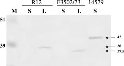
HBL is a membrane lytic system composed of the antigenically distinct proteins B, L1, and L2, encoded by hblA, hblD, and hblC, respectively. Strains secreting complete HBL, as demonstrated by the formation of a discontinuous zone of hemolysis around colonies on sheep blood agar plates (3), were 59.5% (n = 25) of the total number of strains (Table 1). Non-HBL-producing strains (n = 17) were subjected to PCR amplification to evaluate the presence of hbl genes (see Table S1 in the supplemental material) (18, 20) and to immunoblot analysis with rabbit antisera to the individual HBL components (9) for detecting HBL proteins released into culture supernatants (1% tryptone-0.5% NaCl [TrB]). Twelve out of the 17 strains lacked one to three hbl genes and the corresponding encoded proteins (Table 2). Among the remaining HBL-negative isolates, ATCC 33018 and D33 secreted L2 and B plus L1, respectively, whereas the corresponding genes were not detected with two PCR primer sets. This result was interpreted as a consequence of variations at the primer annealing sites, as already reported for other B. cereus isolates secreting HBL proteins but giving negative results for hbl genes (20). In contrast, S2-8, R-12, and F3502/73 gave positive PCR results for all hbl genes, while not all proteins were detected (Table 2). Cell lysates of strain S2-8 never gave a positive signal for L1, thus suggesting that this component was not produced or underwent such rapid and extensive intracellular proteolysis as to be undetectable. When cell lysates of R-12 and F3502/73 were subjected to immunoblotting with anti-B antibodies, one reactive band appeared at a molecular mass lower than that of the extracellular B component of strain ATCC 14579 (41 kDa) (Fig. 2). Since no internal stop codon was detected in hblA of the two strains (sequencing performed with FHA2 and BR1; see Table S1 in the supplemental material), these results suggested that the B component was synthesized but not exported and was partially proteolyzed inside the cell. Failure to secrete intracellularly produced HBL has been reported to occur in a B. thuringiensis mutant lacking flagella (9) and explained by the function of flagella as secretion systems in addition to locomotion organelles (16).
TABLE 2.
Characterization of discontinuous non-hemolysis-producing B. cereus strains
| Strain | FLAa | PCR detection of HBL geneb:
|
HBL protein(s) in culture supernatantsc | ||
|---|---|---|---|---|---|
| hblA | hblD | hblC | |||
| F3748/75 | + | − | − | − | None |
| 296 | + | − | − | − | None |
| Sme1 | + | − | − | − | None |
| S2-8 | + | + | + | + | B, L2 |
| F1589/77 | + | − | − | − | None |
| D5 | + | − | − | − | None |
| D21 | + | + | + | − | B, L1 |
| D26 | + | + | + | − | B, L1 |
| D31 | + | − | + | − | L1 |
| D33 | + | − | − | − | B, L1 |
| R-6 | + | − | + | + | L1, L2 |
| R-12 | − | + | + | + | None |
| F4429/73 | + | − | − | − | None |
| F4431/73 | + | − | − | − | None |
| F3502/73 | +/− | + | + | + | None |
| F4810/72 | + | − | − | − | None |
| ATCC 33018 | + | − | + | − | L1, L2 |
Cell flagellation analyzed by flagellar staining.
PCR analysis was performed with two different primer pairs for each gene.
Protein bands detected by Western blot analysis using antibodies against B, L1, and L2.
FIG. 2.
Immunoblot assay with a polyclonal antibody to the B component of HBL of culture supernatants (S) and cell lysates (L) prepared from the B. cereus strains R12, F3502/73, and ATCC 14579. M, molecular size standards. Arrows indicate estimated molecular masses (in kilodaltons) of B in the different strains.
Interestingly, no flagellum was ever visualized in preparations of strain R-12 and one or two flagella were seen in no more than 15% of the F3502/73 cells (Table 1). This finding strengthens the hypothesis that flagella act as a system for protein export for HBL secretion also in B. cereus. In this context, the observation that nonmotile isolates harboring flagella did secrete HBL (S3-4, 1230-88, and F4433/73 [Table 1]) can also suggest that the functionality of the flagellum as a locomotion organelle is not required for its function as an export apparatus.
Swarm cell differentiation is accompanied by a substantial increase in HBL secretion.
Although HBL-producing strains were either swarmers (n = 15) or nonswarmers (n = 10) and HBL-defective strains could swarm (n = 4) or not (n = 13), a weak but statistically significant association (P = 0.029, Fisher's exact test) between HBL production and swarming was observed. This finding, together with the demonstration that secretion of the L2 component of HBL was detectable only during swarming in a reference B. cereus strain (19), led us to hypothesize that an increase in toxin secretion could occur in swarmers along with swarm cell differentiation. To this end, the amount of toxin secreted by randomly selected HBL-producing strains (six swarming and six nonswarming isolates) was quantified during growth under swarming and nonswarming conditions. Conditions enabling collection of proteins secreted during swarming differentiation were realized by spotting late-exponential-phase TrB cultures (0.5 μl, approximately 2 × 10⁸ cells/ml) onto Anopore membranes (0.2-μm pore size) of 10-mm cell culture inserts (Nalge Nunc International) that were placed into 24-well plates containing 0.5 ml TrB/well. The inserts allowed us to effectively separate the liquid medium from bacteria growing over membranes, thus mimicking bacterial growth atop solid substrates. Well-defined colonies were developed by all strains, and swarm cells, mainly localized at the colony rim, were detected only for the swarming-proficient isolates. After 48 h of incubation at 30°C, the culture inserts were removed, the number of CFU on the membranes was counted, and the culture media were collected to quantify the amount of secreted HBL. Quantification of the B component of HBL was performed by enzyme immunoassay with specific antibodies to purified B (6). The B concentration in samples was calculated by using a calibration curve constructed with purified B protein at concentrations ranging from 0.5 to 10 ng/ml and expressed as the amount of protein for 106 bacterial cells or the total amount of proteins in cell lysates.
The amount of B secreted under nonswarming conditions (liquid cultures in TrB) ranged from 3.8 ± 0.64 to 33.6 ± 3.15 ng/106 cells for nonswarming strains and from 1.25 ± 0.21 to 9.65 ± 1.11 ng/106 cells for swarming strains (Table 3; Table S2 in the supplemental material reports the amount of B as μg/mg of total proteins in cell lysates). When the same strains were propagated over the membranes, significantly higher levels of secreted B, ranging from 28.33 ± 2.44 to 378.35 ± 21.75 ng/106 cells, were detected for swarming-proficient strains. The ratio of the amount of B secreted by cells growing on the membrane surface to that secreted in liquid varied from 9.38 to 44.2 and from 0.74 to 2.40, with a (21.02 ± 14.36)- and a (1.66 ± 0.62)-fold mean increase for the swarming and nonswarming strains, respectively (Table 3). Statistical analysis of the mean ratios for the two groups of strains revealed that the ability to swarm was associated with a significant increase in the secretion of the B component (P = 0.021; two-tailed Welch's t test).
TABLE 3.
Quantification of the B component of HBL secreted by swarming and nonswarming Bacillus cereus strains grown in broth or over a solid surface
| Straina | B component (ng/106 cells [mean ± SD])
|
S/B ratio (C) | C (mean ± SD) | |
|---|---|---|---|---|
| Broth (B) | Solid (S) | |||
| SW+ | 21.02 ± 14.36 | |||
| F837/76 | 8.56 ± 0.99 | 378.35 ± 21.75 | 44.20 | |
| Soc 67 | 3.02 ± 0.52 | 28.33 ± 2.44 | 9.38 | |
| S3-7 | 3.87 ± 0.87 | 60.71 ± 3.51 | 15.69 | |
| FP | 3.18 ± 0.52 | 39.64 ± 4.62 | 12.47 | |
| B-4ac | 1.25 ± 0.21 | 41.82 ± 3.20 | 33.46 | |
| ATCC 14579 | 9.65 ± 1.11 | 105.46 ± 10.05 | 10.93 | |
| SW− | 1.66 ± 0.62 | |||
| PGd3 | 33.60 ± 3.15 | 24.86 ± 5.72 | 0.74 | |
| S1C | 5.55 ± 1.22 | 6.59 ± 1.33 | 1.19 | |
| S3-4 | 4.14 ± 0.70 | 9.23 ± 1.89 | 2.23 | |
| 1230-88 | 3.80 ± 0.64 | 6.46 ± 1.14 | 1.70 | |
| FM-1 | 13.06 ± 2.00 | 22.00 ± 3.31 | 1.68 | |
| F4433/73 | 13.33 ± 1.47 | 31.94 ± 3.90 | 2.40 | |
SW+, swarming; SW−, nonswarming.
Conclusions.
The novelties of this report rely on the demonstration that (i) the ability to swarm is a relatively widespread behavior of B. cereus natural isolates and (ii) swarm cell differentiation in B. cereus is accompanied by a significant increase in HBL secretion. These data highlight the notion that swarming differentiation by B. cereus may contribute to the virulence potential of this opportunistic human pathogen. Moreover, interesting data were derived from the observations that (i) aflagellate B. cereus isolates do not secrete the intracellularly produced HBL, (ii) hyperflagellate swarm cells secrete an increased amount of toxin, and (iii) flagellate but nonmotile strains export HBL. These results support the idea that the flagellum is required for HBL secretion and that its functionality as export machinery is not dependent on its functionality as a locomotion organelle.
Supplementary Material
Acknowledgments
We thank Jean Schoeni for the design of primers Af, Ar, Df, Dr, Cf, and Cr and for immunoblot analysis of some strains.
This work was supported by National Research Project grant 2005058814 from the Ministero dell'Istruzione, dell'Università e della Ricerca.
Footnotes
Published ahead of print on 20 April 2007.
Supplemental material for this article may be found at http://aem.asm.org/.
REFERENCES
- 1.Allison, C., H. C. Lai, and C. Hughes. 1992. Co-ordinate expression of virulence genes during swarm-cell differentiation and population migration of Proteus mirabilis. Mol. Microbiol. 6:1583-1591. [DOI] [PubMed] [Google Scholar]
- 2.Allison, C., N. Coleman, P. L. Jones, and C. Hughes. 1992. Ability of Proteus mirabilis to invade human urothelial cells is coupled to motility and swarming differentiation. Infect. Immun. 60:4740-4746. [DOI] [PMC free article] [PubMed] [Google Scholar]
- 3.Beecher, D. J., and A. C. L. Wong. 1994. Identification of hemolysin BL-producing Bacillus cereus isolates by a discontinuous hemolytic pattern in blood agar. Appl. Environ. Microbiol. 60:1646-1651. [DOI] [PMC free article] [PubMed] [Google Scholar]
- 4.Beecher, D. J., and A. C. L. Wong. 1994. Improved purification and characterization of hemolysin BL: a hemolytic, dermonecrotic vascular permeability factor from Bacillus cereus. Infect. Immun. 62:980-986. [DOI] [PMC free article] [PubMed] [Google Scholar]
- 5.Beecher, D. J., J. L. Schoeni, and A. C. L. Wong. 1995. Enterotoxic activity of hemolysin BL from Bacillus cereus. Infect. Immun. 63:4423-4428. [DOI] [PMC free article] [PubMed] [Google Scholar]
- 6.Beecher, D. J., T. W. Olsen, E. B. Somers, and A. C. L. Wong. 2000. Evidence for contribution of tripartite hemolysin BL, phosphatidylcholine-preferring phospholipase C, and collagenase to virulence of Bacillus cereus endophthalmitis. Infect. Immun. 68:5269-5276. [DOI] [PMC free article] [PubMed] [Google Scholar]
- 7.Belas, M. R., and R. R. Colwell. 1982. Adsorption kinetics of laterally and polarly flagellated Vibrio. J. Bacteriol. 151:1568-1580. [DOI] [PMC free article] [PubMed] [Google Scholar]
- 8.Callegan, M., B. D. Novosad, R. Ramirez, E. Ghelardi, and S. Senesi. 2006. Role of swarming migration in the pathogenesis of Bacillus endophthalmitis. Investig. Ophthalmol. Vis. Sci. 47:4461-4467. [DOI] [PubMed] [Google Scholar]
- 9.Ghelardi, E., F. Celandroni, S. Salvetti, D. J. Beecher, M. Gominet, D. Lereclus, A. C. L. Wong, and S. Senesi. 2002. Requirement of flhA for swarming differentiation, flagellin export, and secretion of virulence associated proteins in Bacillus thuringiensis. J. Bacteriol. 184:6424-6433. [DOI] [PMC free article] [PubMed] [Google Scholar]
- 10.Harshey, R. M. 2003. Bacterial motility on a surface: many ways to a common goal. Annu. Rev. Microbiol. 57:249-273. [DOI] [PubMed] [Google Scholar]
- 11.Henrichsen, J. 1972. Bacterial surface translocation: a survey and a classification. Bacteriol. Rev. 36:478-503. [DOI] [PMC free article] [PubMed] [Google Scholar]
- 12.Hueck, C. J. 1998. Type III protein secretion systems in bacterial pathogens of animals and plants. Microbiol. Mol. Biol. Rev. 62:379-433. [DOI] [PMC free article] [PubMed] [Google Scholar]
- 13.Kim, W., and M. G. Surette. 2005. Prevalence of surface swarming behavior in Salmonella. J. Bacteriol. 187:6580-6583. [DOI] [PMC free article] [PubMed] [Google Scholar]
- 14.Kirov, S. M., M. Castrisios, and J. G. Shaw. 2004. Aeromonas flagella (polar and lateral) are enterocyte adhesins that contribute to biofilm formation on surfaces. Infect. Immun. 72:1939-1945. [DOI] [PMC free article] [PubMed] [Google Scholar]
- 15.Macfarlane, S., M. J. Hopkins, and G. T. Macfarlane. 2001. Toxin synthesis and mucin breakdown are related to swarming phenomenon in Clostridium septicum. Infect. Immun. 69:1120-1126. [DOI] [PMC free article] [PubMed] [Google Scholar]
- 16.Pallen, M. J., and N. J. Matzke. 2006. From the origin of species to the origin of bacterial flagella. Nat. Rev. Microbiol. 4:784-790. [DOI] [PubMed] [Google Scholar]
- 17.Rasko, D. A., M. R. Altherr, C. S. Han, and J. Ravel. 2005. Genomics of the Bacillus cereus group of organisms. FEMS Microbiol. Rev. 29:303-329. [DOI] [PubMed] [Google Scholar]
- 18.Schoeni, J. L. 2002. Protein and deoxyribonucleic acid heterogeneity in hemolysin BL, a tripartite enterotoxin produced by Bacillus cereus. Ph.D. thesis. University of Wisconsin—Madison, Madison.
- 19.Senesi, S., F. Celandroni, S. Salvetti, D. J. Beecher, A. C. L. Wong, and E. Ghelardi. 2002. Swarming motility in Bacillus cereus and characterization of a fliY mutant impaired in swarm cell differentiation. Microbiology 148:1785-1794. [DOI] [PubMed] [Google Scholar]
- 20.Thaenthanee, S., A. C. L. Wong, and W. Panbangred. 2005. Phenotypic and genotypic comparisons reveal a broad distribution and heterogeneity of hemolysin BL genes among Bacillus cereus isolates. Int. J. Food Microbiol. 105:203-212. [DOI] [PubMed] [Google Scholar]
Associated Data
This section collects any data citations, data availability statements, or supplementary materials included in this article.


