Abstract
Purpose
To determine the potential application of contrast-enhanced ultrasound in the characterisation of focal liver lesions encountered in radiological practice at a district general hospital.
Materials & Methods
Retrospective analysis of 68 sequential patients undergoing contrast-enhanced ultrasound (CEUS) of liver. All patients were referred for CEUS following identification of 1 or more focal liver lesions on conventional ultrasound or CT imaging. After baseline US examination (Acuson), a bolus of 1.0-2.4 ml of SonoVue (Bracco, UK) was administered intravenously. CEUS images were obtained during arterial, portal venous and delayed phases. Patients were followed up for a mean period of 6 months. The CEUS diagnosis was compared to that indicated by other imaging modalities, histopathology, and clinical follow up.
Results
CEUS correctly identified malignant liver lesions in 19 patients, with the final diagnosis confirmed by histopathology in 5 cases and clinico-radiological follow up in 14 cases. 47 patients were correctly identified with benign liver lesions on CEUS imaging, with all these cases confirmed on clinico-radiological follow up. In the detection of malignancy, the sensitivity was 95.0% and the specificity was 97.9%
Conclusions
In our experience to date, contrast-enhanced ultrasound imaging is highly accurate in characterising malignant and benign focal liver lesions. It therefore has significant potential for utilisation in most general radiology departments.
Keywords: Ultrasound, Liver, Contrast
INTRODUCTION
The effective non-invasive detection and characterisation of focal liver lesions (FLL) can significantly alter patient management1,4. Early detection of primary or secondary liver malignancies increases the possibility of curative surgical resection or successful percutaneous ablation. It is becoming increasingly evident that contrast-enhanced ultrasonography (CEUS) using non-destructive low-acoustic-power ultrasound scanning with second generation contrast agents, such as perfluorocarbon or sulphur hexafluoride-filled microbubbles, allows improved characterisation of solid focal liver lesions5. CEUS has high sensitivity in the detection and characterisation of hyper-and hypovascular liver malignancies with an accuracy comparable, and in some cases superior to, helical CT1. CEUS may also enable definitive diagnosis of haemangiomas and focal nodular hyperplasia (FNH)3.
The aim of this study was to determine the potential application of contrast-enhanced ultrasound in the characterisation of focal liver lesions encountered in radiological practice at a district general hospital.
METHODS
This study retrospectively reviewed the radiological yield and clinical outcome of 68 sequential patients who underwent CEUS of the liver in Antrim Hospital, a district general hospital in Northern Ireland. The patients were found to have one or more FLLs on conventional ultrasound or contrast-enhanced spiral CT before being referred for CEUS. Information was collated by review of ultrasound examinations, case-notes, and CT and/or MRI investigations. After baseline ultrasound (Acuson, MountainView, USA), continuous ultrasound images were obtained with the “Coherent Contrast Imaging” setting after the administration of a bolus intravenous injection of 1-2.4 ml of SonoVue (Bracco, UK), followed by a 5 ml saline flush. Images were obtained during arterial (15 – 25 seconds following injection), portal venous (45 – 90 seconds), and “late” (180 seconds onward) phases.
Patients were followed up for a mean period of 6.3 months, using case-notes and further Ultrasound, CT and/or MRI scans as evidence of disease progression or diagnosis confirmation. Comparison was made between the working diagnosis as indicated by clinical follow-up and further imaging, and the original CEUS diagnosis.
RESULTS
Of the 68 patients, 41 were female and 27 male. Ages ranged between 17 and 83 years, with a mean age of 56.5 years. CEUS correctly identified malignant liver lesions in 19 patients, with the final diagnosis confirmed by histopathology in 5 cases and clinico-radiological follow up in 14 cases. One patient with a metastatic liver deposit confirmed on clinico-radiological follow up was incorrectly diagnosed by CEUS as a benign lesion (haemangioma). 47 patients were correctly identified with benign liver lesions on CEUS imaging, with all these cases confirmed on clinico-radiological follow up. One patient with a haemangioma confirmed on clinico-radiological follow up was incorrectly diagnosed as having a metastasis on CEUS. The different diagnoses encountered are listed in tables I (benign) and II (malignant). In the 68 cases where focal liver lesions were characterised, CEUS demonstrated a sensitivity of 95.0% and a specificity of 97.9% in the detection of malignancy. The positive predictive value was 95.0%, and the negative predictive value was 97.9%.
Table I.
Benign focal liver lesions
| Diagnosis | Number of patients |
|---|---|
| Haemangioma | 27 |
| Focal fatty sparing | 13 |
| Focal fatty infiltration | 2 |
| Simple cyst | 4 |
| Regenerating nodule | 1 |
| Focal nodular hyperplasia | 1 |
| Total = 48 |
Table II.
Malignant focal liver lesions
| Diagnosis | Number of patients |
|---|---|
| Metastasis | 16 |
| Hepatocellular carcinoma | 2 |
| Cholangiocarcinoma | 1 |
| Lymphoma deposit | 1 |
| Total=20 |
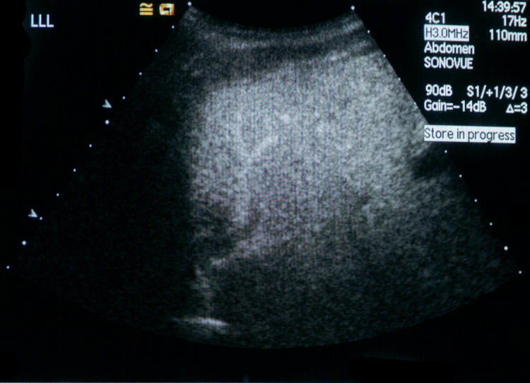
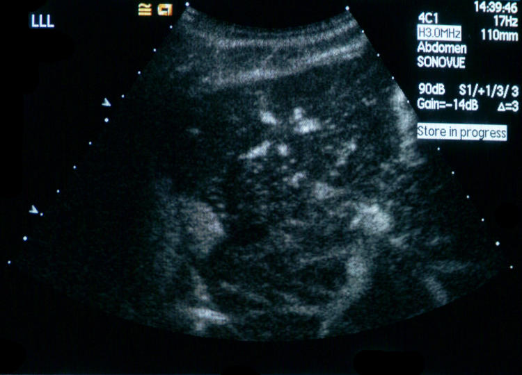
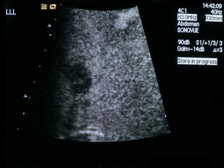
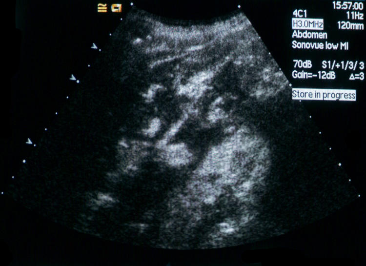
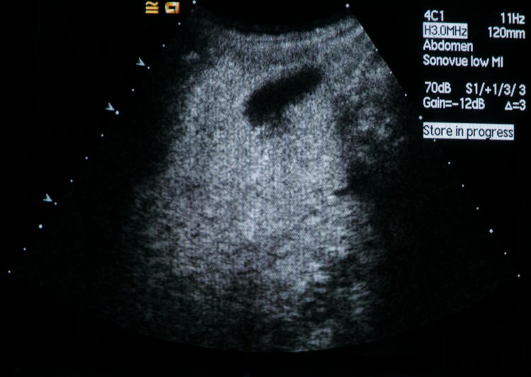
There were 16 cases of metastases, most of which appeared as hypoechoic nodules in contrast to the enhanced background of normal liver parenchyma (fig 1). One case of metastasis demonstrated diffuse enhancement in the arterial phase, suggesting that it was hypervascular, and showed subsequent rapid wash-out of contrast, the lesion becoming hypoechoic to the surrounding liver in the late phase (fig 2a). CEUS identified one patient as having metastatic disease, when CT and biopsy confirmed the diagnosis was hepatocellular carcinoma with metastatic liver disease. In the one case of metastasis misdiagnosed as haemangioma, there was a hypoechoic mass which appeared to gradually fill-in during the late phase.
Fig 1.
Typical hypoechoic appearance of a metastatic deposit on a background of enhanced normal liver parenchyma.
Fig 2a.
Diffuse enhancement of the lesion during the arterial phase.
Fig 2b.
Contrast wash-out occurred in the same lesion in the late phase.
Of the two hepatocellular carcinomas, one demonstrated peripheral enhancement during the arterial phase, followed by isoechoic enhancement with the liver during the portal venous phase, and finally became hypoechoic in the late phase. The other demonstrated isoechoic enhancement with the remainder of the liver in the arterial and portal venous phases, and became hypoechoic during the late phase. The lymphomatous deposit, demonstrated in one patient in whom there was a previous history of lymphoma, remained hypoechoic throughout all phases of CEUS. In the final patient with malignancy, a histologically-proven cholangiocarcinoma was erroneously reported as a metastatic deposit, demonstrating hypoechogenicity throughout all phases of CEUS.
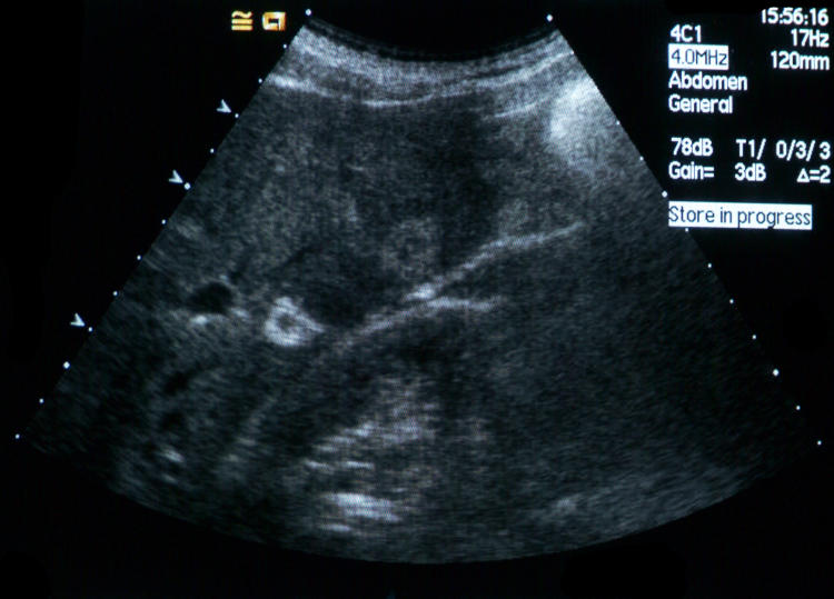
Of the 27 haemangiomas detected, 19 (70%) demonstrated typical appearances of hyperechoic focal lesions on conventional B mode ultrasound (fig 3a), showed rapid peripheral filling-in during the arterial phase of contrast enhancement (figure 3b), and subsequently became isoechoic with the surrounding liver in the portal venous and late phases (figure 3c). A further haemangioma was initially hypoechoic, but demonstrated rapid peripheral filling-in to become isoechoic with the surrounding liver in later phases of the examination. Seven haemangiomas demonstrated atypical behaviour, and were reported as being likely atypical haemangiomas but further follow up was recommended to confirm or exclude metastases. Of these, six haemangiomas demonstrated persistent hypoechoic areas with circumferential filling-in. The other atypical haemangioma demonstrated slow filling-in of peripheral enhancement, and was shown to be unchanged on ultrasound six months later.
Fig 3a.
Typical hyperechoic appearance of a haemangioma on conventional B mode Ultrasound.
Fig 3b.
Rapid peripheral contrast enhancement in the arterial phase.
Fig 3c.
The lesion becomes isoechoic in the portal venous phase.
Another of the haemangiomas demonstrated peripheral enhancement in the arterial phase with rapid wash-out of contrast in the portal venous and late phases, and was therefore thought to be a metastasis. However, MRI and clinical follow up confirmed this lesion to be a haemangioma.
Focal fatty sparing was shown in 13 patients to be a hypoechoic, well-defined area on B mode which became isoechoic with the surrounding liver during all phases of CEUS. Focal fatty infiltration was shown in two patients to be a hyperechoic lesion on B mode which became isoechoic with the surrounding liver during all phases of CEUS. There were four cases of simple hepatic cysts. A focal regenerating nodule was seen to be a well-defined hyperechoic lesion on B mode, and was obscured when CEUS was performed. The diagnosis was confirmed with repeated ultrasonography and CT. The one case of focal nodular hyperplasia demonstrated early central spoke wheel-shaped contrast enhancement, followed by diffuse homogenous enhancement in the arterial phase.
DISCUSSION
Accurate characterisation of focal liver lesions is essential for the utilisation of new treatment strategies in the management of focal liver malignancies.
Ultrasound is a widely used modality for imaging liver pathology. It is relatively inexpensive, does not expose the patient to ionising radiation, and is widely available. However there are limitations to conventional grey scale B mode ultrasound in the detection of focal liver lesions, especially when the lesions are small (<2cm), in the setting of cirrhosis, or in patients undergoing chemotherapy2. Colour and power Doppler has increased sensitivity for focal lesion detection compared to conventional B mode, but sensitivity is still inferior to contrast-enhanced spiral CT and MRI1.
Ultrasound examination with intravenous contrast agents allows dynamic assessment of focal liver lesions, improving the diagnostic performance of conventional sonography5. Perfluorocarbon or sulphur hexafluoride-filled microbubble contrast agents, such as SonoVue, can be used with non-destructive low acoustic power ultrasound scanning. In this way, real-time assessment of contrast enhancement in focal liver lesions is possible. CEUS therefore has the potential to provide firm diagnostic information without the need for other imaging modalities such as CT or MRI. In district general hospitals where imaging resources may be limited, CEUS can be incorporated into radiological practice with a relatively small increase in equipment and operator expenditure.
The results of this study indicate that CEUS in our practice has high sensitivity and specificity in determining if focal liver lesions are malignant. Most of the lesions exhibited definite enhancement patterns on dynamic scanning. As demonstrated in previous studies, the principal difference between benign and malignant liver lesions is their appearance during the late phase of contrast enhancement1,5. Malignant lesions are usually hypoechoic compared to normal liver parenchyma in this phase, whereas benign lesions are usually hyperechoic or isoechoic. In this study, nearly all of the metastases remained hypoechoic throughout all phases, although one case showed arterial enhancement with rapid wash-out. The two hepatocellular carcinomas showed variable enhancement characteristics in arterial and portal venous phases, but were also markedly hypoechoic relative to normal liver parenchyma in the late phase. The hepatic lymphoma and cholangiocarcinoma encountered in our study were correctly identified as malignant, although the enhancement characteristics were indistinguishable from hepatic metastases.
The majority of haemangiomas in our series demonstrated the usual pattern of peripheral enhancement in the arterial phase, followed by central filling-in on the delayed images (fig 3). A few haemangiomas exhibited slightly atypical enhancement patterns, but were correctly identified as benign. CEUS enabled the accurate characterisation of focal fatty sparing and focal fatty infiltration as the cause of liver lesions in several patients. Focal nodular hyperplasia was identified in only one patient in our series, with the classic features of early central spoke wheel-shaped enhancement and isoechoic appearance on late phase imaging.
CONCLUSION
Contrast-enhanced non-destructive ultrasonography using a low mechanical index is the sonographic modality of choice for the detection of liver malignancy1. In our experience, contrast-enhanced ultrasound imaging is highly accurate in characterising malignant and benign focal liver lesions. The equipment and expertise required for this investigation can be incorporated into most general radiology departments. Contrast-enhanced ultrasonography therefore has significant potential for utilisation in district general hospitals as well as specialist centres.
Conflict of Interest – none declared.
REFERENCES
- 1.Solbiati L, Tonolini M, Cova L, Goldberg SN. The role of contrast-enhanced ultrasound in the detection of focal liver lesions. Eur Radiol. 2001;11(Suppl 3):E15–26. doi: 10.1007/pl00014125. [DOI] [PubMed] [Google Scholar]
- 2.Bartolozzi C, Lencioni R. Contrast-specific ultrasound imaging of focal liver lesions. Eur Radiol. 2001;11(Suppl 11):E13–4. doi: 10.1007/pl00014126. [DOI] [PubMed] [Google Scholar]
- 3.Leen E. The role of contrast-enhanced ultrasound in the characterisation of focal liver lesions. Eur Radiol. 2001;11(Suppl 3):E27–34. doi: 10.1007/pl00014128. [DOI] [PubMed] [Google Scholar]
- 4.Sobiati L, Cova L, Ierace T, Marcelli P, Dellanoce M. Liver cancer imaging: the need for accurate detection of intrahepatic disease spread. J Comput Assist Tomogr. 1999;23(Suppl 1):S29–37. doi: 10.1097/00004728-199911001-00005. [DOI] [PubMed] [Google Scholar]
- 5.Quaia E, Calliada F, Bertolotto M, Rossi S. Characterization of focal liver lesions with contrast-specific US modes and a sulphur hexafluoride-filled microbubble contrast agent: diagnostic performance and confidence. Radiology. 2004;232(2):420–30. doi: 10.1148/radiol.2322031401. [DOI] [PubMed] [Google Scholar]


