
Fig. 1.
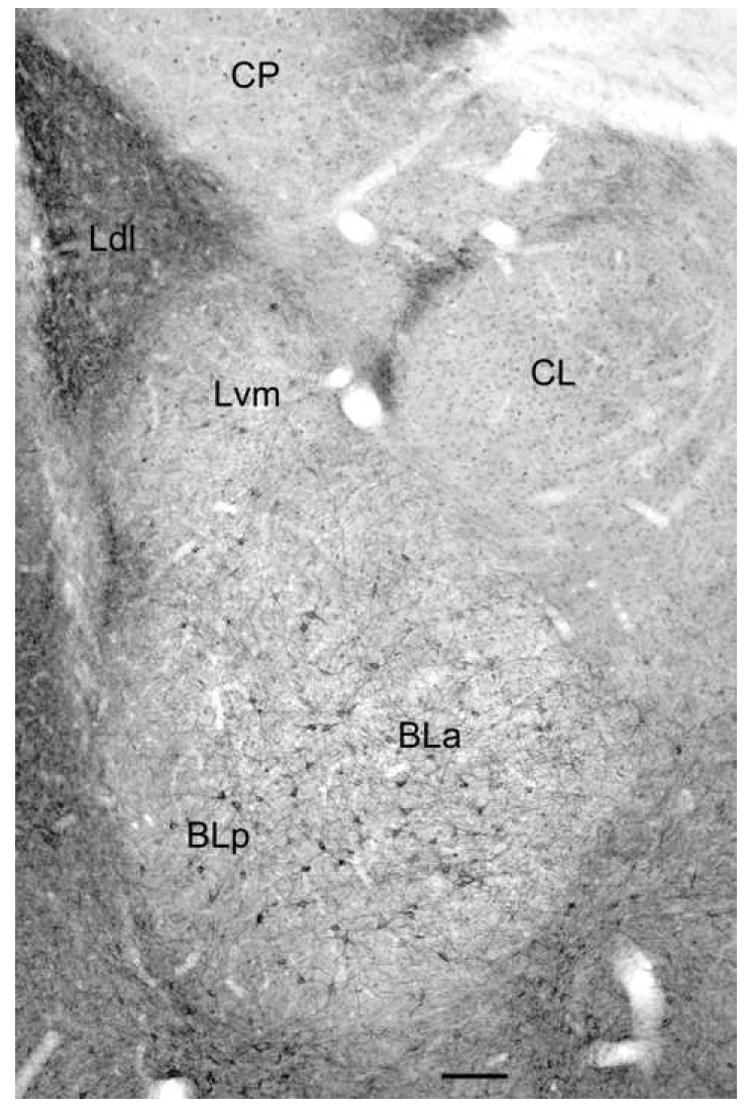
Digital photomicrograph of 5-HT2AR immunoreactivity in the rat amygdala demonstrated with the Oncogene/Calbiochem polyclonal antibody (bregma -2.6 level of Paxinos and Watson, 1997). Note staining of numerous nonpyramidal neurons in the anterior and posterior subdivisions of the basolateral nucleus (BLa and BLp) and the ventromedial subdivision of the lateral nucleus (Lvm). There are similar neurons in the dorsolateral subdivision of the lateral nucleus (Ldl; see Fig. 4), but these are obscured at this low magnification by the dense neuropilar staining. There is also light staining of glial cell nuclei that is mainly evident in the lateral subdivision of the central nucleus (CL) and the caudatoputamen (CP). Scale bar = 100 μm.