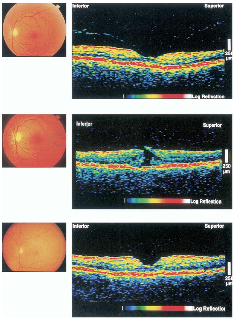
Figure 3.

Evolution of a stage 0 macular hole to a stage 2 macular hole and subsequent surgical repair (case report). Top, Normal color photograph. Optical coherence tomography (OCT) showing normal foveal contour and thickness, a partially separated posterior hyaloid with persistent adherence at the fovea. Middle, Six months later, a color photograph reveals an eccentric full-thickness defect. Optical coherence tomography shows a new disruption in the inner layer of the retina, constituting a stage 2 macular hole. Bottom, Two months after macular hole surgery, a color photograph reveals resolution of the macular hole. The postoperative OCT image demonstrates reapproximation of the edges of the macular hole.
