Full text
PDF

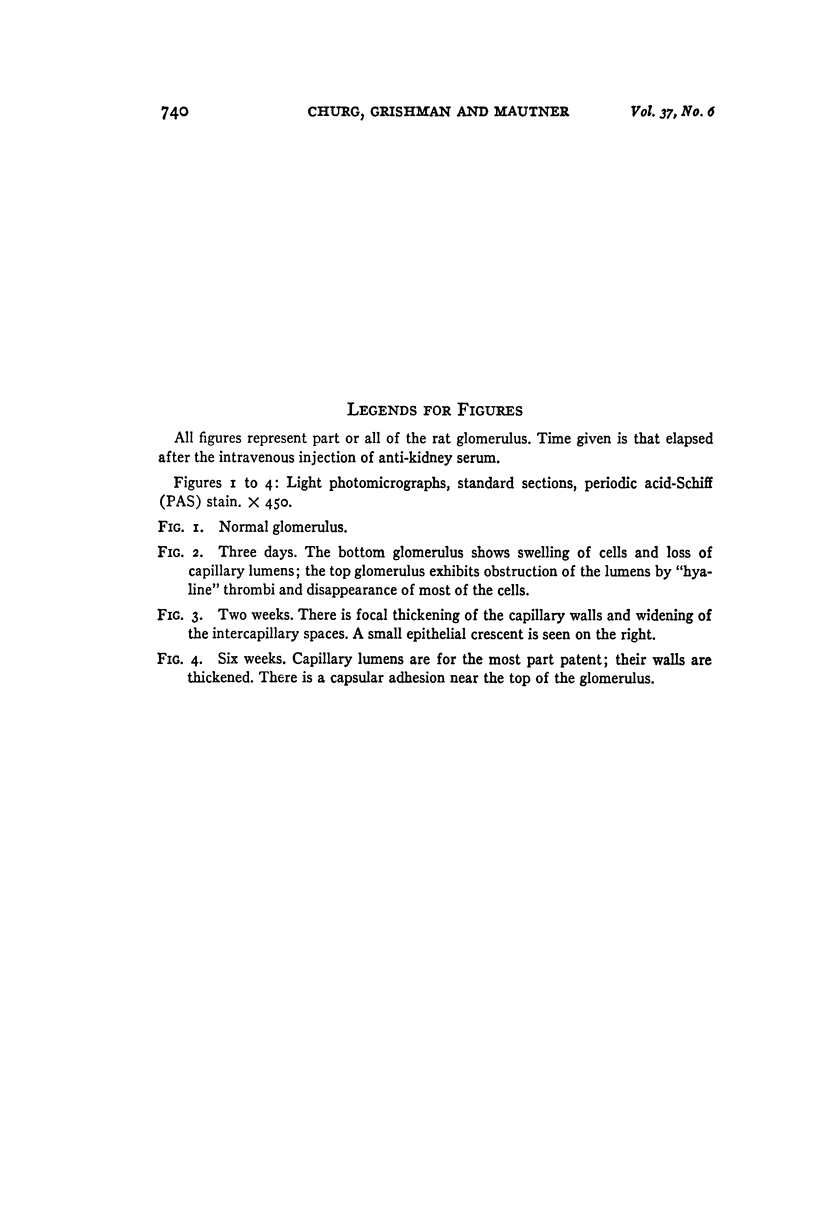
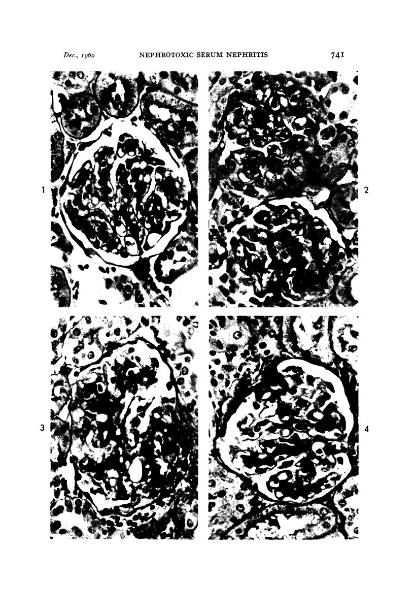
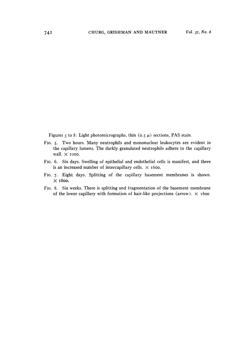
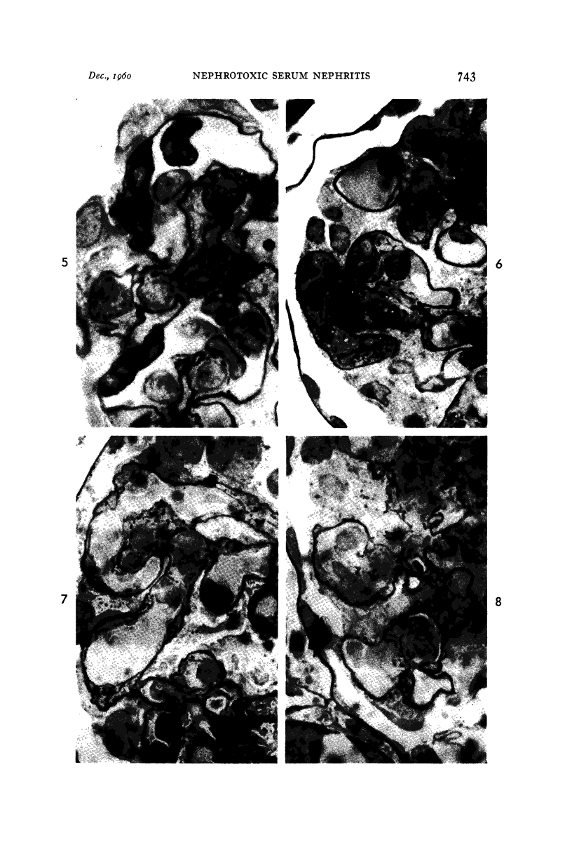
Images in this article
Selected References
These references are in PubMed. This may not be the complete list of references from this article.
- BOHLE A., KRECKE H. J. Zur Frage der Basalmembranen der Glomerulumschlingen in der Niere des Menschen. Virchows Arch Pathol Anat Physiol Klin Med. 1955;327(6):663–696. doi: 10.1007/BF00954932. [DOI] [PubMed] [Google Scholar]
- CHURG J., GRISHMAN E. Application of thin sections to the problems of renal pathology. J Mt Sinai Hosp N Y. 1957 Nov-Dec;24(6):736–744. [PubMed] [Google Scholar]
- CHURG J., GRISHMAN E. Phase microscope studies of renal glomeruli; glomerular deposits of hyaline substances. Am J Pathol. 1953 Mar-Apr;29(2):199–215. [PMC free article] [PubMed] [Google Scholar]
- CHURG J., GRISHMAN E. Subacute glomerulonephritis. Am J Pathol. 1959 Jan-Feb;35(1):25–45. [PMC free article] [PubMed] [Google Scholar]
- CHURG J., MAUTNER W., GRISHMAN E. Silver impregnation for electron microscopy. J Biophys Biochem Cytol. 1958 Nov 25;4(6):841–842. doi: 10.1083/jcb.4.6.841. [DOI] [PMC free article] [PubMed] [Google Scholar]
- EHRICH W. E., FORMAN C. W., SEIFER J. Diffuse glomerular nephritis and lipid nephrosis; correlation of clinical, morphological, and experimental observations. AMA Arch Pathol. 1952 Nov;54(5):463–503. [PubMed] [Google Scholar]
- FARQUHAR M. G., VERNIER R. L., GOOD R. A. An electron microscope study of the glomerulus in nephrosis, glomerulonephritis, and lupus erythematosus. J Exp Med. 1957 Nov 1;106(5):649–660. doi: 10.1084/jem.106.5.649. [DOI] [PMC free article] [PubMed] [Google Scholar]
- FARQUHAR M. G., VERNIER R. L., GOOD R. A. Studies on familial nephrosis. II. Glomerular changes observed with the electron microscope. Am J Pathol. 1957 Jul-Aug;33(4):791–817. [PMC free article] [PubMed] [Google Scholar]
- GRISHMAN E., CHURG J. Acute glomerulonephritis; a histopathologic study by means of thin sections. Am J Pathol. 1957 Sep-Oct;33(5):993–1007. [PMC free article] [PubMed] [Google Scholar]
- HARTMAN M. E. The role of vascular spasm in the etiology of nephrotoxic nephritis in rats. AMA Arch Pathol. 1957 Dec;64(6):679–685. [PubMed] [Google Scholar]
- HEYMANN W. III. Nephrotic syndrome induced by injection of anti-kidney serum. Methods Med Res. 1952;5:264–267. [PubMed] [Google Scholar]
- HEYMANN W., LUND H. Z. Nephrotic syndrome in rats. Pediatrics. 1951 May;7(5):691–706. [PubMed] [Google Scholar]
- JONES D. B. Nephrotic glomerulonephritis. Am J Pathol. 1957 Mar-Apr;33(2):313–329. [PMC free article] [PubMed] [Google Scholar]
- MILLER F., BOHLE A. Elektronenmikroskopische Untersuchungen am Glomerulum bei der Masugi-Nephritis der Ratte. Virchows Arch Pathol Anat Physiol Klin Med. 1957;330(5):483–497. doi: 10.1007/BF00956744. [DOI] [PubMed] [Google Scholar]
- MOVAT H. Z., McGREGOR D. D. The fine structure of the glomerulus in membranous glomerulonephritis (lipoid nephrosis) in adults. Am J Clin Pathol. 1959 Aug;32(2):109–127. doi: 10.1093/ajcp/32.2.109. [DOI] [PubMed] [Google Scholar]
- PAK POY R. K. Electron microscopy of the mammalian renal glomerulus; the problems of intercapillary tissue and the capillary loop basement membrane. Am J Pathol. 1958 Sep-Oct;34(5):885–895. [PMC free article] [PubMed] [Google Scholar]
- PAPPAS G. D., ROSS M. H., THOMAS L. Studies on the generalized Shwartzman reaction. VIII. The appearance, by electron microscopy, of intravascular fibrinoid in the glomerular capillaries during the reaction. J Exp Med. 1958 Mar 1;107(3):333–340. doi: 10.1084/jem.107.3.333. [DOI] [PMC free article] [PubMed] [Google Scholar]
- PIEL C. F., DONG L., MODERN F. W., GOODMAN J. R., MOORE R. The glomerulus in experimental renal disease in rats as observed by light and electron microscopy. J Exp Med. 1955 Nov 1;102(5):573–580. doi: 10.1084/jem.102.5.573. [DOI] [PMC free article] [PubMed] [Google Scholar]
- REID R. T. Electron microscopy of glomeruli in nephrotoxic serum nephritis. Aust J Exp Biol Med Sci. 1956 Apr;34(2):143–150. doi: 10.1038/icb.1956.17. [DOI] [PubMed] [Google Scholar]
- SPIRO D. The structural basis of proteinuria in man; electron microscopic studies of renal biopsy specimens from patients with lipid nephrosis, amyloidosis, and subacute and chronic glomerulonephritis. Am J Pathol. 1959 Jan-Feb;35(1):47–73. [PMC free article] [PubMed] [Google Scholar]
- WACHSTEIN M., LANGE K. Histologic and histochemical studies of the rabbit glomerulonephritis produced by specific antikidney duck serum. Am J Pathol. 1958 Sep-Oct;34(5):835–861. [PMC free article] [PubMed] [Google Scholar]
- YAMADA E. The fine structure of the renal glomerulus of the mouse. J Biophys Biochem Cytol. 1955 Nov 25;1(6):551–566. doi: 10.1083/jcb.1.6.551. [DOI] [PMC free article] [PubMed] [Google Scholar]
- ZOLLINGER H. U. Untersuchungen an Nierenpunktaten: Phasenmikroskopie am Dünnschnitt. Schweiz Z Pathol Bakteriol. 1957;20(1):42–46. [PubMed] [Google Scholar]


