Abstract
Background
Proteins linking intermediate filaments to other cytoskeletal components have important functions in maintaining tissue integrity and cell shape.
Results
We found a set of monoclonal antibodies raised against specific human sarcomeric myosin heavy chain (MyHC) isoforms labels cells in distinct regions of the mammalian epidermis. The antigens co-localize with intermediate filament-containing structures. A slow MyHC-related antigen is punctate on the cell surface and co-localizes with desmoplakin at desmosomal junctions of all suprabasal epidermal layers from rat fœtal day 16 onwards, in the root sheath of the hair follicle and in intercalated disks of cardiomyocytes. A fast MyHC-related antigen occurs in cytoplasmic filaments in a subset of basal cells of skin epidermis and bulb, but not neck, of hair follicles. A fast IIA MyHC-related antigen labels filaments of a single layer of cells in hair bulb. This 230 000 Mr antigen co-purifies with keratin. No obvious candidate for any of the antigens appears in the literature.
Conclusions
We describe a set of molecules that co-localize with intermediate filament in specific cell subsets in epithelial tissues. These antigens presumably influence intermediate filament structure or function.
Keywords: gut, heart, cardiomyocyte, intercalated disk, intermediate filaments, desmosomes.
Background
The diversity of molecules that generate the forces associated with tissue morphogenesis and function is large. Three major classes of motor proteins are known: kinesins and dyneins associated with microtubule-based movements and myosins associated with actin-based motility [1]. The myosin family is the best characterised among motor proteins to date (for review see [2]). The archetypal myosins are the myosin IIs that effect muscle contraction [3] and perform a variety of functions in non-muscle cells, including the regulation of cell motility [4]. Members of the myosin II family consist of two heavy chains and four light chains and function as actin-based ATP-driven motors. The myosin II heavy chains (MyHCs) have a globular ATP- and actin-binding head which is capable of independent force generation, and an elongated coiled coil tail that allows MyHCs to dimerise and then aggregate to form myosin filaments [1]. A variety of MyHC isoforms exist which are generated from distinct genes, as in the case of many skeletal muscle MyHCs, or by alternative splicing or post-translational modification (e.g. [3,5,6] and references therein). In vertebrate skeletal muscle, and probably elsewhere, MyHC heterogeneity contributes to the ability of cells to generate forces of different magnitude and kinetics. Slow skeletal muscle fibres contract slowly because they contain slow MyHC isoforms that hydrolyse ATP slowly. In contrast, fast fibres generate greater forces and contain fast MyHCs which hydrolyse ATP rapidly [7]. Skeletal muscle cells also contain so-called non-muscle MyHCs, which may be involved in regulating muscle differentiation and muscle cell shape [8]. Thus, individual cells express several MyHCs with specific functions, and similar cell types often contain distinct sets of MyHCs that help confer differences in cell behaviour.
MyHCs show high amino acid sequence conservation both between functionally-related isoforms within a species and in homologous isoforms across species. Sequence divergence occurs predominantly at a small number of loci within MyHC that are presumed to confer particular contractile or other properties upon the MyHC isoforms. Evidence in favour of this view has come from the use of antibodies to demonstrate that MyHCs of similar function in skeletal muscles of organisms from fish to man contain the same isoform-specific epitopes (see, for example, [9,10]). In addition, antibodies exist that will apparently detect all members of one MyHC II sub-class, but no others. For example, antibody A4.1025 recognises an epitope near the ATP-binding site of all sarcomeric MyHCs, but the epitope is not found in smooth or non-muscle MyHCs [6,11]. Again, antibody N3.36 recognizes most, if not all, adult fast skeletal MyHCs by binding to a conserved N-terminal epitope, but does not normally recognize any slow skeletal MyHCs [11,12]. Consistent with the view that this epitope conservation reflects conservation of function, exchange of variable domains between MyHC isoforms has demonstrated that particular regions of the MyHC molecule are responsible for its contractile properties [13]. Thus, even the variable domains of MyHC are frequently conserved within sub-classes of myosins.
Skin is noted for its tensile strength combined with suppleness, properties conferred by the extensive cytoskeleton and intercellular connections that maintain tissue integrity. Epidermis contains many morphologically distinct cell types that are readily identifiable based upon their position within the tissue. Rodent epidermis undergoes a series of differentiative and morphogenetic events during development and in the repeating hair cycle during adult life. The initially simple epithelium of embryonic epidermis becomes stratified before birth, leading to cell heterogeneity between epidermal layers. There is also cell diversity within a single layer. Epidermal stem cells have specific locations in the basal layer [14]. Hair follicles are induced perinatally through a mesenchymal-epithelial interaction that instructs a particular region of the surface epidermis to undergo changes in cell shape and gene expression [15]. The epidermal surface undergoes an involution to form a multi-layered hair follicle, best compared in structure to an epidermal leek embedded in dermal soil. It is thought that sheets of hair precursor cells move down in the outer layers of the follicle away from stem cells located at the 'bulge' region [16,17], and then turn inward and move up the centre of the follicle, differentiating as they go, to form the hair proper and its supporting inner root sheath. Thus, a complex series of proliferative, differentiative and morphological changes are co-ordinated during epidermal development.
Here we report the discovery of a number of molecules with MyHC epitopes that are found in a variety of epithelial tissues and mark distinct cell types. These proteins have the unusual feature of sharing structural similarities with distinct isoforms of fast or slow sarcomeric muscle MyHC, yet they associate with intermediate filaments. We suggest that the several proteins with structural similarity to MyHC isoforms could form links between the actin and intermediate filament cytoskeletons.
Results
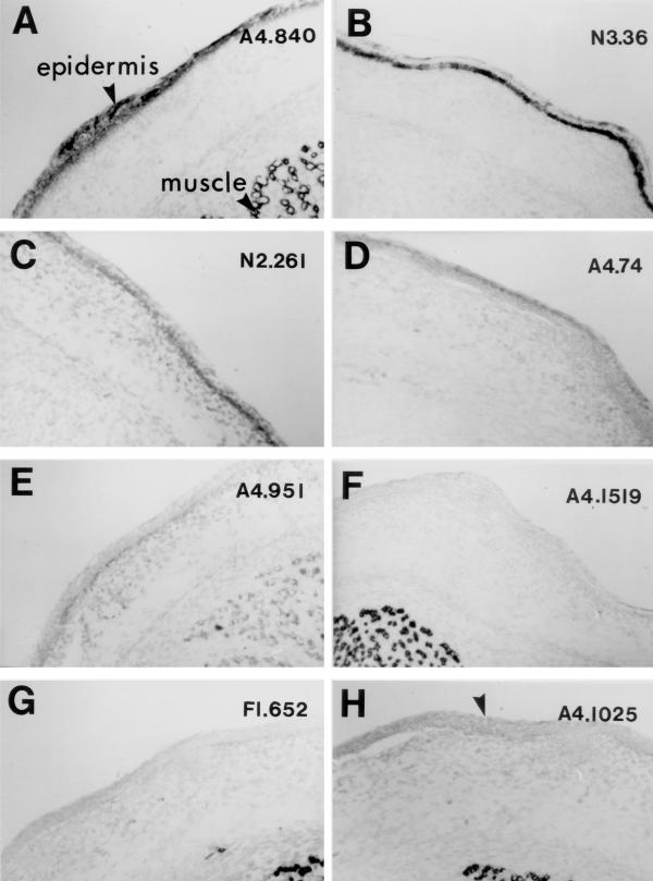
Embryonic day 18 (E18) rat epidermis contains antigens for a number of antibodies previously thought to be specific for particular sarcomeric MyHC isoforms (Fig. 1, see also Table 1 for a summary of antibodies, their known sarcomeric MyHC epitopes and their reactivity with non-muscle tissue). Among a set of eight monoclonal antibodies raised against purifed human skeletal muscle myosin [6,12,18] we found that A4.840, N3.36 and N2.261 detected subsets of cells in E18 epidermis. The A4.840-antigen could be detected in all layers of epidermal cells in cryosections of E18 rat skin (Fig. 1A). In contrast, N3.36-antigen was present only within a subset of cells in the basal layer (Fig. 1B). A third anti-skeletal muscle MyHC antibody, N2.261 also weakly detected cells in basal epidermis (Fig. 1C). At this stage of development, A4.74- and A4.951-antigens were barely detectable (Fig. 1D,1E). Control IgM- and IgG-class anti-sarcomeric MyHC antibodies A4.1519 and F1.652 showed no reaction to epidermis, indicating that binding of A4.840, N3.36 and N2.261 was specific (Fig. 1F,1G, respectively). Skin antigens are not identical to skeletal muscle MyHCs because monoclonal antibodies A4.1025 and MF20 did not react with epidermis (Fig. 1H and data not shown), even though each is reported to detect all isoforms of sarcomeric MyHC [12]. Ten further mouse monoclonal antibodies against sarcomeric MyHC isoforms did not react with rat epidermis at any age examined. In particular, BA-D5, BA-F8 and NA8 antibodies, all of which detect slow skeletal MyHCs in a variety of vertebrate species [6], failed to show any reactivity with epidermis (data not shown). Considering that A4.840- and N3.36-epitopes are normally expressed on the products of different sarcomeric MyHC genes, the data above show that epidermis contains molecules antigenically-related to, but distinct from, sarcomeric MyHC. Hereafter we refer to these molecules as 'MyHC-like' simply to denote this antigenic similarity.
Figure 1.
Distinct MyHC-like proteins are expressed in different subsets of embryonic rat epidermal cells. Transverse cryosections of E18 rat lower hindlimbs were allowed to react with monoclonal antibodies raised against human skeletal muscle myosin and detected by immunohistochemistry. A. Antibody A4.840, which detects slow MyHC isoforms in developing skeletal muscle, reacts throughout the epidermis. B. Antibody N3.36, which detects several neonatal and adult fast MyHC isoforms in skeletal muscle, also detects antigen(s) in the basal cell layer of the epidermis. C. Antibody N2.261, which detects epitopes present on both neonatal slow and adult fast IIa skeletal muscle MyHC, reacts weakly in the basal cell layer of skin, but not in muscle at this age. D. Antibody A4.74, which detects adult fast IIa MyHC in muscle, is not reactive at this age in muscle or epidermis. E. Antibody A4.951, which detects adult slow skeletal MyHC and is therefore not expressed in muscle at this stage, is very weakly-reactive in basal epidermal cells. F. Antibody A4.1519, which detects MyHC(s) that are expressed in all fetal rat myotubes, does not detect epidermis. G. Antibody F1.652, that reacts with rat embryonic skeletal MyHC, is not reactive with embryonic epidermis. H. Antibody A4.1025, that detects all sarcomeric MyHCs, does not react with epidermis (arrow). All panels same scale. Muscle is visible in panels A, E-H and fibres are approximately 20 μm in diameter.
Table 1.
Summary of antibodies, their known MyHC epitopes and epithelial reactivity.
| Antibody | Muscle reactivity | Epithelial reactivity | ||||||
| MyHC Gene | Epitope location | Expression timing | Surface epidermis | Hair¶follicle | Gut | Non-neural retina | Bladder | |
| A4.840 | Slow/β | Rod | Embryo onwards | All | IRS/ ORS | All | + | + |
| HRMC§ | H | HRMC | HRMC | HRMC* | R | R | R | |
| N3.36 | Neo/adult fast | N-terminal 25 aa | Fetal/ postnatal | Some basal cells | ORS | Crypt base | + | + |
| HRM | H | HRM | HR (not M) | HR (not M) | R | R | R | |
| N2.261 | Slow/β + Fast IIA | N-terminal 25 aa | Neonatal onwards | Basal | - | - | + | + |
| HRM | H(β) | RM | HR | HR | R | R | R | |
| A4.74 | Fast IIA | Unknown | Postnatal onwards | - | IRS | - | - | - |
| HRM | RM | HRM | HRM | R | R | R | ||
| N1.551 | Neonatal + Adult Fast | Unknown | Fetal onwards | - | Bulge? neck cell cluster | - | ||
| HRM | HRM | R | R | R | ||||
| A4.951 | Adult slow/β | N-terminal 25 aa | Postnatal onwards | Weak in fetus only | Bulge? neck cell cluster | - | ||
| HRM | H | RM | R | R | R | |||
| A4.1025 | All known | 20 kd / 25 kd junction/ ATP site | Embryo onwards | - | - | - | - | - |
| HRMCDZX | H | HRMCDZX | HRM | HRM | R | R | R | |
Note that three antibodies (N3.36, N2.261 and A4.951) that detect MyHC-like antigens in epithelia have MyHC epitopes in the N terminus of the S1 fragment of the head. X-ray crystallography does not reveal a coiled coil (or any other conserved structure) in this region. §Letters under each entry indicate the species in which reactivity is observed: H human, R rat, M mouse, C chick, D Drosophila, Z zebrafish, X Xenopus. Absence of a species indicates data not available unless specifically noted otherwise. ¶IRS inner root sheath, ORS Outer root sheath, * in feather germ.
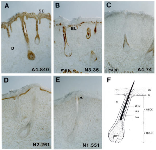
To determine when and in which cells the MyHC-like molecules are expressed, we analysed cryosections throughout postnatal epidermal development when the skin thickens and matures and hair follicles are formed, and found additional sarcomeric MyHC antibody cross-reactivities. By postnatal day 1 (P1) A4.840-antigen was present in the full thickness of the suprabasal surface epidermis and also within many cell layers of the hair follicle (Fig. 2A, shown diagrammatically in 2F). N3.36-antigen was restricted to a subset of basal cells in both surface epidermis and an outer layer of the hair follicle (Fig. 2B). In contrast to younger skin, in which hair follicles had not yet begun to mature, we observed an A4.74-antigen in a single layer of cells near the base of the growing hair follicles (Fig. 2C). By P1, N2.261-antigen detected all basal layer cells in surface epidermis, both those that express N3.36-antigen and those that do not (Fig. 2D). However, N2.261-antigen was not detected in the hair follicle (Fig. 2D). The N1.551-antigen was found restricted to a small cluster of cells in the neck of hair follicles (Fig. 2E). Many other anti-MyHC antibodies, such as A4.1025, F1.652 and A4.1519, do not show any reactivity with skin (data not shown). So, the patterns of expression of the MyHC-like molecules change with development in ways that suggest each may have specific functions in particular epidermal cell types.
Figure 2.
MyHC-like molecules are developmentally regulated in skin. Cryosections of P1 rat back skin was reacted with A4.840 (A), N3.36 (B), A4.74 (C), N2.261 (D) or N1.551 (E) and the antibody detected by peroxidase immunohistochemistry. Hair follicles were sectioned longitudinally, as illustrated diagramatically (F). A4.840 labelled all upper layer epidermal cells, whereas N3.36 labelled only some basal cells of the surface epidermis and an outer layer of the hair bulb. A4.74 labelled part of the root sheath at the base of the hair shaft. N2.261 detected the whole basal layer of surface epidermis, but not the hair follicle. N1.551 labelled only a small group of cells on one side of the neck of the hair bulb (arrow, E). Muscle (mus) showed the expected MyHC immunoreactivity for this stage of development. Dermis (D), surface epidermis (SE), basal epidermis (BL), inner root sheath (IRS), outer root sheath (ORS) and dermal papilla (DP). All panels same scale. Muscle is visible in panel B and fibres are approximately 25 μm in diameter.
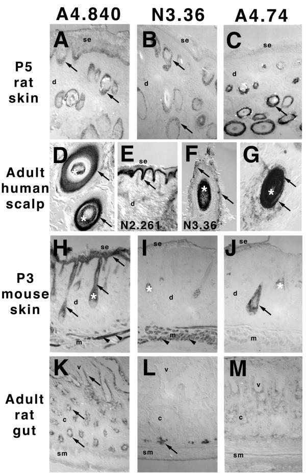
We examined the epidermis of species other than rat, on the rationale that evolutionarily conserved epitopes are likely to have functional significance. A4.840, N3.36 and N2.261 showed similar staining in P5 rat back and adult human scalp epidermis (Fig. 3). A4.840 detected most cells in epidermis, although more weakly as the skin cornified, and in the hair follicle (Fig. 3A,3D). In contrast, N3.36 detected a few cells of the basal layer of P5 rat epidermis, but not adult human surface epidermis. However, the outer layers of the hair follicle were labelled in both species (Fig. 3B,3F). As in rat, in human tissue A4.74 only reacted with cells in the inner root sheath of hair follicles, and N2.261 reacted with basal cells (compare Fig. 2D and 3C,3E,3G). In mouse skin, A4.840 and A4.74 reacted with similar, if not identical, sets of cells to those detected in the rat (Fig. 3H,3J). However, N3.36 did not detect proteins in mouse skin (Fig. 3I). This again suggests that the molecules in skin are not the same as those in muscles, because N3.36 reacts to mouse sarcomeric MyHC, just as to rat and human. We have also observed A4.840-reactivity in chicken skin and feather germs (data not shown). Taken together, these data suggest a family of MyHC-related molecules with substantial evolutionary conservation are expressed in epidermis.
Figure 3.
The MyHC-like molecules are evolutionarily conserved and found in a variety of epithelial tissues. Cryosections of P5 rat tail skin (A-C), adult human scalp skin (D-G), P3 mouse back skin (H-J) and adult rat jejunum (K-M) were reacted with A4.840 (A,D,H,K), N3.36 (B,F,I,L), N2.261 (E) or A4.74 (C,G,J,M) and the antibodies detected by peroxidase-based immunohistochemistry. A4.840-antigen is found in all tissues (arrows, A,D,H,K), but is most readily detected in lower suprabasal layers as skin cornifies in the older tissue (arrow, A). N2.261-reactivity was observed in the basal layer of human skin (arrow, E). N3.36-antigen is present in a subset of cells in a single layer in human and rat epidermis (arrows B,F), although neither the dl DBA mice shown nor two other wild type strains (Balb/c, SwissWebster) showed any reaction (I). A4.74-reactivity was found in the inner root sheath of human, rat and mouse hair follicles (arrows, C,G,J). Note that the pigmented hair shaft also appears dark (asterisks, D-J). Dermal muscle provided positive controls for antibody reactivity (arrowheads, H,I). In the gut, A4.840-antigen was present on most surface epithelial cells of both crypt and villus (arrows, K). N3.36 detected an antigen in cells at the base of crypts, similar to Paneth cells in location (arrow L). se, surface epidermis; m, muscle; d, dermis; c, crypt region; v, villus region; sm, smooth muscle. All panels same scale. Muscle is visible in panels H-J and fibres are approximately 30 μm in diameter.
The MyHC-like proteins are also expressed in other epithelial tissues
To shed light on the potential functions of the MyHC-like molecules in epidermis, we analysed a variety of epithelial and non-epithelial tissues for the presence of the MyHC-like molecules. We never observed any reaction with any non-epithelial tissues other than skeletal or cardiac muscle, consistent with the fact that the monoclonal antibodies to specific skeletal muscle isoforms were originally screened for lack of reaction with several non-muscle tissues [19]. However, we did find reactivity with several epithelial tissues. A4.840 reacted broadly with all cells of the gut epithelium, whereas N3.36 reacted in a small cluster of cells at the base of each crypt (Fig. 3K,3L). A4.74-antigen was undetectable in gut (Fig. 3M). N3.36 reacted with the simple epithelium of the bladder (data not shown). N3.36, N2.261 and A4.840 each reacted with distinct cell populations in the non-neural retina (data not shown). None of these tissues reacted with the general anti-sarcomeric MyHC antibody A4.1025 (data not shown). Thus, the new antigens may perform similar functions in several epithelial cell populations.
Sub-cellular localisation of the MyHC-like molecules
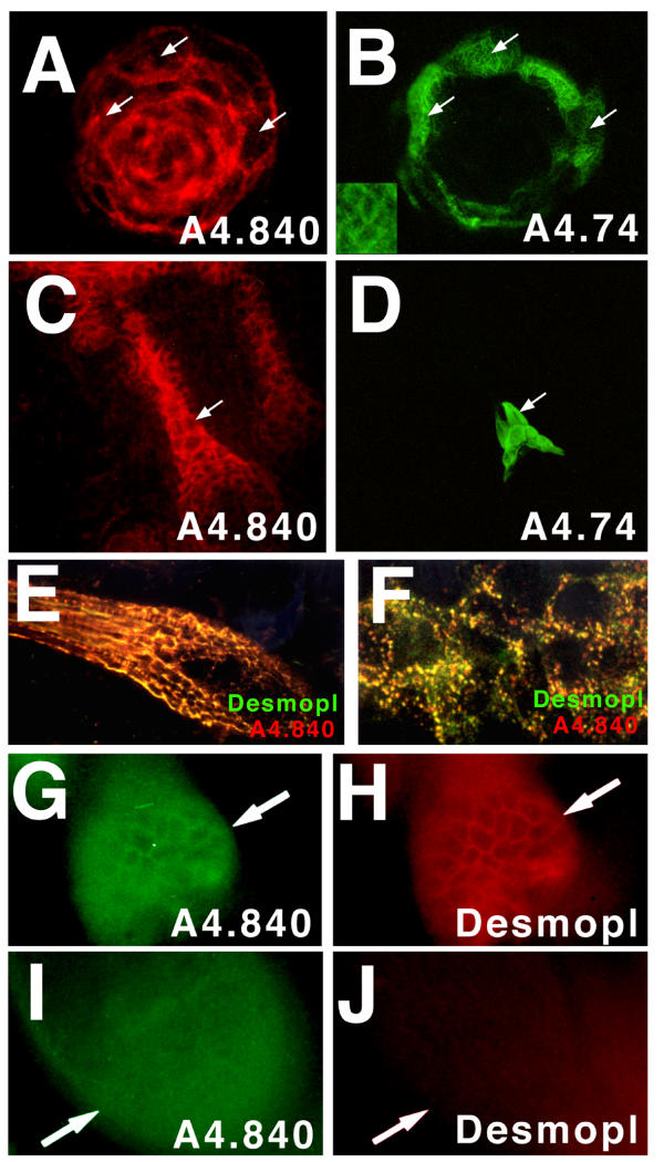
Myosin IIs are generally cytoplasmic proteins that can form filaments and generate force in concert with actin. To determine whether the antigens we detected might have a similar role, we examined the sub-cellular location of each antigen. Initial analyses of cryosections using immunohistochemistry and fluorescence showed that A4.840-reactive molecules were at the cell periphery, whereas A4.74- and N3.36-reactive molecules seemed to accumulate more widely within the cytoplasm (Figs. 2,3). To obtain better spatial resolution of molecules within epidermal cells, we developed a method of partial trypsinisation which loosened the connections between cells while retaining their relative positions within the tissue. Cryosections of this trypsinised material gave good spatial resolution and clearly revealed that A4.840-reactivity was located at the plasma membrane in a punctate distribution (Figs. 4A,4C). The punctate distribution of A4.840-reactive material close to the plasma membrane was highly reminiscent of the abundant desmosomes of epidermis [20,21]. We employed anti-desmoplakin antibodies in dual immunofluorescence confocal microscopy to examine the possibility of co-localization more closely (Fig. 4E,4F). Using fresh frozen cryosections which had not been subjected to trypsinisation, we confirmed the punctate distribution of A4.840-antigen and found it to be strongly associated with desmoplakin immunoreactivity at cell borders (Fig. 4E). We compared images of surface epidermis A4.840-labelling with those of desmoplakin-labelling (Fig. 4F) and scored the number of puncta of fluorescence that labelled with one, the other or both antibodies. Greater than 90% of puncta showed co-localization of A4.840-antigen and desmoplakin. We obtained two pieces of evidence in favour of an intracellular location for the A4.840-antigen: a) A4.840 showed no punctate staining in wholemount stains of unpermeabilised P7 rat skin but readily labelled the surface of hair follicle cells in punctate distribution in wholemount skin fragments permeabilised with Triton X100 (Fig. 4G,4H,4I), b) punctate A4.840-reactivity could be detected in permeabilized cultured skin cells (see Fig. 5A), but were not detected when cells were stained without fixation or permeabilization (data not shown). Attempts to immunolocalise the A4.840-antigen by electron microscopy have so far been unsuccessful, probably due to fixation sensitivity of the A4.840 epitope (unpublished observations kindly provided by Alison North and David Garrod). We conclude that A4.840-antigen is co-localized with desmosomes.
Figure 4.
Cellular and sub-cellular localisation of A4.840-antigen. Immunofluorescent detection of A4.840 (A,C,E,F red; G,I green), A4.74 (B,D, green), and desmoplakin (E,F, green; H,J, red) in rat epidermis. A-D. A4.840-epitope is distributed at the cell periphery of most cells of trypsinised epidermis (A,B a hair follicle bulb in high mag transverse section; C,D surface and hair follicle epidermis in low mag longitudinal section). The A4.74-epitope is in fibrillar structures within the outermost layer of A4.840-reactive cells in the root sheath (arrows, B,D). E,F. Confocal microscopy using A4.840 (red) and anti-desmoplakin (green) on cryosections of P7 rat back skin. Desmoplakin co-localises with A4.840-antigen labelling at the surface of cells in many layers of the root sheath of the hair follicle (E). At higher magnification, desmoplakin and A4.840-antigen co-localise in a punctate array at cell borders in surface epidermis (F). G-J. Intact pieces of P7 rat epidermis were stained for A4.840 (green G,I) and desmoplakin (red H,J). A network of cell surface staining is detected with both antigens after permeabilisation with 1% Triton X100 (G,H arrows), but not without such permeabilisation, when only background fluorescence was detected (I,J arrows).
Figure 5.
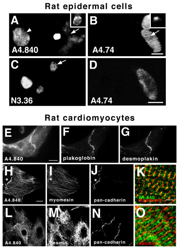
Distribution of MyHC-like antigens is retained in epidermal cells in tissue culture and mimicked in cardiomyocytes. Primary dissociates of P1 rat skin (A-D) were grown in culture and stained for A4.840 (A) or N3.36 (C) and A4.74 (B,D) by dual immunofluorescence. After two days in culture, fibrillar A4.74-reactive material is visible in large spindle-shaped cells, some of which did not contain A4.840-reactive material (arrows, A,B). Other A4.74-reactive cells do contain A4.840-antigen (insets A,B). Large 'donut' cells contain abundant A4.840-reactive dots near the cell surface (arrowhead, A). These donut cells are probably from the prickle cell layer of the surface epidermis based on a) the appearance of cells of similar morphology in the prickle cell layer of cryosections of near-dissociated trypsinised skin and b) the presence of these cells in dissociated 'epidermal' layer cultures, but not in 'dermal' layer cultures that contain numerous dermis and hair follicle cells but little surface epidermis (data not shown). After one day in culture, N3.36-labelled material is also fibrillar (arrow, C) but is co-expressed in A4.74-reactive cells extremely rarely (C,D). Confocal microscopy of primary cultures of neonatal rat cardiomyocytes (E-O) revealed staining for A4.840 (E,H,K,L,O), plakoglobin (F), pan-cadherin (J,N), desmoplakin (G), myomesin (I,K) and desmin (M,O) by triple immunofluorescence. Significantly enhanced A4.840-antigen is detected at the junctions between cells that are marked by plakoglobin and desmoplakin (E-G). Cytoplasmic A4.840-reactivity is also present in some cells, and co-localises in M-lines detected by myomesin (H-J, shown in higher magnification in K,O). Other cells containing sarcomeric striations do not contain detectable A4.840-antigen (asterisks, H-J). The A4.840-antigen is not localised in the Z line marked by desmin (L-O). Bars = 2 μm in K,O, 10 μm in rest.
Desmosomes are not only present in epithelia but also in other tissues under tensile stress. For example, in the myocardium, desmosomes are found at specialised cell-cell contacts, the intercalated disks, where cardiomyocytes are tightly connected to ensure mechanical coupling during the contraction process. Primary cultures of cardiomyocytes continue to beat and intercalated disk-like structures are re-established. The A4.840 antigen co-localized with desmoplakin, plakoglobin and pan-cadherin antigens at these intercalated disk-like structures (Figure 5E,5F,5G,5H,5J,5L,5N). Additional A4.840 reactivity was found in the myofibrils (Figure 5H,5I,5J,5K), localized in the region of the M-band of the sarcomere, as demonstrated by double-labelling with antibodies to myomesin, an integral component of the M-band that is responsible for integrating myosin and titin filaments in the sarcomere [22]. However, only some cardiomyocytes showed these sarcomeric striations with the A4.840 antibody and there are myomesin-stained M-bands that do not show the A4.840 signal (asterisk in H-J). Double staining of the A4.840 antigen with the muscle intermediate filament protein desmin, which is localized around the Z-disks of the sarcomere, revealed alternating striations of the myofibrils (Figure 5L,5M,5N,5O). Taken together, these data raise the possibility that in cardiac muscle, in addition to A4.840-reactive slow sarcomeric MyHC, the novel A4.840 antigen is also expressed and associated mainly with the intercalated disk.
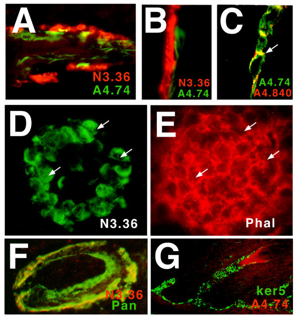
Unlike the A4.840-antigen, A4.74-reactivity was distributed in cells of the hair follicle in a fibrillar arrangement (Fig. 4B,4D). Dual stained sections showed that A4.74-epitope was present in the outermost layer of the A4.840-epitope-containing cells of the hair follicle just above the bulb (Fig. 4A,4B). Similar comparisons between N3.36- and A4.74-antigens showed each to be present within a distinct concentric layer of cells in the follicle, with N3.36 reacting with cells further from the hair itself (Fig. 6A). Both antigens were cytoplasmic (Fig. 4B,4D and Fig. 6). No extracellular or nuclear labelling was detected with any of the three antibodies. Thus, the MyHC-like molecules are distributed distinctly within cells.
Figure 6.
Association of the MyHC-like molecules with intermediate filament-containing structures. N3.36-antigen (A,B,F red; D green), A4.74-antigen (A-C, green; G, red), A4.840-antigen (C, red), phalloidin (E), pan-keratin (F, green) and keratin 5 (G, green) in cryosections of rat epidermis. Longitudinal section of hair follicle from partially-trypsinised P7 rat skin showing the single layer of N3.36-labelled cells of the hair bulb (A, red), immediately external to the A4.74-labelled layer (A, green). At higher magnification, the same distribution is observed in confocal cryosection of an intact rat hair follicle with A4.74-antigen present in the layer internal to the N3.36-antigen (B). A4.74-labelled fibrils (C, green) appear to course through the cytoplasm of cells of the root sheath linking to regions of A4.840-labelled plasma membranes (C, red). Dual localisation of N3.36-reactive fibrils by immunofluorescence (D) and actin microfibrils with rhodaminated-phalloidin (E) show that the major location of the MyHC-like molecule is not on actin cytoskeleton. Similar results were obtained with A4.74 and phalloidin. The N3.36-antigen (red, F) is associated with the outermost layer of keratin-containing cells (green, F). These cells also contain keratin 5 (green, G), based on their position immediately external to the A4.74-reactive cell layer (red, G).
The A4.74- and N3.36-antigens were localized in fibrillar structures within the cytoplasm of fibres (Fig. 4B,4D and Fig. 6C,6D). To characterize their distribution in more detail we tested whether these fibrillar structures represented one of the described cytoskeletal fibrillar systems. Neither N3.36- nor A4.74-reactive material was co-localized with actin filaments labelled with rhodaminated-phalloidin (Fig. 6D,6E and data not shown). On the other hand, A4.74-antigen localized to filaments within the cytoplasm of the hair follicle cells that appeared to connect desmosomes, revealed by abundant A4.840-antigen (Fig. 6C). As desmosomes are connected in this manner by keratin filaments in epidermis [23], this finding suggested that A4.74-antigen may be keratin intermediate filament-associated. The sub-cellular distribution of N3.36-antigen was very similar to that revealed by an anti-pan-keratin antibody in the outermost layer of the root sheath (Fig. 6F), although not all keratin-reactive material co-localized with N3.36-antigen within these cells. Based on dual staining for A4.74-antigen and keratin 5, it appears that N3.36-antigen is expressed in a subset of cells that express keratin 5 (Fig. 6G). Taken together, these data suggest that both the A4.74-antigen and N3.36-antigen are co-localized with intermediate filament proteins in the hair follicle.
To analyse antigen location in more detail, we examined primary dissociated cells from P1 rat skin that had been grown in culture for two days. A4.840-antigen was often, but not exclusively, localized in plasma membrane-associated dots in the cultured cells (Fig. 5A). Both the A4.74- and N3.36-antigens remained in fibrillar structures within distinct populations of these dissociated and fixed cultured epidermal cells (Fig. 5B,5C,5D). Whereas A4.74-antigen was in filaments in either large oval or small round cells in these primary dissociates, most A4.840-antigen that we could detect was located in large cells with a morphology reminiscent of cells in the prickle cell layer of our partially trypisinised tissue (Fig. 5A,5B). In contrast, N3.36-antigen was in a distinct population of cultured cells not labelled with A4.74 (Fig. 5C,5D), just as we observe in vivo. The numbers of cells expressing the MyHC-like antigens appeared to decline with time in dissociated cell culture, suggesting the loss of tissue integrity or cell-cell contact may lead to loss of the antigens. However, due to the small numbers of antigenically-reactive cells, we could not eliminate the possibility of cell death accounting for this observation (data not shown). Consistent with the idea of loss of MyHC-like antigens in culture, after two days in vitro we observed A4.74-reactive cells that did not contain A4.840-antigen (Fig. 5A,5B). Such cells were never observed in vivo. Examination of differentiating human epidermal keratinocyte cultures also failed to reveal any of the MyHC-like antigens, even though differentiation-marker keratins were expressed and desmosomes were formed (data not shown). These results suggest that expression or accumulation of the MyHC-like antigens, requires some aspect of the in vivo environment that is not mimicked in the culture systems examined.
Molecular characterisation of A4.74-antigen
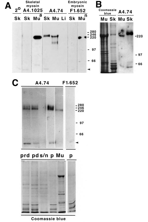
To gain further insight into the nature of the MyHC-like antigens, we performed a partial purification. Western analysis of P9 rat tail skin homogenates revealed a protein of around Mr 230000 that could be detected by antibody A4.74 (Fig. 7A). Antibodies A4.840 and N3.36 did not detect any protein in skin homogenates, despite detecting slow and fast MyHCs (respectively) from muscle tissue, and so could not be analysed further (data not shown). However, the lack of reactivity of these antibodies on skin Western blots supports the notion that the skin and muscle antigens are distinct. The detected skin A4.74-antigen was not a conventional skeletal muscle MyHC because it migrated more slowly than muscle MyHCs and it did not react with antibodies, such as A4.1025, that detect all isoforms of skeletal muscle MyHC (Fig. 7A). Although no obvious band was visible on Coomassie Blue-stained gels at the position of the A4.74-antigen, the antibody reacted strongly with this Mr 230 000 band, even in comparison to its reactivity with muscle MyHC (Fig. 7B). Moreover, the skin sample did not contain skeletal muscle, as antibody A4.1025 does not detect any protein in tail skin, although it readily detects MyHC in skeletal muscle (Fig. 7A). A4.1025 does detect MyHC in proteins isolated from P7 rat back or head skin because these tissues are usually contaminated during isolation with the underlying thin dermal muscle layer, the paniculus carnosus, which is not present in the tail (data not shown and Fig. 3). Other antibodies that detect either all skeletal MyHCs or subsets of these proteins do not react with the skin sample. For example, F1.652, an antibody which recognizes embryonic mammalian MyHC, does not detect proteins in rat skin (Fig. 7A). Thus, skin contains a skeletal muscle fast IIA MyHC-related protein that is not identical to the muscle myosin.
Figure 7.
Skin A4.74-antigen is distinct from fast skeletal myosin IIA and co-purifies with keratin. A. Western analysis of P9 rat tail skin proteins (Sk) shows that proteins around Mr 230000 are detected by A4.74, but not by F1.652 or A4.1025. A4.74 detects a protein of Mr 220000 typical of skeletal muscle MyHC fast IIA in samples of P29 rat extensor digitorum longus (edl) muscle (Mu) but no proteins in P7 liver (Li). A similar Mr 220000 band was detected with anti-embryonic skeletal myosin antibody F1.652 in neonatal P7 lower hindlimb muscle (Mun) and with the general anti-sarcomeric myosin antibody A4.1025 in P35 lower hindlimb muscle (Mua). The faint Mr~45 000 band (arrowhead) in the skin sample is a non-specific cross-reaction, as the control secondary antibody alone detects this band (2°). Comparable total protein (~50 μg) was loaded per lane and checked by Coomassie stain of a duplicate gel. B. The A4.74-antigen in skin (Sk) was more readily detected than fast IIA MyHC in P29 edl (Mu) muscle in Western blots, even when more muscle protein was loaded as indicated with Coomassie blue staining in identical lanes. C. When skin is dissociated in 6 M urea, the A4.74-reactive proteins (upper panel) are fully dissolved and isolated quantitatively in a high speed supernatant (prd) which contains a wide range of proteins including abundant keratins (bands below Mr 66000, lower panel, Coomassie blue stain of a replicate gel). In this experiment, absolute loaded protein was not determined but a precisely similar proportion of the total sample was run in each lane, as can be seen from the Coomassie stain. After dialysis into PBS to remove urea, no protein or antigenic reactivity is lost (pd). Upon re-centrifugation, all detectable A4.74-reactive protein is found in the pellet (p) associated with keratins, whereas many other proteins remain in the supernatant (s/n). Control antibody F1.652 demonstrates the non-specificity of the low Mr band (arrowhead), but shows no reactivity to A4.74-antigen, whereas A4.74 detects the Mr 220000 skeletal IIA fast MyHC in P29 edl muscle extract (Mu). Each skin lane contains an equivalent proportion of the original sample.
We obtained further evidence that the A4.74-antigen in skin was not skeletal muscle fast IIA MyHC. When skeletal muscle is homogenised in high salt solutions myosin is readily extracted. Similar homogenisation of skin failed to release the A4.74-antigen (data not shown). Indeed, homogenisation by mechanical, manual or sonication procedures in solutions containing high salt with or without various detergents failed to release the A4.74-antigen from material that could be pelleted by centrifugation at 100,000 × g for 15 minutes. In contrast, solubilisation of tissue in buffer containing 6 M urea did release the A4.74-antigen to the high speed supernatant (Fig. 7C). Subsequent dialysis against PBS caused a re-precipitation of various highly insoluble skin proteins, including keratins, and this procedure also quantitatively precipitated the A4.74-antigen (Fig. 7C). Dialysis into 0.6 M KCl-containing buffer, which would retain sarcomeric myosin in the supernatant, did not affect the precipitation of the A4.74-reactive protein in skin (data not shown). Therefore, the A4.74-reactive 230 kDa protein in skin co-purifies with skin keratins, and is not skeletal fast IIA MyHC.
Discussion
We have found several molecules which, although antigenically-related to sarcomeric MyHCs, are distinct from them. These evolutionarily conserved molecules have distinct cellular and sub-cellular distributions within epithelial tissues that indicate their involvement in cytoskeletal organisation and/or dynamics.
Molecular nature of the MyHC-like proteins
The single MyHC-like molecule in skin of which we have been able to determine the size had an apparent Mr around 230 000, the size range of non-muscle myosin IIs. This, together with its sub-cellular location and affinity for keratin shows that the protein is not sarcomeric fast IIA MyHC. The co-incidence of size and antigenicity argues against a chance cross-reactivity of A4.74 to an otherwise unrelated protein in skin. Although various described proteins share some characteristics with the A4.74-antigen, we have been unable to find proteins in the literature whose characteristics and expression patterns match with any of the newly-described molecules. For example, both the A4.74-antigen and each of the other MyHC-like molecules in epidermis, show different non-overlapping cellular distributions from that of any of seven keratin antibodies tested (against keratins 1,5,6,10,14,16 and pan-keratin; unpublished results). Similarly, although synemin, paranemin and BPAG1 are ~Mr 230 000 proteins associated with intermediate filaments, they do not have the same cellular distribution as any of the novel MyHC-like antigens [24,25]. Moreover, the presence of five distinct sarcomeric MyHC epitopes in different skin cells raises the possibility that the A4.74-antigen in skin constitutes one member of a family of MyHC-like proteins performing subtly different functions in distinct skin cell populations.
Particular anti-MyHC antibodies that react with skin detect primary amino acid sequence epitopes spread throughout the length of sarcomeric myosin IIs (see Table 1). For example, A4.840 recognizes slow β/cardiac MyHC rod polypeptides expressed in transfected bacteria or COS cells [6,18]. On the other hand, the N2.261 epitope is located at the N-terminal end of the head of slow β/cardiac MyHC, and may depend on post-translational modifications [6]. N3.36 detects the N-terminus of many fast MyHCs and can also be detected on COS cell-expressed sarcomeric perinatal MyHC [6,11,12]. The location of the A4.74 epitope on fast IIA sarcomeric MyHC is unknown. In our view, therefore, it is likely that the evolutionarily conserved epitopes on the new proteins have some element of common function with the evolutionarily conserved epitopes on distinct isoforms of skeletal muscle MyHC. As three of the skin-reactive antibodies detect epitopes in the N-terminus of the sarcomeric MyHC S1 head region, it is highly unlikely that these cross-reactions represent chance similarity of a coiled-coil structure. A4.840 and A4.74, however, may detect coiled coil epitopes which might contribute to their co-localization with intermediate filament that are rich in such structure. Nevertheless, evolutionary conservation argues that these epitopes structures are not chance cross-reactivities. We envisage three possible mechanisms by which such duplication of epitopes could arise. First, the skin molecules could be derived from the same genes that encode skeletal muscle MyHC isoforms. This is unlikely, as such a model would require extensive alternative splicing of the RNA transcripts of several MyHC genes because no single skeletal muscle MyHC gene encodes all the epitopes [6,12,18]. Splicing would be required a) to remove the many conserved skeletal muscle MyHC epitopes which we do not detect in the skin proteins and b) to replace them with sufficient material to maintain the apparent Mr of the protein above 220 000, at least in the case of the A4.74-antigen. Second, there could be a parallel MyHC gene family formed by duplication of several muscle MyHC genes followed by retention of many of the epitopes due to functional demands on the new gene family. Such a model might suggest that the distinct MyHC-like proteins in skin cells could generate distinct forces or develop force at different rates or in response to different activators. Again this is unlikely as the complete murine genome sequence contains no obvious candidate gene set. Third, there could have been extensive convergent evolution of some other family of myosin or non-myosin proteins to allow each to contain one of these MyHC-like epitopes – presumably either due to interaction with as yet unknown partner proteins or to some other conserved function. One possibility is that the new proteins contain MyHC-like epitopes to control structural changes within the molecule that relate to their mechanical properties.
Sub-cellular location of the MyHC-like proteins and their putative function
All three MyHC-like molecules examined in detail are co-localize with cytoskeletal structures. The A4.74-antigen co-purifies with keratin intermediate filament proteins and co-localises to intermediate filaments in skin tissue, and in cultured epidermal cells. The N3.36-antigen also localises to filamentous structures in the basal layer cells of epidermis. Similarly, the A4.840-antigen is present at desmosomes in a variety of cell types, and is also found in the intercalated discs of cardiomyocytes. Desmosomes are the target plasma membrane anchorage site for keratin filaments in epithelial tissues [23], including epidermis and gut, so A4.840-antigen in skin cells is co-localised with the ends of the intermediate filaments in this tissue. This location for the MyHC-like antigens suggests that the function of these proteins may be concerned with the structure and/or dynamics of the intermediate filament network.
The presence of epitopes found on the N terminus of the MyHC head (N3.36, N2.261 and possibly A4.74) on the new molecules raises the possibility that the molecules may function either as molecular motors or as actin binding proteins. It also makes the idea that these antibodies cross-react to intermediate filaments due to the chance recurrence of epitopes in coiled-coil protein structure, as found in the rod regions of MyHC, unlikely to be correct. A number of proteins that cross-link microtubules to microfilaments have been described (see, for example, [26-28]), but fewer are known to cross-link intermediate filament cytoskeleton and microfilaments [25,29-31]. The MyHC-like proteins are new candidates for such cross-links in epithelia. It is striking that in cardiac muscle the A4.840 antibody reacts at the intercalated disks, where myofibrils are anchored in adherens-type junctions, in close proximity to desmosomes that contain the cardiac intermediate filament protein desmin. Since the intercalated disks are the sites of mechanical coupling between cardiomyocytes, specialised cross-links might be required there to ensure proper function. Plectin, a cytoskeletal protein that crosslinks intermediate filaments with microfilaments and microtubules is also concentrated at intercalated disks [32] and its absence leads to partial disintegration of intercalated disk structure at the ultrastructural level [33]. Our results suggest that the A4.840 antigen also acts as an additional cross-linker in the intercalated disks in order to cope with the immense mechanical stress at this site.
On the other hand, structural features of MyHC unrelated to its actin binding capacity could also be conserved in evolution. We have suggested that some of the isoform-specific epitopes on sarcomeric MyHCs may regulate protein stability and/or turnover [6]. These N-terminal epitopes (N3.36, N2.261 and A4.951 (which last detects similar skin cells to N1.551, unpublished result)) are also found in the new skin molecules. The need to turn over sarcomeric MyHCs while maintaining an intact sarcomeric framework is a characteristic that is shared with stable cytoskeletal structures like desmosomal intermediate filaments, but not with most non-muscle MyHCs. Resolution of these issues will await the sequencing of the skin molecules.
Cellular location of the MyHC-like molecules defines epidermal cell sub-populations
Regardless of the nature and function of the MyHC-like molecules, they appear to define populations of epidermal cells. Moreover, analysis of the expression of each antigen, reveals common themes in the cell types in which they are expressed. In epidermis, the N3.36-antigen is located within cells of the basal layer, which contains both the slowly-replicating stem cells, and the highly proliferative precursors of the upper layers of the skin (reviewed in [14]). Early in development, almost all basal layer cells are labelled by N3.36, as is the entire simple epithelium of the bladder. But later, as the N2.261-antigen is up-regulated in the basal layer, the N3.36-antigen becomes restricted to a sub-population of basal cells as the skin matures. This sub-population, nevertheless, appears too abundant to be the slowly-dividing stem cells [34]. As cells leave the basal layer and begin to differentiate, N2.261-antigen is lost and A4.840-antigen is up-regulated. These cells do not express N3.36. Thus, in surface epidermis there is a defined developmental progression of three of the MyHC-like antigens.
In the gut epithelium, the location of the N3.36-antigen in the cells at the base of crypts is also suggestive of labelling of cells that are recently derived from, yet are one step more differentiated than, the stem cells [35]. The A4.840-antigen is expressed the vast majority of cells of the rest of the gut epithelium, suggesting a progression from stem cell, to N3.36-reactive cell and then A4.840-reactive cell. In both epidermis and gut, the N3.36- and A4.840-reactive cell populations have distinct location and geometry.
In the specialised epidermis of hair follicles, a similar, but more complex, sequence of expression is observed. As epidermal buds begin to invaginate, regions lacking N3.36- and N2.261-antigens, but expressing A4.840-antigen, appear. N2.261-antigen was not subsequently detected in follicles. However, N3.36-antigen re-appeared in the outermost layer of the maturing follicle. This suggests location in proliferative early precursors of the follicle that are thought to be recently-derived from stem cells in the bulge region [16], and give rise to the inner layers of the follicle, including the hair [17]. A4.840-antigen showed a complementary pattern, as in surface epidermis, with expression extending throughout the follicle from the surface epidermis, except for the outermost layer of cells that contain keratin 5 and the N3.36-antigen. The N1.551-antigen detects a small group of cells in a similar vicinity to the bulge region described in human follicles, which is the location of follicular stem cells [16]. If N1.551-antigen were present within follicular stem cells, its late appearance in rodent hair would suggest that this cell type is only formed after the initiation of the first anagen. The A4.74-antigen appears late and is only apparent as follicular cells begin to generate the hair proper. This, together with its location in inner root sheath cells, suggests that the cells that express A4.74-antigen may be terminally-differentiating cells destined to support the hair in its outgrowth from the follicle. However, as the lineage of various cell layers within the hair follicle has not been precisely determined, we are unsure of the exact nature of these cells. Nevertheless, it is clear that in the hair follicle there is a characteristic progression of the new MyHC-like antigens.
So the N3.36-, N2.261- and N1.551-antigens appear to mark distinct subsets of precursor cells of epithelial tissues, whereas the A4.840-antigen is expressed in a wide variety of more mature, although not necessarily terminally-differentiated, epithelial and non-epithelial cell types that require the tensile strength conferred by desmosomal junctions. In contrast to the widespread expression of A4.840-antigen, the A4.74-antigen is so far observed only in late-stage cells in the inner root sheath of the hair follicle, a highly specialised cell subset. It is striking that the order of expression of the new antigens is very similar to the expression of the same epitopes on skeletal muscle MyHC isoforms. The two most broadly expressed of the new antigens react with A4.840 and N3.36, the two antibodies that mark general slow and general fast sarcomeric MyHCs, respectively, that are expressed early in development and widely [6,12,18]. The N2.261-antigen is the next most frequent, and also the next to appear developmentally, in epidermis. Similarly, the N2.261 epitope appears in slow muscle fibres after the A4.840 epitope and is present on both slow and fast IIA sarcomeric MyHCs [6,18]. And the last adult sarcomeric MyHC isoforms to appear in rodent skeletal muscle are A4.951- and A4.74-reactive adult slow and adult fast IIA MyHCs, respectively [6,18]. This is paralleled by the late appearance of A4.74- and A4.951-antigens in specific subsets of cells in the hair follicle. Thus, the timing of developmental expression of the new antigens supports the notion of some parallel function(s) with sarcomeric MyHCs. Further understanding of the antigens may shed light not only on epithelial biology, but also the role of MyHC isoform transitions in skeletal muscle.
Conclusions
The chief significance of this work is the identification of a set of apparently novel intermediate-filament co-localized proteins in epithelial tissues. The full nature and sequence of the molecules remains unknown. Nevertheless, our antibody reagents should permit further identification or manipulation of these molecules.
Methods
Biological materials
Wistar rats were bred in the KCL animal facility, and dl DBA mice obtained from Jackson Labs. Human skin samples were generously provided by Niels Krejci and Irene Leigh. Keratinocyte cultures were a gift from Fiona Watt. Irene Leigh also kindly provided a range of anti-keratin antibodies. All MyHC antibodies used in this study can be purchased form Alexis, Switzerland.
Immunohistology
Small pieces of fresh postnatal day 1–6 (P1-6) rat back skin were rapidly frozen and cryo-sectioned at 10–16 μm onto gelatinized slides. Where indicated, fresh skin fragments were incubated in 2% trypsin in PBS for 4 hours at room temperature before freezing to loosen the skin structure. All steps were performed in a humidified box at room temperature. Sections were blocked with PBS, 5% HS for 30 min. Slides were then incubated with A4.1025, F1.652, A4.951, N2.261, A4.74, pan-keratin (IgG) and A4.840, N3.36, A4.1519, N1.551, anti-keratin 5 (IgM) monoclonal antibodies for 1 hr, washed for 1, 5, 15 min in phosphate buffered saline (PBS) 0.5% Tween 20, and IgM or IgG detected with either Texas red- or biotin-conjugated goat anti-mouse IgM (μ-specific)(Vector) or FITC- or biotin-conjugated goat anti-mouse IgG (γ-specific)(Vector) diluted in PBS 5% HS, respectively. Slides were washed three times in PBS 0.5% Tween 20 for a total of 30 min and then mounted under coverslips in polyvinylalcohol anti-fade, or reacted with the ABC kit (Vector) and diaminobenzidene/H2O2. Immuno-labelling was analysed using a Zeiss Axiophot fluorescence microscope. For confocal microscopy, IgM was detected with biotin-conjugated goat anti-mouse Ig (μ-specific), followed by rhodamine-labelled streptavidin diluted in PBS, 5% HS.
Antigen purification
Muscle-free skin peeled from P6-9 rat tail was mechanically homogenised on ice in PBS 0.2 mM EDTA containing protease inhibitors 1 mM PMSF and 1 μg/ml each aprotinin, leupeptin and pepstatin A. Homogenate was centrifuged at 1000 × g for 10 min and the supernatant discarded. To solubilize skin proteins, pellet was re-suspended in 6 M urea, PBS, 0.1% Tween, 0.2 mM EDTA, 2 mM dithiothreitol, protease inhibitors and incubated in a water bath sonicator (Sonomatic, Langford Ultrasonics) for 2 hrs at 4°C. To remove insoluble material the solution was centrifuged at 100,000 × g (Beckman TL-100) for 15 min. The solubilization buffer was removed by dialysis of <1 ml sample against two changes of 800 ml PBS, 0.2 mM EDTA, 0.1 mM PMSF at 4°C for 4 hr. To separate proteins soluble and insoluble in PBS after dialysis the material was re-spun at 100,000 × g for 15 min.
Western Blotting
Crude muscle and liver extracts were prepared by homgenization in 10% SDS, 50 mM Tris/HCl pH 7.4, 5 mM EDTA. After a brief spin to remove tissue, crude supernatant was used in Fig. 7A. Samples were diluted 1:1 with SDS sample buffer (0.5 M Tris/HCl pH 6.8, 20% glycerol, 5% SDS, 10% β-mercaptoethanol), boiled for 2–5 min, electrophoresed through standard 0.1% SDS, 7.5% polyacrylamide separation gel [36]. After electrophoresis some gels were stained with Coomassie Brilliant Blue. For immunoblotting, the electrophoresed proteins were transferred in a semi-dry apparatus [37] to nitrocellulose membranes (Hybond C-super, Amersham) for 75 min at 220 V. Membranes were stained with Ponceau red and photocopied to record the position of major bands. Membranes were blocked with 5% fat free milk (Marvel) overnight at 4°C. All other procedures were done at room temperature. After brief rinsing in PBS, strips of membrane were incubated with monoclonal antibodies diluted 1:10 in PBS, 2% BSA, then rinsed for 1, 5, 15 min in PBS and incubated with goat anti-mouse IgG (whole molecule) peroxidase conjugate antibody (Sigma) diluted 1:100 in PBS, 2% BSA. Proteins detected by the antibodies were visualized by incubation of the membranes with ECL luminescent peroxidase substrate (Amersham) for 1 min and exposure to Hyperfilm ECL (Amersham) until bands were visible. For molecular mass standards BioRad Low Molecular Weight markers, spectrin (246 kD and 280 kD, a kind gift of Jeni Fordham) and MyHC (220 kD) were used.
Primary culture of rat skin cells
Newborn rat (P0–P3) skin was stretched and flattened dermis-side down and incubated in 2% trypsin, PBS at 37°C for 1 hr and then for 4 hr at room temperature. Epidermis was separated from dermis and each part of the dissected skin was minced finely with scissors in Eagle's minimal essential medium, 1.8 mM Ca2+, 10%FCS at 4°C and stirred for 15 min to release cells from the epidermal and dermal sheet mechanically. The cell suspension was filtered through Nytex gauze, centrifuged at 500 rpm (Beckman TJ-6) for 5 min at 4°C and re-suspended in MEM 1.3 mM CaCl2, 10%FCS. Cells were plated at about 105cells/well on Nunc chamber slides which had been coated with poly D-lysine (10 μg/ml, 100 μl/well 2 hours at room temperature) followed by laminin (5 μg/ml, 100 μl/well 2 hours at 37°C) and BSA (1%, 100 μl/well for 15 min at 37°C). After 1–2 days culture, cells were fixed with -20°C methanol for 2 min prior to immuno-detection of antigens.
Neonatal rat cardiomyocyte culture
Hearts from newborn rats were digested with collagenase (108 U/ml, Worthington Biochemical Corp., Freehold, NJ, USA) in ADS buffer (116 mM NaCl, 20 mM HEPES, 0.8 mM NaH2PO4, 1 g/l glucose, 5.4 mM KCl, 0.8 mM MgSO4; pH 7.35) and cultured as described [38]. Cells were plated into dishes coated with 10 μg/ml fibronectin in plating medium (67% Dulbecco's MEM (Amimed AG, Basel, Switzerland), 17% Medium M199 (Amimed AG), 10% horse serum (Gibco, Life Technologies, Basel, Switzerland), 5% fetal calf serum (Gibco) and 1% penicillin/streptomycin (Gibco)). After one day the medium was replaced by maintenance medium (78% Dulbecco's MEM, 20% Medium M199, 1% penicillin/streptomycin, 1% horse serum and 10-4M phenylephrine (Sigma)). Immunofluorescent stainings were performed as described previously [22] using mouse monoclonal anti-plakoglobin (γ-catenin) from Transduction Laboratories, distributed by Maechler, Basel, Switzerland; polyclonal rabbit anti-desmoplakin generously donated by Alison J North, University of Manchester, UK; polyclonal rabbit anti-pan-cadherin and FITC anti-mouse IgM (μ-chain specific) from Sigma, Buchs, Switzerland; TRITC anti-mouse IgG (gamma-chain specific) from Cappel, distr. Organon Technika, Pfaeffikon, Switzerland and Cy5 anti-rabbit IgG from Jackson laboratories, distr. Milan, La Roche, Switzerland. Confocal images were recorded using a Leica PL APO 63x/1.4 oil immersion lens on a Leica inverted microscope DM IRB/E equipped with a Leica true confocal scanner TCS NT and an argon/krypton mixed gas laser. Image processing was performed on a Silicon Graphics workstation using "Imaris" (Bitplane AG, Zurich, Switzerland).
Author's Contributions
SMH made the original observation of reactivity to epithelial tissues, performed the immunohistochemical experiments, conceived the study and wrote the manuscript. AJ performed most of the experiments. EE performed the heart analysis. All authors read and approved the final manuscript.
Acknowledgments
Acknowledgements
We thank Charlotte Peterson and John Mulligan, in whose labs this work was initiated. Special thanks to Niels Krejci, Fiona Watt and Irene Leigh for advice on human dermatology and reagents, Evelyne Perriard for cardiomyocyte preparation, Alan Entwistle for patient help with skin confocal microscopy, David Garrod and Alison North for desmosome advice and permitting us to quote their unpublished work and Jeni Fordham and members of the Hughes laboratory. The work was funded by SMH, the MRC, European Union QLK6-2000-530 and Muscular Dystrophy Campaign (SMH) and an EU Tempus Award (AJ). EE thanks J.-C. Perriard for support.
Contributor Information
Ania Jazwinska, Email: A.Jazwinska@unibas.ch.
Elisabeth Ehler, Email: elisabeth.ehler@kcl.ac.uk.
Simon M Hughes, Email: simon.hughes@kcl.ac.uk.
References
- Kreis T, Vale R, eds Guidebook to the cytoskeletal and motor protiens. Oxford: Oxford University Press. 1993.
- Mooseker M. A multitude of myosins. Curr Biol. 1993;3:245–248. doi: 10.1016/0960-9822(93)90346-p. [DOI] [PubMed] [Google Scholar]
- Schiaffino S, Reggiani C. Molecular diversity of myofibrillar proteins: gene regulation and functional significance. Physiol Rev. 1996;76:371–423. doi: 10.1152/physrev.1996.76.2.371. [DOI] [PubMed] [Google Scholar]
- Maciver SK. Myosin II function in non-muscle cells. BioEssays. 1996;18:179–82. doi: 10.1002/bies.950180304. [DOI] [PubMed] [Google Scholar]
- Bernstein SI, Milligan RA. Fine tuning a molecular motor: the location of alternative domains in the Drosophila myosin head. J Mol Biol. 1997;271:1–6. doi: 10.1006/jmbi.1997.1160. [DOI] [PubMed] [Google Scholar]
- Maggs AM, Taylor-Harris P, Peckham M, Hughes SM. Evidence for differential post-translational modifications of slow myosin heavy chain during murine skeletal muscle development. J Muscle Res Cell Motil. 2000;21:101–13. doi: 10.1023/A:1005639229497. [DOI] [PubMed] [Google Scholar]
- Bàràny M. ATPase activity of myosin correlated with speed of muscle shortening. J Gen Physiol. 1967;50:197–218. doi: 10.1085/jgp.50.6.197. [DOI] [PMC free article] [PubMed] [Google Scholar]
- Wells C, Coles D, Entwistle A, Peckham M. Myogenic cells express multiple myosin isoforms. J Muscle Res Cell Motil. 1997;18:501–15. doi: 10.1023/A:1018607100730. [DOI] [PubMed] [Google Scholar]
- Blagden CS, Currie PD, Ingham PW, Hughes SM. Notochord induction of zebrafish slow muscle mediated by Sonic Hedgehog. Genes Dev. 1997;11:2163–2175. doi: 10.1101/gad.11.17.2163. [DOI] [PMC free article] [PubMed] [Google Scholar]
- Robson LG, Hughes SM. Local signals in the chick limb bud can override myoblast lineage commitment: induction of slow myosin heavy chain in fast myoblasts. Mech Dev. 1999;85:59–71. doi: 10.1016/S0925-4773(99)00060-X. [DOI] [PubMed] [Google Scholar]
- Dan-Goor M, Silberstein L, Kessel M, Muhlrad A. Localization of epitopes and functional effects of two novel monoclonal antibodies against skeletal muscle myosin. J Muscle Res Cell Motil. 1990;11:216–226. doi: 10.1007/BF01843575. [DOI] [PubMed] [Google Scholar]
- Cho M, Hughes SM, Karsch-Mizrachi I, Travis M, Leinwand LA, Blau HM. Fast myosin heavy chains expressed in secondary mammalian muscle fibres at the time of their inception. J Cell Sci. 1994;107:2361–2371. doi: 10.1242/jcs.107.9.2361. [DOI] [PubMed] [Google Scholar]
- Lauzon AM, Tyska MJ, Rovner AS, Freyzon Y, Warshaw DM, Trybus KM. A 7-amino-acid insert in the heavy chain nucleotide binding loop alters the kinetics of smooth muscle myosin in the laser trap. J Muscle Res Cell Motil. 1998;19:825–37. doi: 10.1023/A:1005489501357. [DOI] [PubMed] [Google Scholar]
- Watt FM. Epidermal stem cells: markers, patterning and the control of stem cell fate. Philos Trans R Soc Lond B Biol Sci. 1998;353:831–7. doi: 10.1098/rstb.1998.0247. [DOI] [PMC free article] [PubMed] [Google Scholar]
- Jahoda CA, Horne KA, Oliver RF. Induction of hair growth by implantation of cultured dermal papilla cells. Nature. 1984;311:560–2. doi: 10.1038/311560a0. [DOI] [PubMed] [Google Scholar]
- Cotsarelis G, Sun TT, Lavker RM. Label-retaining cells reside in the bulge area of pilosebaceous unit: implications for follicular stem cells, hair cycle, and skin carcinogenesis. Cell. 1990;61:1329–37. doi: 10.1016/0092-8674(90)90696-c. [DOI] [PubMed] [Google Scholar]
- Rochat A, Kobayashi K, Barrandon Y. Location of stem cells of human hair follicles by clonal analysis. Cell. 1994;76:1063–73. doi: 10.1016/0092-8674(94)90383-2. [DOI] [PubMed] [Google Scholar]
- Hughes SM, Cho M, Karsch-Mizrachi I, Travis M, Silberstein L, Leinwand LA, Blau HM. Three slow myosin heavy chains sequentially expressed in developing mammalian skeletal muscle. Dev Biol. 1993;158:183–199. doi: 10.1006/dbio.1993.1178. [DOI] [PubMed] [Google Scholar]
- Silberstein L, Webster SG, Travis M, Blau HM. Developmental progression of myosin gene expression in cultured muscle cells. Cell. 1986;46:1075–81. doi: 10.1016/0092-8674(86)90707-5. [DOI] [PubMed] [Google Scholar]
- Matoltsy AG. Desmosomes, filaments, and keratohyaline granules: their role in the stabilization and keratinization of the epidermis. J Invest Dermatol. 1975;65:127–42. doi: 10.1111/1523-1747.ep12598093. [DOI] [PubMed] [Google Scholar]
- Kurzen H, Moll I, Moll R, Schafer S, Simics E, Amagai M, Wheelock MJ, Franke WW. Compositionally different desmosomes in the various compartments of the human hair follicle. Differentiation. 1998;63:295–304. doi: 10.1007/s002580050254. [DOI] [PubMed] [Google Scholar]
- Ehler E, Rothen BM, Hammerle SP, Komiyama M, Perriard JC. Myofibrillogenesis in the developing chicken heart: assembly of Z-disk, M-line and the thick filaments. J Cell Sci. 1999;112:1529–39. doi: 10.1242/jcs.112.10.1529. [DOI] [PubMed] [Google Scholar]
- Kowalczyk AP, Bornslaeger EA, Norvell SM, Palka HL, Green KJ. Desmosomes: intercellular adhesive junctions specialized for attachment of intermediate filaments. Int Rev Cytol. 1999;185:237–302. doi: 10.1016/s0074-7696(08)60153-9. [DOI] [PubMed] [Google Scholar]
- Granger BL, Lazarides E. Synemin: a new high molecular weight protein associated with desmin and vimentin filaments in muscle. Cell. 1980;22:727–38. doi: 10.1016/0092-8674(80)90549-8. [DOI] [PubMed] [Google Scholar]
- Ruhrberg C, Watt FM. The plakin family: versatile organizers of cytoskeletal architecture. Curr Opin Genet Dev. 1997;7:392–7. doi: 10.1016/S0959-437X(97)80154-2. [DOI] [PubMed] [Google Scholar]
- Henriquez JP, Cross D, Vial C, Maccioni RB. Subpopulations of tau interact with microtubules and actin filaments in various cell types. Cell Biochem Funct. 1995;13:239–50. doi: 10.1002/cbf.290130404. [DOI] [PubMed] [Google Scholar]
- Fujii T, Hiromori T, Hamamoto M, Suzuki T. Interaction of chicken gizzard smooth muscle calponin with brain microtubules. J Biochem (Tokyo) 1997;122:344–51. doi: 10.1093/oxfordjournals.jbchem.a021759. [DOI] [PubMed] [Google Scholar]
- Goode BL, Wong JJ, Butty AC, Peter M, McCormack AL, Yates JR, Drubin DG, Barnes G. Coronin promotes the rapid assembly and cross-linking of actin filaments and may link the actin and microtubule cytoskeletons in yeast. J Cell Biol. 1999;144:83–98. doi: 10.1083/jcb.144.1.83. [DOI] [PMC free article] [PubMed] [Google Scholar]
- Houseweart MK, Cleveland DW. Intermediate filaments and their associated proteins: multiple dynamic personalities. Curr Opin Cell Biol. 1998;10:93–101. doi: 10.1016/S0955-0674(98)80091-4. [DOI] [PubMed] [Google Scholar]
- Allen PG, Shah JV. Brains and brawn: plectin as regulator and reinforcer of the cytoskeleton. BioEssays. 1999;21:451–4. doi: 10.1002/(SICI)1521-1878(199906)21:6<451::AID-BIES1>3.3.CO;2-2. [DOI] [PubMed] [Google Scholar]
- Dalpe G, Mathieu M, Comtois A, Zhu E, Wasiak S, De Repentigny Y, Leclerc N, Kothary R. Dystonin-deficient mice exhibit an intrinsic muscle weakness and an instability of skeletal muscle cytoarchitecture. Dev Biol. 1999;210:367–80. doi: 10.1006/dbio.1999.9263. [DOI] [PubMed] [Google Scholar]
- Wiche G, Krepler R, Artlieb U, Pytela R, Denk H. Occurrence and immunolocalization of plectin in tissues. J Cell Biol. 1983;97:887–901. doi: 10.1083/jcb.97.3.887. [DOI] [PMC free article] [PubMed] [Google Scholar]
- Andrä K, Lassmann H, Bittner R, Shorny S, Fässler R, Propst F, Wiche G. Targeted inactivation of plectin reveals essential function in maintaining the integrity of skin, muscle, and heart cytoarchitecture. Genes Dev. 1997;11:3143–3156. doi: 10.1101/gad.11.23.3143. [DOI] [PMC free article] [PubMed] [Google Scholar]
- Jensen UB, Lowell S, Watt FM. The spatial relationship between stem cells and their progeny in the basal layer of human epidermis: a new view based on whole-mount labelling and lineage analysis. Development. 1999;126:2409–18. doi: 10.1242/dev.126.11.2409. [DOI] [PubMed] [Google Scholar]
- Potten CS. Stem cells in gastrointestinal epithelium: numbers, characteristics and death. Philos Trans R Soc Lond B Biol Sci. 1998;353:821–30. doi: 10.1098/rstb.1998.0246. [DOI] [PMC free article] [PubMed] [Google Scholar]
- Laemmli UK. Cleavage of structural proteins during the assembly of the head of bacteriophage T4. Nature. 1970;227:680–5. doi: 10.1038/227680a0. [DOI] [PubMed] [Google Scholar]
- Kylise-Andersen J. Electroblotting of multiple gels: a simple apparatus without buffer tank for rapid transfer of proteins from polyacrylamide to nitrocellulose. J Biochem Biophys Methods. 1984;10:203–209. doi: 10.1016/0165-022X(84)90040-X. [DOI] [PubMed] [Google Scholar]
- Sen A, Dunnmon P, Henderson SA, Gerard RD, Chien KR. Terminally differentiated neonatal rat myocardial cells proliferate and maintain specific differentiated functions following expression of SV40 large T antigen. J Biol Chem. 1988;263:19132–6. [PubMed] [Google Scholar]


