Abstract
Background
Bezoars usually present as a mass in the stomach. The patient often has a preceding history of some psychiatric predisposition. Presentation could be in the form of trichophagy followed by trichobezoar (swallowing of hair leading to formation of bezoar), orphytobezoar (swallowing of vegetable fibres). Rapunzel syndrome is a condition where the parent bezoar is in the stomach and a tail of the fibres or hair extends in to the jejunum. Presentation as intestinal obstruction due to a bezoar in the intestine without a parent bezoar in the stomach is rare, therefore we report it here.
Case report
A 35 year old lady tailor with a previous history of receiving treatment for depression on account of being infertile- years after her marriage, presented to the surgical emergency department with features of acute intestinal obstruction. Exploratory laparotomy and enterotomy revealed a cotton bezoar in the terminal ileum without a parent bezoar in the stomach. She was managed by resection of the affected segment of the ileum and end-to-end anastomosis of the bowel. In the postoperative period the patient gave a history of ingesting cotton threads whenever she was depressed.
Conclusion
Presence of cotton bezoar is rare and an intestinal bezoar in the absence of parent bezoar in the stomach is still rarer.
Keywords: cotton bezoar, ileum
Background
Bezoars are rare and are often reported in patients with some psychiatric ailment [1-3] They usually present with signs and symptoms due to a mass in the stomach and may rarely extend in to the jejunum as a tail (Rupenzel syndrome)[3-5]. Isolated presentation in the ileum without a parent bezoar in the stomach is rare. Stomach bezoars if detected in time may be treated by endoscopic retrieval but if presentation is in the form of intestinal obstruction with or without perforation management is by a formal exploratory laparotomy followed up by treatment for the underlying psychiatric disorder. [5-8]
Case Report
A 35 year old lady, who was a tailor by occupation was admitted in the emergency surgical wing with acute pain abdomen, distension, vomiting and absolute constipation of 24 hrs duration. She had no cough, no hemetemesis and no malaena but there was significant weight loss along with loss of appetite for the past year. She had been married for the last seven years, had miscarriages three times and had no children. There was a history of receiving psychiatric consultation for depression on account of infertlity.
Examination revealed an anxious patient in distress with marked pallor, tachycardia (pulse: 110/mt, BP 100/60 mm Hg) and tachypnoea. The abdomen was tense tender and distended with peritoneal signs and a vague mass in the right lumbar region extending on to the right iliac fossa, which was dull on percussion. Liver dullness was not obliterated and rectal examination revealed an empty and ballooned rectum.
She was found to be severely anemic (Hb 6 gm%) with marked leucocytosis (Total count 15000/cmm). The blood chemistry was also deranged (blood urea 70 mg% along with hyponatremia and hypokalemia). X ray of the abdomen erect and supine showed multiple air fluid levels without any gas under the diaphragm. Ultrasound of the abdomen showed a bowel mass in the right iliac fossa with a mixed echogenic pattern there was minimal free fluid in the peritoneal cavity.
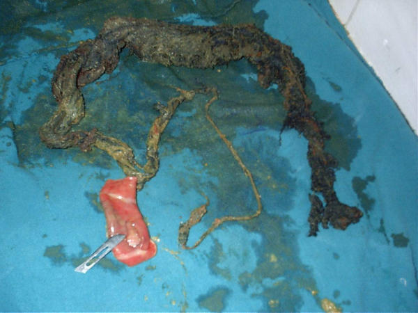
Patient was taken up for exploratory laparotomy through a midline incision, which revealed grossly dilated, and distended small bowel loops with an indentable bowel mass in the ileum measuring 10 × 6 × 6 cm about 2 ft from the ileocaecal junction. The mass could be palpated extending and tailing off in to the transverse colon through the ileocaecal junction and ascending colon. Enterotomy was performed and a foul smelling mass of cotton threads in the form of a bunch (including the tail in the ascending and transverse colon) was negotiated and retrieved (fig). Since the segment including the site of enterotomy was of doubtful viability about 4 inches of the ileum was resected and an ileo-ileal anastomosis performed. There was no bezoar in the stomach or duodenum and proximal jejunum. After a thorough peritoneal lavage the abdomen was closed with drains in situ. The patient made a satisfactory postoperative recovery and gave a history of swallowing cotton threads whenever she was depressed on account of being issueless. She had a similar episode of acute pain abdomen two years back when she was relieved after vomiting out a bunch of cotton threads. She has been referred to the psychiatry outpatients for treatment of her depression and six months of follow up reveal a satisfactory recovery. Post-operative upper gastrointestinal endoscopic studies did not reveal any concomitant bezoar in the stomach.
Conclusion and discussion
The term Bezoar comes from the Arabic "badzehr" or from the Persian "panzer" both meaning counterpoison or antidote [1,3]. Hindus used bezoars in the twelfth century BC for rejuvenating the old, neutrallizing snake venom and other poisons, treating vertigo, epilepsy, melancholia and even plague. A genuine bezoar was recognized by its failure to smoke when a red-hot needle was plunged into it [2-5] Causes of bezoar include the presence of indigestible material in the lumen, gastric dysmotility (including previous surgery like vagotomy and partial gastrectomy etc.) and certain other substances that encourage stickiness and concretion formation. Around 400 cases of trichobezoar and a larger number of phytobezoars have been reported in the literature but many go unreported[1,5]. They occur mainly in the young women who chew and swallow their hair (trichobezoar) or phytobezoar (vegetable fibres) or diospyrobezoar (persimmon fibres) or pharmacobezoar (tablets/semi liquid masses of drugs) [2,4-8]. With time these are retained by mucus and become enmeshed, creating a mass in the shape of the stomach where they are usually found. They may attain large sizes owing to the chronicity of the problem and delayed reporting by the patients. The clinical presentation may be a palpable, firm, non-tender epigastric mass, which is either discovered, on routine physical examination in an asymptomatic patient or as an operative surprise as in our patient. Bezoars have been reported between the ages of 1 and 56 yrs, most presenting between the ages of 15–20 yrs and 90% are in females. Approximately 10%show psychiatric abnormalities or mental retardation[5].
Bezoars mostly originate in the stomach probably related to high fat diet causing non-specific symptoms like epigastric pain, dyspepsia and post-prandial fullness, the stomach is not able to push the hair or other substance out of the lumen because the friction surface is in-sufficient for propulsion by peristalsis [2-4]. They may also present with gastrointestinal bleeding (6%) and intestinal obstruction or perforation (10%) [2,4,5]. Diagnosis at an early stage is important since conservative treatment (fragmentation and endoscopic extraction, enzymatic destruction) is possible for gastric bezoars. Rarely the bezoars may extend in to the small intestine as a tail (Rapunzel syndrome after "Rapunzel" the fair, long haired maiden in the Grimm brother's fairy tail who lowered her tresses to allow Prince charming to climb up to her prison tower to rescue her) or may get broken lodging in the intestine to cause intestinal obstruction, ulceration, bleeding and perforation. Small intestinal bezoars have also been reported after truncal vagotomy and with compression of the duodenum by the superior mesenteric artery [6]
Treatment is removal of the mass by a single enterotomy or resection of the bowel if not viable [7,8] Duncan et al. recommended bezoar extraction by multiple enterotomies in the Rapunzel syndrome [9]. DeBakey and Oschner reported an operative mortality of 10.4% [10]. It is mandatory to do a thorough exploration of the rest of the small intestine and the stomach to look for retained bezoars. If available endoscopic examination of the stomach is the preferred method of exploring the stomach for the concomitant bezoar while managing a case of intestinal bezoar. Exploration may reveal concomitant gastric bezoar which may be retrieved endoscopically or by gastrotomy[8]. Escamilla et al reported twenty three cases of concomitant gastric bezoars (extracted by gastrotomies) out of eighty seven cases of intestinal bezoars[8]. If detected in the intestine they may be milked down to the enterotomy site for retrieval through one opening or they may require multiple enterotomies..
It is rare for a cotton bezoar to present so low down in the terminal ileum with extension in to the ascending and transverse colon without a concomitant parent bezoar in the stomach. This patient was an operative surprise since the patient did not give any contributory history and the presentation was essentially as a case of acute intestinal obstruction The author had earlier reported a case of a trichobezoar obstructing the terminal ileum, which also did not have any parent bezoar in the stomach [1].
Pre-publication history
The pre-publication history for this paper can be accessed here:
Figure 1.

Shows the retrieved cotton bezoar. The widest part was in the terminal ileum and the tail was extending in to the ascending colon and transverse colon also seen is the resected ileum.
Acknowledgments
Acknowledgement
Written consent was taken from the husband of the patient regarding the publication of this interesting case report. The patient herself is illiterate hence could not give the desired written consent.
Contributor Information
Chintamani, Email: chintamani7@rediffmail.com.
Rakesh Durkhure, Email: chintamani@doctor.com.
Singh JP, Email: chintamani@doctor.com.
Vinay Singhal, Email: chintamani@doctor.com.
References
- Sharma RD, Chintamani , Bhatnagar D. Trichobezoar obstructing the terminal ileum. Trop Doct. 2002;32:99–100. doi: 10.1177/004947550203200217. [DOI] [PubMed] [Google Scholar]
- Charles AndrusH, Jeffrey PonskyL. Bezoars: Classification, Pathophysiology, and Treatment. Am J Gastroenterol. 1988;83:476–478. [PubMed] [Google Scholar]
- Allred-Crouch AL, Young EA. Bezoars: When the "knot in the stomach" is real. Postgrad Med. 1985;78:261–5. doi: 10.1080/00325481.1985.11699169. [DOI] [PubMed] [Google Scholar]
- Goldstein SS, Lewis JH, Rothstein R. Intestinal obstruction due to bezoars. Am J Gastroenterol. 1984;79:313–8. [PubMed] [Google Scholar]
- Senapati MK, Subramanian S. Rapunzel syndrome. Trop Doct. 1997;27:53–54. doi: 10.1177/004947559702700122. [DOI] [PubMed] [Google Scholar]
- Doski JJ, Priebe CJ, Jr, Smith T, et al. Duodenal trichobezoar caused by compression of the superior mesenteric artery. J Pediatr Surg. 1995;30:1598–9. doi: 10.1016/0022-3468(95)90165-5. [DOI] [PubMed] [Google Scholar]
- Santiago Sanchez CA, Garau Diaz P, Lugo Vicente HL. Trichobezoar in a 11 y-year old girl: A case report. Bol Asoc Med PR. 1996;88:8–11. [PubMed] [Google Scholar]
- Escamilla C, Robles-Campos R, Parrilla-Paricio P. Intestinal obstruction and bezoars. J Am Coll Surg. 1994;179:285–8. [PubMed] [Google Scholar]
- Duncan ND, Altken R, Venugopal S, et al. The Rapunzel syndrome. Report of a case and review of literature. West Indian Med J. 1994;43:63–5. [PubMed] [Google Scholar]
- De Bakey M, Oschner A. Bezoars and concretions: comprehensive review of literature, with analysis of 303 collected cases and presentation of eight additional cases. Surgery. 1939;5:132–160. [Google Scholar]


