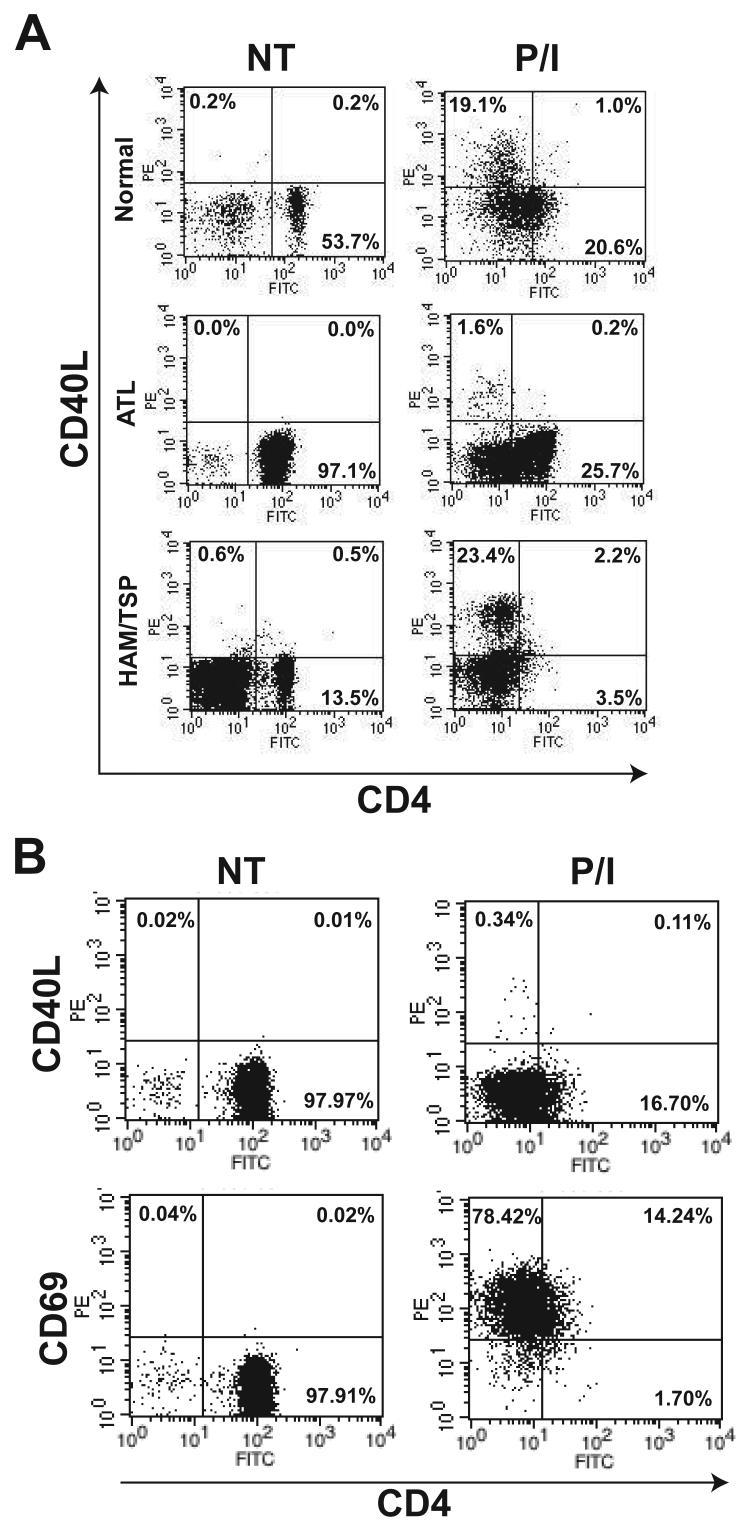
Figure 4. CD40L, but not CD69, induction is defective in peripheral blood mononuclear cells of ATL patients.
(A) Flow cytometry analysis of CD40L and CD4 cell surface expression in PBMCs obtained from normal and ATL or HAM/TSP patients. Cells were either untreated or treated overnight with PMA (10 ng/ml) and ionomycin (2μM). Cells were stained with PE-conjugated anti-CD40L antibody, PE-Cy5-conjugated anti-CD25 and FITC-conjugated anti-CD4 antibody. The data are presented as a two-dimensional dot plot depicting cells that were within the gated region of the light scattering plot. Gating was done using FSC and SSC properties and for each test, at least 10,000 gated cells were analyzed. The numbers in each panel denote the percentage of cells falling within the indicated regions. (B) Flow cytometry analysis of PBMCs from an acute ATL patient. Cells were treated as in panel A and stained with either PE-conjugated anti-CD40L or anti-CD69 together with PE-Cy5-conjugated anti-CD25 and FITC-conjugated anti-CD4 antibody. Analysis was performed as described in panel A.

