

Full text
PDF
Images in this article
Selected References
These references are in PubMed. This may not be the complete list of references from this article.
- BAKER R. F., GOODMAN J. R., MOORE R. E. Electron microscopic study of phagocytosis of staphylococcus by human leukocytes. II. Virulent and non-virulent staphylococci. J Bacteriol. 1956 Dec;72(6):736–745. doi: 10.1128/jb.72.6.736-745.1956. [DOI] [PMC free article] [PubMed] [Google Scholar]
- BRODOFF M., HOFFMAN W. A., DELUCA V. A., Jr, SPIRO H. M. Intestinal lipodystrophy (Whipple's disease); diagnosis by small-intestine biopsy tube. J Am Med Assoc. 1959 Sep 12;171:154–157. doi: 10.1001/jama.1959.03010200022006. [DOI] [PubMed] [Google Scholar]
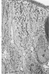
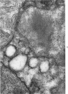
- CHEARS W. C., Jr, ASHWORTH C. T. Electron microscopic study of the intestinal mucosa in Whipple's disease. Demonstration of encapsulated bacilliform bodies in the lesion. Gastroenterology. 1961 Aug;41:129–138. [PubMed] [Google Scholar]
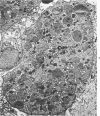
- HAUBRICH W. S., WATSON J. H., SIERACKI J. C. Unique morphologic features of Whipple's disease. A study by light and electron microscopy. Gastroenterology. 1960 Oct;39:454–468. [PubMed] [Google Scholar]
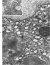
- PALADE G. E. The endoplasmic reticulum. J Biophys Biochem Cytol. 1956 Jul 25;2(4 Suppl):85–98. doi: 10.1083/jcb.2.4.85. [DOI] [PMC free article] [PubMed] [Google Scholar]
- ROWLANDS D. T., Jr, LANDING B. H. Colonic histiocytosis in children. Report of a form resembling that seen in Whipple's disease. Am J Pathol. 1960 Feb;36:201–211. [PMC free article] [PubMed] [Google Scholar]
- YARDLEY J. H., FLEMING W. H., 2nd Whipple's disease: a note regarding PAS-positive granules in the original case. Bull Johns Hopkins Hosp. 1961 Aug;109:76–79. [PubMed] [Google Scholar]
- YOUNGMAN R. A., ZEMAN E. D. Whipple's disease. Report of case with favorable response to treatment. Nebr State Med J. 1961 Jan;46:3–9. [PubMed] [Google Scholar]