

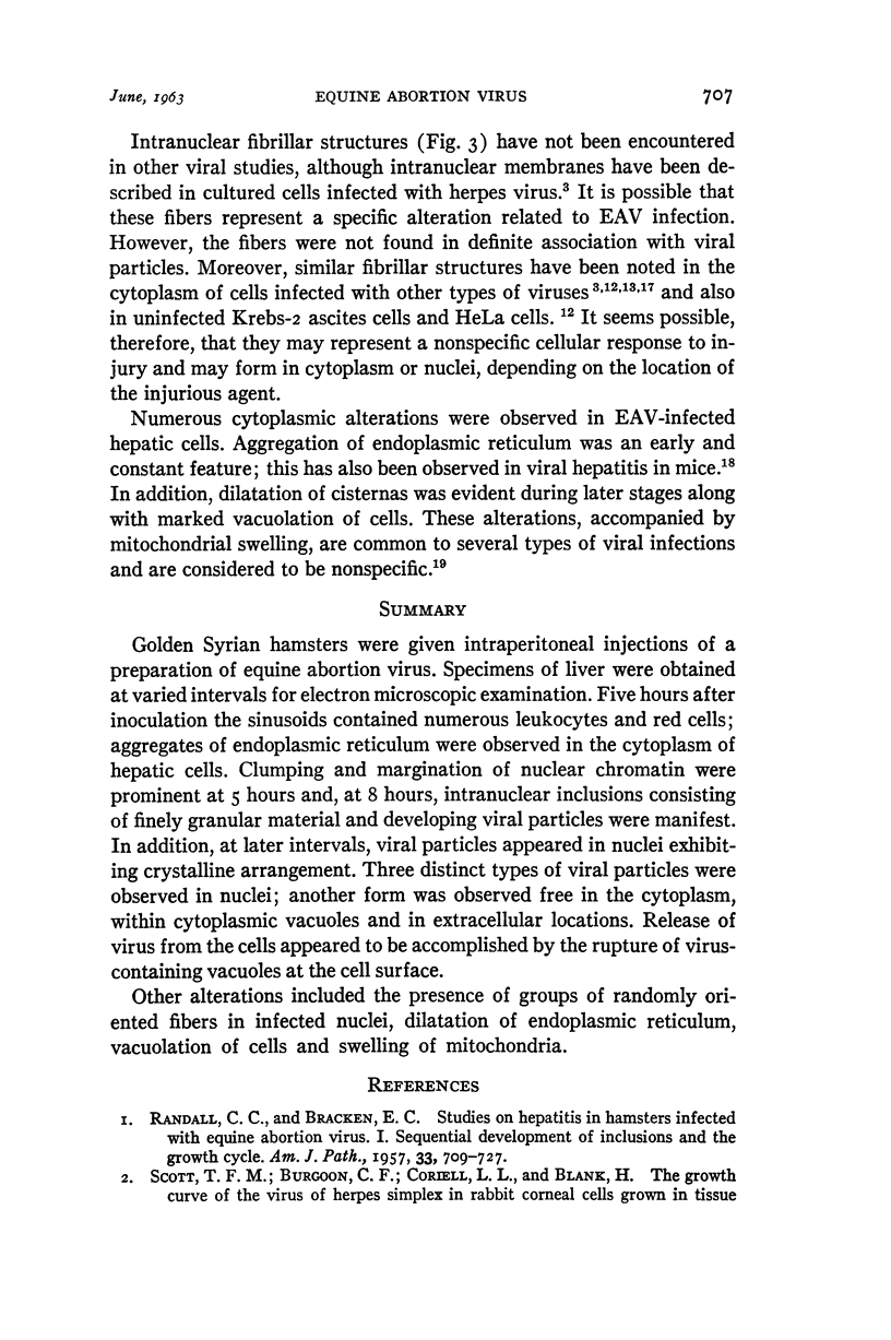
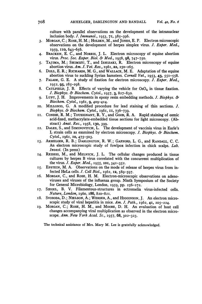
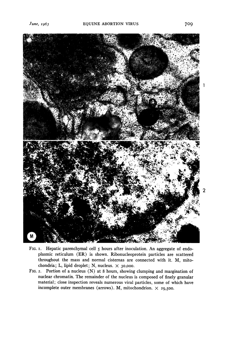
Full text
PDF
Images in this article
Selected References
These references are in PubMed. This may not be the complete list of references from this article.
- BRACKEN E. C., NORRIS J. L. Electron microscopy of equine abortion virus. Proc Soc Exp Biol Med. 1958 Aug-Sep;98(4):747–750. doi: 10.3181/00379727-98-24171. [DOI] [PubMed] [Google Scholar]
- CAULFIELD J. B. Effects of varying the vehicle for OsO4 in tissue fixation. J Biophys Biochem Cytol. 1957 Sep 25;3(5):827–830. doi: 10.1083/jcb.3.5.827. [DOI] [PMC free article] [PubMed] [Google Scholar]
- DALES S., SIMINOVITCH L. The development of vaccinia virus in Earle's L strain cells as examined by electron microscopy. J Biophys Biochem Cytol. 1961 Aug;10:475–503. doi: 10.1083/jcb.10.4.475. [DOI] [PMC free article] [PubMed] [Google Scholar]
- DOLL E. R., RICHARDS M. G., WALLACE M. E. Adaptation of the equine abortion virus to suckling Syrian hamsters. Cornell Vet. 1953 Oct;43(4):551–558. [PubMed] [Google Scholar]
- EPSTEIN M. A. Observations on the mode of release of herpes virus from infected HeLa cells. J Cell Biol. 1962 Mar;12:589–597. doi: 10.1083/jcb.12.3.589. [DOI] [PMC free article] [PubMed] [Google Scholar]
- LUFT J. H. Improvements in epoxy resin embedding methods. J Biophys Biochem Cytol. 1961 Feb;9:409–414. doi: 10.1083/jcb.9.2.409. [DOI] [PMC free article] [PubMed] [Google Scholar]
- MILLONIG G. A modified procedure for lead staining of thin sections. J Biophys Biochem Cytol. 1961 Dec;11:736–739. doi: 10.1083/jcb.11.3.736. [DOI] [PMC free article] [PubMed] [Google Scholar]
- MORGAN C., ROSE H. M., HOLDEN M., JONES E. P. Electron microscopic observations on the development of herpes simplex virus. J Exp Med. 1959 Oct 1;110:643–656. doi: 10.1084/jem.110.4.643. [DOI] [PMC free article] [PubMed] [Google Scholar]
- MORGAN C., ROSE H. M., MOORE D. H. An evaluation of host cell changes accompanying viral multiplication as observed in the electron microscope. Ann N Y Acad Sci. 1957 Oct 21;68(2):302–323. doi: 10.1111/j.1749-6632.1957.tb56087.x. [DOI] [PubMed] [Google Scholar]
- PALADE G. E. A study of fixation for electron microscopy. J Exp Med. 1952 Mar;95(3):285–298. doi: 10.1084/jem.95.3.285. [DOI] [PMC free article] [PubMed] [Google Scholar]
- RANDALL C. C., BRACKEN E. C. Studies on hepatitis in hamsters infected with equine abortion virus. I. Sequential development of inclusions and the growth cycle. Am J Pathol. 1957 Jul-Aug;33(4):709–727. [PMC free article] [PubMed] [Google Scholar]
- REISSIG M., MELNICK J. L. The cellular changes produced in tissue cultures by herpes B virus correlated with the concurrent multiplication of the virus. J Exp Med. 1955 Mar 1;101(3):341–352. doi: 10.1084/jem.101.3.341. [DOI] [PMC free article] [PubMed] [Google Scholar]
- SCOTT T. F. M., BURGOON C. F., CORIELL L. L., BLANK H. The growth curve of the virus of herpes simplex in rabbit corneal cells grown in tissue culture with parallel observations on the development of the intranuclear inclusion body. J Immunol. 1953 Dec;71(6):385–396. [PubMed] [Google Scholar]
- SIEGEL B. V. Filamentous-structures in ectromelia virusinfected cells. Nature. 1960 Jun 4;186:820–821. doi: 10.1038/186820a0. [DOI] [PubMed] [Google Scholar]
- SVOBODA D., NIELSON A., WERBER A., HIGGINSON J. An electron microscopic study of viral hepatitis in mice. Am J Pathol. 1962 Aug;41:205–224. [PMC free article] [PubMed] [Google Scholar]
- TAJIMA M., SHIMIZU T., ISHIZAKI R. Electron microscopy of equine abortion virus. Am J Vet Res. 1961 Mar;22:250–265. [PubMed] [Google Scholar]