Abstract
Bacillus thuringiensis mosquitocidal toxin Cry4Ba has no significant natural activity against Culex quinquefasciatus or Culex pipiens (50% lethal concentrations [LC50], >80,000 and >20,000 ng/ml, respectively). We introduced amino acid substitutions in three putative loops of domain II of Cry4Ba. The mutant proteins were tested on four different species of mosquitoes, Aedes aegypti, Anopheles quadrimaculatus, C. quinquefasciatus, and C. pipiens. Putative loop 1 and 2 exchanges eliminated activity towards A. aegypti and A. quadrimaculatus. Mutations in a putative loop 3 resulted in a final increase in toxicity of >700-fold and >285-fold against C. quinquefasciatus (LC50 ≅ 114 ng/ml) and C. pipiens (LC50 ⩬ 37 ng/ml), respectively. The enhanced protein (mutein) has very little negative effect on the activity against Anopheles or Aedes. These results suggest that the introduction of short variable sequences of the loop regions from one toxin into another might provide a general rational design approach to enhancing B. thuringiensis Cry toxins.
The ultimate goal of protein engineering of the insecticidal crystal proteins from Bacillus thuringiensis is to be able to design any Cry toxin to possess toxic activity against any insect. A more immediate goal is to introduce activity into a toxin that does not possess it. We chose to modify Cry4Ba, a toxin expressed by B. thuringiensis var. israelensis (B. thuringiensis subsp. israelensis), which has toxicity toward mosquitoes of the genera Anopheles and Aedes but has no activity against Culex (14).
B. thuringiensis has been used successfully in forestry and agriculture, both as spray formulations and as systemic pesticides expressed in plants. The high level of target specificity makes B. thuringiensis a more desirable choice than broad-spectrum chemical insecticides from an environmental perspective, but it limits its market potential (48). B. thuringiensis subsp. israelensis has also been used successfully in control of mosquito vectors. It expresses several genes and has broad control over most important mosquitoes but lacks good persistence in the field (5).
There are various approaches to broadening the insecticidal spectrum of B. thuringiensis, including introduction of toxin genes that code for separate insecticidal activities into a recipient bacterium (4, 7, 26), creating a bacterium that expresses a combination of toxins instead of a single toxin (46), and engineering mutant toxins with new or broadened insecticidal spectra (6, 8). In these approaches, multiple gene constructs with separate mechanisms of action are useful to forestall resistance development. It is also important that the genes used are as toxic as possible against the target pests.
B. thuringiensis subsp. israelensis-based biopesticides are used worldwide to control mosquito and black fly populations (5). The mosquitocidal toxins of B. thuringiensis subsp. israelensis are due to parasporal toxins deposited during sporulation as crystals. These insecticidal crystal proteins are composed of Cry4Aa, Cry4Ba, Cry11Aa, and Cyt1Aa (51). Toxic protein genes from this bacterium have been inserted into other suitable hosts to confer mosquitocidal activity on the new hosts (24, 31, 32, 36). These approaches hoped to increase the efficacy of the toxins over that for the original host.
Genetic studies of coleopteran- and lepidopteran-active B. thuringiensis insecticidal crystal proteins indicated that domain II loop regions are involved in receptor binding (12, 29). Domain II loop mutations may have either a negative or a positive effect on binding and toxicity (44). The loop residues are generally considered to be involved with receptor binding, but the functional interactions of individual residues may differ significantly between target insects or receptors (40). A mutation in loop 2 of Cry1Ab decreases the toxicity to Manduca sexta by affecting irreversible binding, but the same mutant affects neither toxicity nor binding affinity to Heliothis virescens (37). On the other hand, alanine substitutions of loop 3 residues G439 and F440 in Cry1Ab affect the initial receptor binding and toxicity in both M. sexta and H. virescens (41). These loops are proposed to be exceptional targets for genetic engineering to create more potent toxins with diverse insect specificity (38). For a more general review, see reference 42.
The present study explored the domain II loop regions of Cry4Ba in search of sites involved in mosquitocidal activities. Putative loop residues from Cry4Aa were exchanged with putative loop residues from Cry4Ba. The results of the exchanges were tested on four different species of important human disease vectors, Aedes aegypti (dengue and yellow fever), Anopheles quadrimaculatus (malaria), Culex quinquefasciatus (West Nile virus), and Culex pipiens (West Nile virus). Our results showed that the putative loop 3 exchange caused significant increases in toxicity of greater than 700-fold against C. quinquefasciatus and greater than 285-fold against C. pipiens while still maintaining wild-type levels against the other two mosquitoes. The significant increase in toxicity, or its lack thereof, was surprisingly not related to either receptor binding or irreversible binding. Putative loop 1 and 2 switches, however, abolished toxicity towards both Aedes and Anopheles but did not establish toxicity towards Culex.
MATERIALS AND METHODS
Cloning and construction of a trypsin-site deletion mutant of Cry4Ba.
The cry4Ba gene was a kind gift from A. Delécluse (Pasteur Institute, Paris, France). The gene was amplified by PCR with a set of primers (forward primer FW4AB, 5′ GAT Tgg atc cAA TGT AAT ATG GGA G 3′ [lowercase letters indicate BamHI site], and reverse primer RE4AB, 5′ TAT TTT Tgg tac cAG AAT TAA TAA ATG CAG 3′ [lowercase letters indicate KpnI site]) and subcloned into plasmid pTZ19R (Fermentas), which was double digested with BamHI and KpnI. The cry4Ba gene was put under the control of the lac promoter of the vector in this construct. This construct was transformed into DH5α Escherichia coli cells for DNA isolation and protein expression. The wild-type Cry4Ba protoxin produced a ∼46-kDa toxin fragment when digested with trypsin (data not shown). This fragment was found to be inactive (25). The trypsin site was removed by mutating R203 to A by site-directed mutagenesis, as previously reported by Angsuthanasombat et al. (3). The mutated toxin was called 4BRA, and it produced a ∼66-kDa toxin fragment when digested by trypsin (data not shown).
Mutating Cry toxins by site-directed mutagenesis.
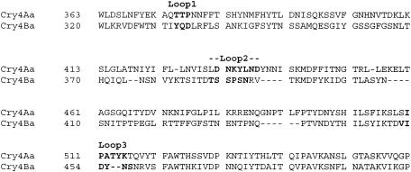
Location of the putative loop regions was made by homology modeling the structure of Cry4Aa on Cry3Aa with Swiss Model (http://www.expasy.org/swissmod/SWISS-MODEL.html) (data not shown) and aligning the amino acid sequences of Cry4Aa and Cry4Ba with Swiss PDB-Viewer (18, 35) (Fig. 1). The putative loops 1, 2, and 3 of 4BRA were mutated to mimic the corresponding putative loops of Cry4Aa. In the putative loop 1, 331IYQ333 were mutated to QTT to yield 4BL1QTT. In the putative loop 2, NDY was inserted between V393 and T394 to yield 4BL2NDY. In the putative loop 3, D454 was replaced with P and AT was inserted after position 454 to yield 4BL3PAT. In further mutations of the putative loop 3, D454 was replaced with G and AV was inserted after position 454 to yield 4BL3GAV. P454 in the 4BL3PAT construct was mutated to A to yield 4BL3AAT. P454 was mutated to G to yield 4BL3GAT. Mutation of T456 in the 4BL3PAT background to A yielded 4BL3PAA. Both P454 and T456 were mutated to A's to yield 4BL3AAA. Site-directed mutagenesis was performed by the modified Quick Change (Stratagene) method. DNA templates were purified (Qiagen), methylated (dam methylase; New England BioLabs), and replicated by PCR with Expand long-template polymerase (Roche) with the following cycling parameters: step 1, 94°C, 2 min; step 2, 94°C, 10 s; step 3, 48°C, 30 s; step 4, 68°C, 4 min (repeat steps 2 to 4 nine times); step 5, 94°C, 15 s; step 6, 48°C, 30 s; step 7, 68°C, 4 min plus 20 s every successive cycle (repeat steps 5 to 7 15 times); step 8, 68°C, 7 min; step 9, 4°C, unlimited. The sequences of the primers are listed in Table 1. The reaction product was digested with DpnI (Roche) to remove the methylated template DNA. The digested PCR product was used to transform E. coli DH5α competent cells. Mutations were confirmed by automated DNA sequencing (Plant-Microbe Genomics Facility, The Ohio State University).
FIG. 1.
Sequence alignments based on the model structures made with Swiss-Pdb Viewer (18) of Cry4Aa with Cry4Ba. Loop positions are indicated above the sequences, while the amino acid residues involved are in boldface.
TABLE 1.
Sequences of primers used in site-directed mutagenesis
| Primera | Sequence (5′→3′) | Mutant |
|---|---|---|
| Fw4BR203A | GGTCTTTAGCAGCTAGTGCTGGTGACC | 4BRA |
| Re4BR203A | GGTCACCAGCACTAGCTGCTAAAGACC | |
| Fw4BL1QTT | ACCAATACTCAAACTACAGATTTAAGATTTTTATC | 4BL1QTT |
| Re4BL1QTT | TCTTAAATCTGAAGTTTGAGTATTGGTCCAGAAATC | |
| Fw4BL2NDY | CTAATCGAGTTAATGATTATACAAAAATGGATTTC | 4BL2NDY |
| Re4BL2NDY | CATTTTTGTATAATCATTAACTCGATTAGAGGGTATTC | |
| Fw4BL3PAT | GATGTTATACCTGCGACTTATAACAGTAACAGGGTTTC | 4BL3PAT |
| Re4BL3PAT | CTGTTATAAGTCGCAGGTATAACATCAGTTTTTATATAG | |
| Fw4BL3AAT | GATGTTATAGCTGCGACTTATAACAGTAACAGGGTTTC | 4BL3AAT |
| Re4BL3AAT | CTGTTATAAGTCGCAGCTATAACATCAGTTTTTATATAG | |
| Fw4BL3GAT | GATGTTATAGGTGCGACTTATAACAGTAACAGGGTTTC | 4BL3GAT |
| Re4BL3GAT | CTGTTATAAGTCGCACCTATAACATCAGTTTTTATATAG | |
| Fw4BL3GAV | GATGTTATAGGTGCGGTTTATAACAGTAACAGGGTTTC | 4BL3GAV |
| Re4BL3GAV | CTGTTATAAAGCGCACCTATAACATCAGTTTTTATATAG | |
| Fw4BL3PAA | GATGTTATACCTGCGGCTTATAACAGTAACAGGGTTTC | 4BL3PAA |
| Re4BL3PAA | CTGTTATAAGTCGCAGGTATAACATCAGTTTTTATATAG | |
| Fw4BL3AAA | GATGTTATAGCTGCGGCTTATAACAGTAACAGGGTTTC | 4BL3AAA |
| Re4BL3AAA | CTGTTATAAGCCGCAGCTATAACATCAGTTTTTATATAG |
The sets of complementary primers for creating the mutants are grouped together.
Isolating and purifying Cry toxin.
E. coli DH5α cells containing the toxin construct were grown on Luria-Bertani agar plates (43) supplemented with 100 μg of ampicillin/ml at 37°C. Crystal inclusions were expressed in Terrific Broth (24 g of yeast extract/liter, 12 g of tryptone/liter, 2% glycerol, 25.08 g of K2HPO4/liter, 4.62 g of KH2PO4/liter), supplemented with 100 μg of ampicillin/ml, and grown for 72 h at 37°C in an incubator-shaker at 250 rpm. Cells were harvested and lysed, and crystal inclusions were washed as previously described (27).
Crystal inclusion protein was solubilized in carbonate buffer (30 mM Na2CO3, 20 mM NaHCO3, pH 10.0), and the protein concentration was measured using the Coomassie protein assay reagent (Pierce) with bovine serum albumin (BSA) as standard. For binding assays, solubilized toxin was incubated with 1/20 (vol/vol) 10-mg/ml trypsin (Sigma) at 37°C for 3 h. The activated toxin was purified by high-pressure liquid chromatography with a Superdex 200 (Pharmacia) column.
Secondary structure analysis by circular dichroism (CD) spectroscopy.
Column-purified toxins were concentrated to at least 1 mg/ml with a Centricon instrument (YM-30; Millipore). Concentrated toxins were diluted in a phosphate buffer (10 mM KH2PO4-K2HPO4, 40 mM NaCl, pH 7.4) prepared in Milli-Q (Millipore) water. CD data were collected at room temperature with a 1-cm-path-length quartz cell (Hellma) on an AVIV Model 62A DS spectrophotometer, scanning from 250 to 200 nm at 1.0-nm steps. Data shown were based on the averages of 10 scans.
Determining toxicity of Cry toxins by mosquito larva bioassay.
Mosquitoes were reared in an environment-controlled room at 28°C and 85% humidity, with a photoperiod of 14 h of light and 10 h of dark. The A. quadrimaculatus culture was from Peggy Hodges (University of Notre Dame), A. aegypti and C. quinquefasciatus cultures were from A. Yousten (Virginia Polytechnic Institute), and C. pipiens (recently isolated from nature in Ohio) was from R. Robish and W. Foster (Ohio State University). Adult mosquitoes were maintained on heparin-treated cow blood, sugarcane cubes (Domino Dots), and dechlorinated tap water. Aedes and Culex larvae were maintained on fish food pellets (Koi Floating Blend; Aquaricare), while Anopheles larvae were maintained on a 2:1 ratio of ground fish food flakes (Vitapro Plus Cichlid Power Flakes; Mike Reed Enterprises) and brewer's yeast (M. Q. Benedict, Centers for Disease Control and Prevention). Second-instar larvae were used for all bioassays. Bioassays were performed on different days after hatching due to the different growth rates of the mosquito larvae. A. aegypti, C. quinquefasciatus, and C. pipiens larvae were tested 2 days after hatching, while A. quadrimaculatus larvae were tested 3 days after hatching. A total of six larvae per 2.5 ml of water with one replicate in a 24-well Costar cell culture plate (Corning) were fed a serial dilution of Cry toxins (as inclusions), and the number of mortalities was counted after a 24-h incubation at 28°C. The bioassay was repeated to obtain a concentration range on Cry toxin inclusions yielding 10 to 90% mortality. The 50% lethal concentration was calculated by a Probit method using SoftTOX version 1.1 (WindowChem).
Preparing mosquito brush border membrane vesicles (BBMV).
Fourth-instar mosquito larvae were filtered with a nylon mesh, washed in distilled water, separated from large residual food particles, and dried briefly on a filter paper (Fisher) under vacuum suction. Harvested larvae were frozen at −70°C until needed. About 4 to 6 g of frozen larvae was homogenized in 8 to 12 ml of cold buffer A (300 mM mannitol, 5 mM EGTA, 17 mM Tris-HCl, pH 7.5). Larvae were homogenized by 40 strokes of a Potter-Elvehjem polytetrafluoroethylene pestle in a glass tube at speed number 5 (∼6,000 rpm). The homogenized sample was centrifuged at 11,000 rpm × g for 5 min at 4°C in a JA-17 rotor. The pellet was discarded while the supernatant was kept for the next step. The supernatant was filtered through a Whatman (no. 1) filter paper under vacuum, and the filtrate was collected on ice. The filtrate (4 ml) was layered on top of a 0 to 45% sucrose gradient and centrifuged at 40,000 × g for 2 h at 4°C in an SW28 rotor. The top layer was removed by suction and discarded, leaving the lowest visible layer or the pellet. This layer was resuspended in cold sterile double-distilled H2O and centrifuged at 35,000 × g for 15 min at 4°C in a JA-17 rotor. The supernatant was discarded, and any loose pellet was rinsed off with binding buffer (60 mM K2HPO4, 5 mM KH2PO4, 150 mM NaCl, 10 mM EGTA, pH 7.00). The BBMV pellet was resuspended in 1 ml of ice-cold binding buffer supplemented with Complete (Roche) protease inhibitor and homogenized by 10 strokes of a small Potter-Elvehjem polytetrafluoroethylene pestle and glass tube. The protein concentration of the BBMV was measured with the Coomassie protein assay reagent (Pierce), with BSA as the standard. The BBMV was distributed into 0.5-ml aliquots and kept at −70°C until needed. Aminopeptidase activity was determined as described previously (15). Aminopeptidase in A. quadrimaculatus BBMV was 5.5-fold enriched over that in the homogenate.
Radioactive labeling of Cry toxins.
Activated toxins were iodinated as previously described (52). Briefly, 0.3 to 0.5 mCi of 125I-Na (Perkin-Elmer) was incubated with one Iodo-bead (Pierce) for 5 min at room temperature. Then, toxin (45 μg) in 0.1 ml of 30 mM Na2CO3-20 mM NaHCO3 (pH 10.0) was added to the bead reaction mixture. After 5 min at room temperature, the mixture was passed through a 2-ml Excellulose column (Pierce) to remove free iodine from the toxin.
Saturation binding assay.
Mosquito BBMV (0.5 to 1.0 μg) were incubated with an increasing concentration of 125I-labeled toxin in 0.1 ml of binding buffer for 1 h at room temperature. The reaction mixture was centrifuged at 26,000 × g for 10 min to separate unbound toxin from BBMV, and the BBMV pellet was washed two times in binding buffer by centrifugation. The final pellet was counted in a gamma counter (Wallac) to measure bound 125I-toxin. Nonspecific binding was the amount of toxin bound in the presence of at least a 250-fold excess of unlabeled toxin. Specific binding was obtained by subtracting the nonspecific binding counts from the total binding counts. Data were plotted with SigmaPlot version 8.0 (SPSS, Inc.).
Competition binding assay.
The course of toxin binding to BBMV was suggested to occur through a two-step process involving reversible (19, 20) and irreversible (21, 37, 50) steps. In this assay, 10 μg of mosquito BBMV was incubated with 1 nM 125I-labeled toxin in 0.1 ml of binding buffer with increasing amounts of unlabeled toxin for 1 h at room temperature. The reaction mixture was centrifuged at 26,000 × g for 10 min to separate unbound labeled toxin from the BBMV. The supernatant was discarded while the pellet was washed twice with binding buffer. Counting and plotting of the data were done as described under “Saturation binding assay.”
Irreversible binding assay.
In the irreversible binding assay, 2 μg of mosquito BBMV was incubated with 2 nM 125I-labeled toxin in 0.1 ml of binding buffer for 1 h at room temperature. Then a 1,000 nM final concentration was added to the binding reaction mixture and was incubated for a different length of time. Unbound labeled toxin was separated from the BBMV, and the resulting data were obtained as described above. Nonspecific binding data were obtained by incorporating the unlabeled toxin into the labeled toxin at the start of the assay and incubating the toxins with the BBMV for the maximum duration of the assay period. Specific binding was obtained by subtracting the nonspecific binding counts from the total binding counts.
Proteinase K protection assay.
In this assay, 5 μg of C. quinquefasciatus BBMV was incubated with a 10 nM concentration of either 125I-labeled 4BRA or 125I-labeled 4BL3PAT in 0.1 ml of binding buffer for 1 h at room temperature. Later, 10 μg of proteinase K (Roche) was added and incubated for a further 20 min. The action of the protease was stopped by 100 μg of Pefabloc SC (Roche). The reaction mixture was centrifuged at 26,000 × g for 10 min at room temperature to separate the remaining toxin bound or inserted in the BBMV. The pellet was washed two times with binding buffer without resuspending the pellet.
Preparation of Culex BBMV for membrane permeation assay.
C. quinquefasciatus larvae were raised to fourth instar as described earlier. Larvae were collected, washed in distilled water, dried briefly on filter paper, and stored at −70°C. BBMV were prepared from thawed whole larvae. Thawed larvae were homogenized in 10 mM HEPES (pH 7.5)-250 mM sucrose containing Complete EDTA-free protease inhibitor (Roche). After separation by sucrose gradient centrifugation (0 to 45% [wt/vol] sucrose), the BBMV were washed once in 10 mM HEPES (pH 7.5), resuspended in 10 mM HEPES (pH 7.5)-Complete EDTA-free protease inhibitor, and stored at 0°C overnight.
BBMV permeation assay.
BBMV were diluted to 0.2 mg/ml in 10 mM HEPES, pH 7.5, at room temperature (22°C), at least 30 min prior to being used in the BBMV permeation assay. BBMV permeation was measured by a Hi-Tech SF-61 stopped-flow spectrophotometer (Hi-Tech Scientific, Salisbury, United Kingdom). Right-angle, light-scattering intensity was measured at 488 nm. Assays were initiated by mixing the BBMV with an equal volume of 150 mM KCl in 10 mM HEPES (pH 7.5) containing toxin. Experiments were repeated by mixing the BBMV either with 150 mM KCl in 10 mM HEPES (pH 7.5) or with 10 mM HEPES (pH 7.5) alone.
Biotinylation of Cry toxins.
Purified toxin was incubated with N-hydroxysuccinimide-biotin (Pierce) at a ratio of 50:1 (biotin to protein) for 1.5 h at room temperature. Free biotin was removed by dialysis against 20 mM Na2CO3 (pH 9.2)-200 mM NaCl for 2 h at 4°C. Protein concentration was determined by the Bio-Rad protein assay with BSA as a standard.
Precipitation of C. quinquefasciatus BBMV proteins.
BBMV proteins were precipitated using the Plus-One 2-D Clean-Up kit (Amersham) according to the manufacturer's instructions. Resulting pellets were resuspended in 5 M urea-2 M thiourea-2% CHAPS {3-[(3-cholamidopropyl)-dimethylammonio]-1-propanesulfonate}-2% SB3 to 10 (Sigma). The protein concentration was determined using the Plus-One 2-D Quant kit (Amersham).
Ligand blots of 4BRA and 4BL3PAT on C. quinquefasciatus BBMV proteins.
BBMV and precipitated BBMV proteins separated by sodium dodecyl sulfate-10% polyacrylamide gel electrophoresis were transferred to polyvinylidene difluoride membranes by the method of Towbin et al. (47). Blots were blocked with 5% BSA in TBST (1× Tris-buffered saline, 0.1% Tween 20) for 2 h followed by incubation with 5 nM biotinylated toxin overnight at 4°C. Blots were washed with three changes of TBST for 20 min each. Washed blots were incubated with a 1:250,000 dilution of a monoclonal antibody to biotin conjugated to horseradish peroxidase for 1 h at room temperature. After another set of washes, blots were developed with ECL+ (Amersham).
RESULTS
Structural analysis.
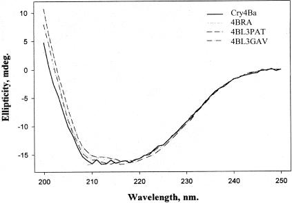
Expression of Cry4Ba protoxins in E. coli and subsequent digestion by trypsin are a presumptive test of global folding fidelity (1). All of the Cry4Ba mutant proteins constructed in this study were stable to trypsin digestion (data not shown). The CD spectra of the Cry4Ba activated toxin mutants indicated that there was no global or significant perturbation in the secondary structure of the toxins due to the mutations (Fig. 2). The CD spectrum of wild-type Cry4Ba was of interest because it was nicked by trypsin at the position between R203 and S204 (determined by N-terminal amino acid sequencing results), and yet it was similar to 4BRA (the trypsin site was removed) and other mutants based on 4BRA.
FIG.2.
CD spectra of purified toxins of Cry4Ba and its mutants.
Mosquito bioassay of Cry4Ba and its muteins.
Mutations in the predicted loop regions of domain II affected toxicities against the three genera of mosquito. Loop 1 of 4BRA was mutated to mimic loop 1 of Cry4Aa, 331IYQ333 to QTT (4BL1QTT). This caused the toxin to lose activity against A. aegypti and A. quadrimaculatus with activity against C. quinquefasciatus remaining nil (Table 2). The mutation in loop 2, where NDY was inserted between V393 and T394 (4BL2NDY), also caused the toxin to lose activity against A. aegypti and A. quadrimaculatus, with activity against C. quinquefasciatus remaining nil (Table 2). In contrast, the mutation in loop 3, where D454 was replaced with P and AT was inserted after position 454, caused the toxin to gain activity more than 219-fold against C. quinquefasciatus and C. pipiens, relative to 4BRA, while still maintaining activity against both Aedes and Anopheles (Table 2). In further mutations in loop 3, D454 was replaced with G and AV was inserted after position 454 (4BL3GAV), modeled after the loop 3 sequence of Cry1Aa. The toxicities of 4BL3GAV and 4BL3GAT against C. quinquefasciatus showed more than 700-fold increases relative to 4BRA and 2-fold increases over 4BL2PAT (Table 2). The same effect was not observed against C. pipiens, as 4BL3GAV was as toxic as 4BL3PAT and 4BL3GAT was twofold less toxic than 4BL3PAT to this mosquito (Table 2).
TABLE 2.
Bioassay results for four species of mosquitoes
| Toxin | LC50 (ng/ml)a
|
|||
|---|---|---|---|---|
| A. quadrimaculatus | A. aegypti | C. quinquefasciatus | C. pipiens | |
| Cry4Aa | ND | 600 (1,300-2,000)b | 980 (680-1,490)c | 400 (300-500)b |
| Cry4Ba | 25 (18-32) | 61 (28-175) | >80,000d | >20,000g |
| 4BwtGAV | 745 (607-962) | 174 (117-280) | >20,000f | ND |
| 4BRA | 21 (15-29) | 21 (5-51) | >80,000e | >20,000d |
| 4BL1QTT | >20,000d | >20,000c | >20,000d | ND |
| 4BL2NDY | >20,000d | >20,000d | >20,000d | ND |
| 4BL3PAT | 44 (40-50) | 53 (19-91) | 365 (267-529) | 95 (69-130) |
| 4BL3AAT | 16 (8-23) | 68 (19-140) | 1,035 (485-8,972) | 229 (142-512) |
| 4BL3GAT | 88 (64-119) | 64 (39-94) | 122 (75-189) | 180 (117-317) |
| 4BL3GAV | 52 (32-74) | 44 (20-68) | 114 (83-150) | 70 (34-129) |
| 4BL3PAA | 197 (136-328) | 144 (75-277) | 4,000 (1,948-14,838) | 481 (44-988) |
| 4BL3AAA | 23 (17-30) | 82 (50-126) | >20,000e | 630 (306-11,328) |
Two-day-old larvae of A. aegypti, C. quinquefasciatus, and C. pipiens and 3-day-old larvae of A. quadrimaculatus were used for bioassays. Mortality was recorded after 24 h of exposure to a serial dilution of the toxins. The 95% confidence limits are indicated in parentheses. Bioassays for the Cry4B constructs used purified inclusion crystal protein produced in E. coli. ND, not determined. LC50, 50% lethal, concentration.
Data from reference 14.
Data from reference 2.
8% mortality was observed at this dose.
No mortality was observed.
17% mortality was observed at this dose.
8% mortality was observed at this dose.
Other mutations in loop 3 caused variable toxicities against the different species of mosquitoes used in this study (Table 2). When P454 in the 4BL3PAT construct was mutated to A (4BL3AAT), toxicity against A. quadrimaculatus improved 2.8-fold over that of 4BL3PAT. The same mutation did not significantly alter its toxicity towards A. aegypti, but it also reduced its toxicity against the two Culex species. When P454 was mutated to G (4BL3GAT), activities against A. quadrimaculatus and C. pipiens were reduced twofold, while the activity against A. aegypti was not significantly different. Mutation of T456, in the 4BL3PAT background, to A (4BL3PAA) reduced toxicity against A. quadrimaculatus, A. aegypti, and C. pipiens. However, when both P454 and T456 were mutated to A's to yield 4BL3AAA, the activity of the mutant against A. quadrimaculatus was twofold more toxic than that of 4BL3PAT and the activity against Culex species was lost, while activity against A. aegypti was not significantly different from that of 4BL3PAT.
Mutation of R203 to A in the loop area between two alpha-helices to remove a trypsin site did not significantly improve toxicities against A. aegypti or A. quadrimaculatus in the Cry4Ba background. However, when the trypsin site was not removed in the 4BwtGAV construct, it caused a significant reduction in the activity against both Aedes and Anopheles compared to that of 4BL3GAV.
Binding assays.
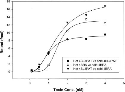
Saturation binding of 4BL3PAT and 4BRA was conducted in the presence of a 250-fold excess of homologous and heterologous toxin to determine specific binding of these toxins to C. quinquefasciatus BBMV. The binding curves indicated a sigmoid response for both toxins, and 4BL3PAT showed slightly better binding than 4BRA did (Fig. 3).
FIG.3.
Specific saturation binding of 4BL3PAT and 4BRA to C. quinquefasciatus BBMV.
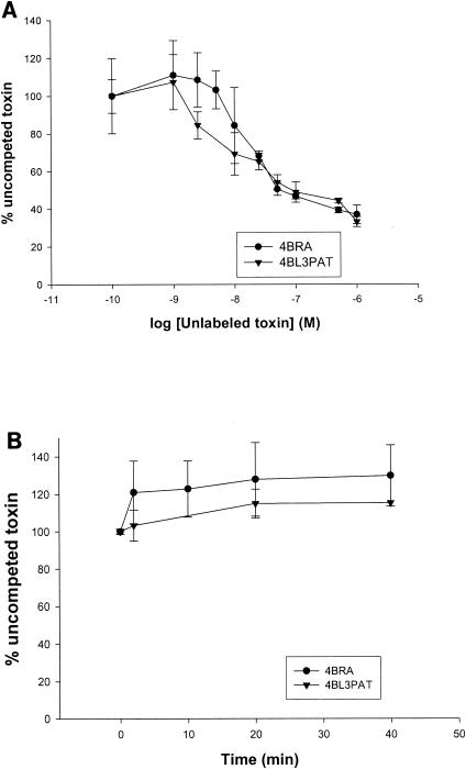
Heterologous competition between labeled 4BRA toxin and 4BL3PAT toxin for binding to C. quinquefasciatus BBMV was conducted, with homologous binding of 4BRA to itself as a comparison (Fig. 4A). The results indicated that there was a slightly better competition of 4BL3PAT for the 4BRA binding site on C. quinquefasciatus BBMV but not sufficient to explain the more than 219-fold better toxicity. Likewise, there was also no significant difference in the ability to irreversibly bind the BBMV for both 4BRA and 4BL3PAT (Fig. 4B).
FIG. 4.
(A) Homologous and heterologous competition binding assays. 125I-labeled 4BRA was incubated with C. quinquefasciatus BBMV with increasing amounts of unlabeled toxin. (B) Irreversible binding studies. C. quinquefasciatus BBMV were preincubated with 125I-labeled toxin for 1 h. Then binding was competed with excess cold toxin for different durations. Data shown are means of three binding experiments.
Proteinase K protection assay.
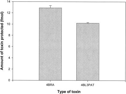
4BRA and 4BL3PAT toxins were partitioned into C. quinquefasciatus BBMV and subsequently digested with proteinase K. The total bound and proteinase K-protected toxins are shown in Fig. 5. More 4BRA than 4BL3PAT was protected.
FIG. 5.
Proteinase K protection assay of 4BRA and 4BL3PAT. 125I-labeled toxin was incubated with C. quinquefasciatus BBMV for 1 h. Free and noninserted toxin was digested with proteinase K, and the reaction was stopped with Pefabloc. BBMV-protected toxin was separated by centrifugation, and the counts were measured.
Pore formation measured by light scattering on mosquito BBMV.
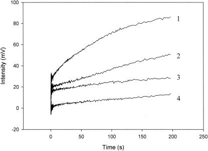
Light-scattering assays were performed according to the technique of Carroll and Ellar (9). 4BL3PAT and 4BRA were tested on C. quinquefasciatus BBMV. Our results (Fig. 6) indicate that 4BL3PAT has more pore-forming ability than 4BRA does.
FIG. 6.
C. quinquefasciatus BBMV light-scattering assay. BBMV (0.2 mg/ml) were coinjected with different samples. All samples were in 10 mM HEPES buffer, pH 7.5. The samples were KCl (150 mM) (line 1), 4BRA (66 pmol of toxin/mg in 150 mM KCl) (line 2), 4BL3PAT (66 pmol of toxin/mg in 150 mM KCl) (line 3), and HEPES buffer alone (line 4). Light scattering was measured using a stopped-flow apparatus at a 488-nm wavelength with the temperature controlled at 20°C. The curves were normalized to begin from the same point. The increasing intensity is indicative of reswelling of the vesicles. The data shown are averages of two experiments.
Ligand blots on C. quinquefasciatus BBMV proteins.
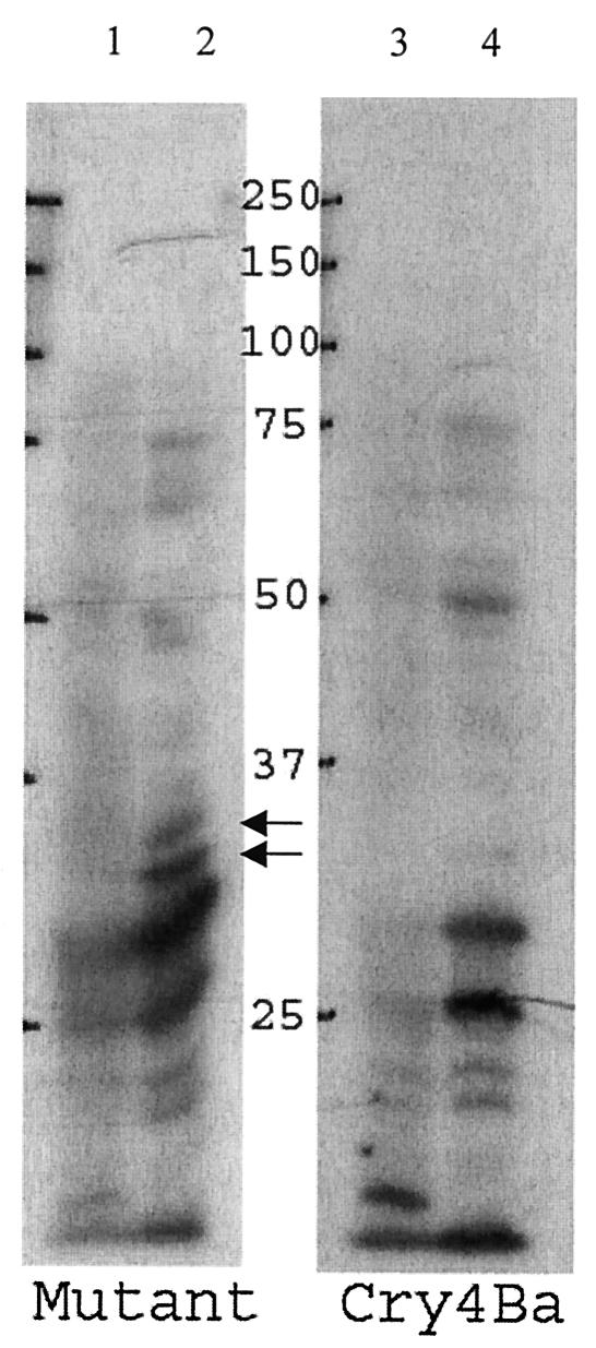
Both toxins bound weakly to proteins in the range of 60 to 75 kDa and strongly to proteins at 18, 20, 25, and 30 kDa (Fig. 7, lanes 2 and 4). 4BRA bound uniquely to a 50-kDa protein with moderate intensity (lane 4), and 4BL3PAT bound uniquely and intensely to two bands at 35 and 36 kDa (lane 2).
FIG. 7.
Ligand blot of biotin-labeled 4BRA (Cry4Ba) and 4BL3PAT (Mutant) to C. quinquefasciatus BBMV. Ten micrograms of BBMV proteins was separated by sodium dodecyl sulfate-polyacrylamide gel electrophoresis, blotted onto a polyvinylidene difluoride membrane, and probed with 5 nM biotinylated toxins. Arrows point to bands that are uniquely bound by 4BL3PAT. Lanes 1 and 3, BBMV; lanes 2 and 4, precipitated BBMV. Numbers between the panels are molecular masses in kilodaltons.
DISCUSSION
Cry4Ba toxin is active against Anopheles and Aedes but has no measurable activity against Culex species (14) (Table 2). We applied present knowledge of the general location of receptor binding epitopes on Cry toxins (22, 42) and have demonstrated that certain mutants in domain II loop 3 of the Cry4Ba toxin specifically increase mosquitocidal toxicity towards Culex species (Table 2). We also observed that mutations of loop 1 or 2 of domain II reduced activity against Aedes and Anopheles, indicating a role for these loop residues in the mechanism of action of Aedes and Anopheles.
As was previously reported, wild-type Cry4Ba is cleaved by gut juice of C. pipiens into 18- and 46-kDa fragments (25). Similar results were obtained by trypsin digestion in this study. We observed that the 18- and 46-kDa fragments were associated with each other by gel filtration chromatography (results not shown), agreeing with the results of Yamagiwa et al. (54), and our secondary structural analysis by CD spectroscopy (Fig. 2) showed that differences between the wild-type Cry4Ba and its mutants were insignificant. The blocking of the trypsin site in domain I of Cry4Ba resulted in an increase in toxicity of 4BRA relative to Cry4Ba of 2.9-fold, in agreement with previous results (3), but our 95% confidence limit of the toxins overlapped, indicating that the increase in toxicity against Anopheles or Aedes in this construct was not significant. However, removal of the trypsin site yielded more pronounced results in comparing two mutants in the putative domain II loop 3. 4BwtGAV, which retained the trypsin cleavage, was 4.0-fold less toxic to Aedes, 14.3-fold less toxic to Anopheles, and more than 175-fold less toxic to C. quinquefasciatus than 4BL3GAV, which had the trypsin cleavage site removed (Table 2). Therefore, the removal of the trypsin site in this construct significantly improved the overall activity of the toxins towards all species of mosquitoes tested.
Bioassays against four species of mosquitoes with the wild-type Cry4Ba toxin, 4BRA toxin, and the loop muteins are shown in Table 2. The data indicate that introducing three residues (PAT) from the putative loop 3 of Cry4Aa into 4BRA caused more than a 219-fold increase in activity against Culex. Variation of this sequence caused variable effects on toxicity to the mosquitoes tested. The 4BL3GAV mutant gave more than a 700-fold increase over 4BRA or wild-type Cry4Ba. While the removal of the trypsin cleavage site aided toxicity against all genera of mosquito, no Culex activity was observed unless the putative domain II loop 3 was modified by insertion and substitution.
Mutations in putative loops 1 and 2 to introduce residues from Cry4Aa significantly disrupted toxicity against both Anopheles and Aedes but did not introduce activity against Culex. The roles that the different loops played in the activity against the different mosquito species were not conclusive. The activity of the toxins seemed to rely on the interactions between the different loops. The specific sequence in the putative loop 3 was not critical for Culex toxicity, as the different combinations of the PAT sequence, e.g., GAT, AAT, PAA, and AAA, all had a certain level of Culex toxicity. However, all the variations in the putative loop 3 that were constructed involved conservative mutations, as no charged residue was used. The results indicated that the putative loop 3 was particularly important for Culex activity but also could affect Aedes and Anopheles activities. The putative loops 1 and 2 were critical for Aedes and Anopheles activities, but their function for Culex activity is not clear. There remains a possibility that other residues could be substituted to optimize the mosquitocidal activity of the toxin.
We explored the mechanistic basis of the improvement of the putative domain II loop 3 mutant 4BL3PAT by several methods. It had previously been shown that loop regions of domain II harbor receptor-binding epitopes in several insecticidal Cry toxins (23, 39, 41, 45, 53). It has also been shown that some mutations in loop regions do not affect initial receptor binding, as measured by competition binding, but affect irreversible binding, suggesting that they affect insertion of the toxin into the membrane (39, 53).
We began our analysis of the 4BL3PAT mechanism of action by examining saturation binding. Saturation binding studies of 4BRA and 4BL3PAT binding to Culex BBMV, with the use of homologous and heterologous competing toxins to reduce nonspecific binding, yielded sigmoid-shaped binding curves (Fig. 3), indicating cooperativity in binding. This has not been observed in previous studies of B. thuringiensis Lepidoptera-active toxins binding to Lepidoptera BBMV (16, 28, 49). Nonlinear regression analysis (Table 3) shows tighter binding of the toxic 4BL3PAT than of the nontoxic 4BRA. However, 4BRA showed a higher concentration of maximum binding sites (Bmax) than did 4BL3PAT, perhaps due to more positive cooperative binding as indicated by the higher Hill coefficient number for 4BRA. When nonspecific binding of 4BL3PAT was blocked with 4BRA, both tight binding and higher Bmax were observed. Cooperativity could be explained in several ways. One possibility is that multiple binding sites exist for 4BRA and 4BL3PAT. The high-affinity sites are detected above background first, and then the low-affinity sites are revealed as more labeled toxin is added to the binding mixture. 4BRA does not displace the high-affinity 4BL3PAT binding. Another possibility may be the oligomerization process as toxins associate with the receptors in the “pre-pore” formation (17) or in the membrane.
TABLE 3.
Nonlinear regression of the binding curves to a Hill equationa
| Toxin | Bmax (fmol/μg) | K′D (nM) | Hill coefficient |
|---|---|---|---|
| 4BRA | 13.1 ± 0.5 | 6.9 ± 2.7 | 4.8 ± 0.8 |
| 4BL3PAT | 9.3 ± 0.4 | 0.9 ± 0.2 | 2.8 ± 0.5 |
| 4BL3PAT* | 18.3 ± 1.6 | 2.6 ± 0.5 | 2.3 ± 0.4 |
Each binding condition was conducted with 250 M excess homologous toxin added to reduce nonspecific binding, with the exception that 4BL3PAT* was conducted with 250 M excess 4BRA.
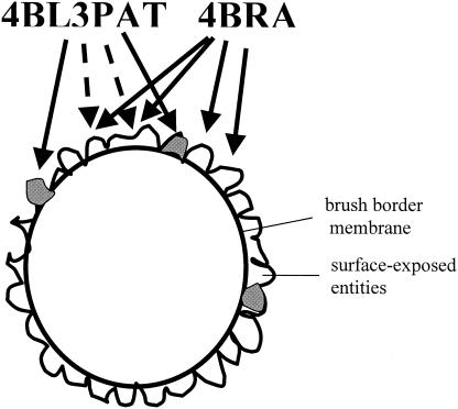
Competition binding of 4BRA toxin and 4BL3PAT to C. quinquefasciatus BBMV indicated that there was little significant difference in the ability to reversibly bind C. quinquefasciatus BBMV (Fig. 4A). Both saturation binding and competition suggest a model where 4BL3PAT binds to both productive binding proteins and more numerous nonproductive sites, while 4BRA binds only to the nonproductive sites (Fig. 8).
FIG. 8.
Model for binding of toxic 4BL3PAT to productive binding sites (gray) and nonproductive binding sites (white). Nontoxic protein 4BRA binds only to the nonproductive binding sites. The nonproductive binding sites are more numerous.
Further experiments with irreversible binding assays indicated that there was also no significant difference in the abilities of the muteins and wild-type toxins to irreversibly bind BBMV (Fig. 4B). Indeed, more 4BRA than 4BL3PAT bound irreversibly, and both toxins continued to associate with BBMV with time rather than be chased off.
Irreversible binding is generally assumed to represent membrane insertion (21, 30, 37, 39, 50) and has been shown to correlate with toxicity. We therefore conducted proteinase K studies to determine if the bound toxins were susceptible to exogenous proteinase. Figure 5 shows that relatively more 4BRA than 4BL3PAT is protected. These results counter the expectation that more of the active toxin would bind and be inserted into the membrane. Whether these toxins, 4BRA in particular, are protected by insertion into the membrane or by association with BBMV proteins remains to be shown.
To examine the pore-forming ability of the active and inactive toxins, we conducted light-scattering assays on Culex BBMV according to the method of Carroll and Ellar (10), previously used for lepidopteran toxins. Our results (Fig. 6) indicate that 4BL3PAT has more pore-forming ability than does 4BRA, but the latter showed greater pore-forming ability than expected from its low toxicity.
In summary, we have demonstrated that relatively few amino acid residue changes in domain II loop 3 of Cry4Ba can introduce a large increase in toxicity toward a genus of mosquito against which it was not active. We have previously reviewed the attempts to improve Cry toxins by rational design (13), but the greater-than-750-fold increase in Culex activity for Cry4Ba far exceeds previous accomplishments. Surprisingly, our studies of the mechanism of action of Cry4Ba indicate that more-active toxins do not bind to BBMV with more affinity than less active toxins, nor are they inserted to a greater extent. We observe that Cry4Ba and the trypsin-stable mutant form, 4BRA, bind (Fig. 4A) and are inserted into (Fig. 4B and 5) Culex BBMV (or are somehow protected from proteinase K), as well as or better than 4BL3PAT but do not form very active pores in vitro (Fig. 6). Since the few amino acid residues that were introduced in domain II loop 3 resulted in a significantly more active toxin, we must speculate on the role of these residues in the increased toxicity. A radical hypothesis would be that these residues directly affect the pore-forming ability of the toxin. Another radical hypothesis for the enhanced toxin, 4BL3PAT, would involve programmed cell death (apoptosis). Apoptosis has been observed to occur in B. thuringiensis subsp. israelensis endotoxin-treated A. aegypti cell cultures (11). Other Cry toxins seem to exert the same apoptotic effect on cultured midgut cells of H. virescens (33, 34). These hypotheses are at variance with the Lepidoptera paradigm of the mechanism of action of Cry toxins, which describes the loops of domain II as receptor-binding epitopes. The more parsimonious hypothesis (and one more in keeping with the Lepidoptera paradigm) is that the residue changes in domain II loop 3 allow 4BL3PAT to a bind to a “functional” receptor (one that leads to proper insertion and activity), as illustrated in Fig. 8. This model is supported by the ligand blots that show that both 4BRA and 4BL3PAT bind to a series of proteins, while 4BL3PAT binds uniquely to two proteins of 35 and 36 kDa (Fig. 7). The model is also supported by the specific saturation binding studies, where 4BRA was used to compete for binding of 4BL3PAT (Fig. 3). Further experiments will be conducted to resolve this paradox.
Acknowledgments
We thank A. Curtiss, F. Ahmed, and K. Nzinga for technical assistance. Special thanks go to A. Valaitis for amino acid sequencing.
This work was supported by an NIH grant to D. H. Dean and M. J. Adang (grant no. R01 AI 29092).
REFERENCES
- 1.Almond, B. D., and D. H. Dean. 1993. Structural stability of Bacillus thuringiensis δ-endotoxin homolog-scanning mutants determined by susceptibility to proteases. Appl. Environ. Microbiol. 59:2442-2448. [DOI] [PMC free article] [PubMed] [Google Scholar]
- 2.Angsuthanasombat, C., N. Crickmore, and D. J. Ellar. 1992. Comparison of Bacillus thuringiensis subsp. israelensis CryIVA and CryIVB cloned toxins reveals synergism in vivo. FEMS Microbiol. Lett. 94:63-68. [DOI] [PubMed] [Google Scholar]
- 3.Angsuthanasombat, C., N. Crickmore, and D. J. Ellar. 1993. Effects on toxicity of eliminating a cleavage site in a predicted interhelical loop in Bacillus thuringiensis CryIVB delta endotoxin. FEMS Microbiol. Lett. 111:255-261. [DOI] [PubMed] [Google Scholar]
- 4.Bar, E., J. Lieman-Hurwitz, E. Rahamim, A. Keynan, and N. Sandler. 1991. Cloning and expression of Bacillus thuringiensis israelensis δ-endotoxin in B. sphaericus. J. Invertebr. Pathol. 57:149-158. [DOI] [PubMed] [Google Scholar]
- 5.Becker, N., and J. Margalit. 1993. Use of Bacillus thuringiensis israelensis against mosquitoes and blackflies, p. 145-170. In P. F. Entwistle, P. F. Cory, M. J. Bailey, and S. Higgs (ed.), Bacillus thuringiensis, an environmental biopesticide: theory and practice. John Wiley & Sons, Inc., New York, N.Y.
- 6.Berry, C., J. Hindley, A. F. Ehrhardt, T. Grounds, I. deSouza, and E. W. Davidson. 1993. Genetic determinants of host ranges of Bacillus sphaericus mosquito larvicidal toxins. J. Bacteriol. 175:510-518. [DOI] [PMC free article] [PubMed] [Google Scholar]
- 7.Bourgouin, C., A. Delecluse, F. De la Torre, and J. Szulmajster. 1990. Transfer of the toxin protein genes of Bacillus sphaericus into Bacillus thuringiensis subsp. israelensis and their expression. Appl. Environ. Microbiol. 56:340-344. [DOI] [PMC free article] [PubMed] [Google Scholar]
- 8.Caramori, T., A. M. Albertini, and A. Galizzi. 1991. In vivo generation of hybrids between two Bacillus thuringiensis insect-toxin-encoding genes. Gene 98:37-44. [DOI] [PubMed] [Google Scholar]
- 9.Carroll, J., and D. J. Ellar. 1993. An analysis of Bacillus thuringiensis δ-endotoxin action on insect-midgut-membrane permeability using a light-scattering assay. Eur. J. Biochem. 214:771-778. [DOI] [PubMed] [Google Scholar]
- 10.Carroll, J., and D. J. Ellar. 1997. Analysis of the large aqueous pores produced by a Bacillus thuringiensis protein insecticide in Manduca sexta midgut-brush-border-membrane vesicles. Eur. J. Biochem. 245:797-804. [DOI] [PubMed] [Google Scholar]
- 11.Charles, J.-F. 1983. Action of the delta-endotoxin of Bacillus thuringiensis var. israelensis on cultured cells from Aedes aegypti L. Ann. Microbiol. (Paris) 134A:365-381. [PubMed] [Google Scholar]
- 12.Dean, D. H., F. Rajamohan, M. K. Lee, S.-J. Wu, X.-J. Chen, E. Alcantara, and S. R. Hussain. 1996. Probing the mechanism of action of Bacillus thuringiensis insecticidal proteins by site-directed mutagenesis. Gene 179:111-117. [DOI] [PubMed] [Google Scholar]
- 13.Dean, D. H., T. H. You, F. Rajamohan, M. K. Lee, J. L. Jenkins, M. Audtho, S.-J. Wu, and X. Liu. 2002. Rational design of Cry toxins, p. 112-117. In R. J. Akhurst, C. E. Beard, and P. Hughes (ed.), Biotechnology of Bacillus thuringiensis and its environmental impact. Proceedings of the 4th Pacific Rim Conference. Scribbly Gum Publications, Canberra, Australia.
- 14.Delécluse, A., S. Poncet, A. Klier, and G. Rapoport. 1993. Expression of cryIVA and cryIVB genes, independently or in combination, in a crystal-negative strain of Bacillus thuringiensis subsp. israelensis. Appl. Environ. Microbiol. 59:3922-3927. [DOI] [PMC free article] [PubMed] [Google Scholar]
- 15.Erlanger, B. E., N. Kokowsky, and W. Cohen. 1961. The preparation and properties of two new chromogenic substrates of trypsin. Biochem. Biophys. 95:271-278. [DOI] [PubMed] [Google Scholar]
- 16.Estada, U., and J. Ferré. 1994. Binding of insecticidal crystal proteins of Bacillus thuringiensis to the midgut brush border of the cabbage looper, Trichoplusia ni (Hübner) (Lepidoptera: Noctuidae), and selection for resistance to one of the crystal proteins. Appl. Environ. Microbiol. 60:3840-3846. [DOI] [PMC free article] [PubMed] [Google Scholar]
- 17.Gómez, I., J. Sánchez, R. Miranda, A. Bravo, and M. Soberón. 2002. Cadherin-like receptor binding facilitates proteolytic cleavage of helix α-1 in domain I and oligomer pre-pore formation of Bacillus thuringiensis Cry1Ab toxin. FEBS Lett. 513:242-246. [DOI] [PubMed] [Google Scholar]
- 18.Guex, N., and M. C. Peitsch. 1997. SWISS-MODEL and the Swiss-Pdb Viewer: an environment for comparative protein modeling. Electrophoresis 18:2714-2723. [DOI] [PubMed] [Google Scholar]
- 19.Hofmann, C., and P. Lüthy. 1986. Binding and activity of Bacillus thuringiensis delta-endotoxin to invertebrate cells. Arch. Microbiol. 146:7-11. [DOI] [PubMed] [Google Scholar]
- 20.Hofmann, C., P. Lüthy, R. Hütter, and V. Pliska. 1988. Binding of the delta-endotoxin from Bacillus thuringiensis to brush-border membrane vesicles of the cabbage butterfly (Pieris brassicae). Eur. J. Biochem. 173:85-91. [DOI] [PubMed] [Google Scholar]
- 21.Ihara, H., E. Kuroda, A. Wadano, and M. Himeno. 1993. Specific toxicity of δ-endotoxins from Bacillus thuringiensis to Bombyx mori. Biosci. Biotechnol. Biochem. 57:200-204. [DOI] [PubMed] [Google Scholar]
- 22.Jenkins, J. L., and D. H. Dean. 2000. Exploring the mechanism of action of insecticidal proteins by genetic engineering methods, p. 33-54. In J. K. Setlow (ed.), Genetic engineering: principles and methods, vol. 22. Plenum Press, New York, N.Y. [DOI] [PubMed]
- 23.Jenkins, J. L., M. K. Lee, A. P. Valaitis, A. Curtiss, and D. H. Dean. 2000. Bivalent sequential binding model of a Bacillus thuringiensis toxin to gypsy moth aminopeptidase N receptor. J. Biol. Chem. 275:14423-14431. [DOI] [PubMed] [Google Scholar]
- 24.Khampang, P., W. Chungjatupornchai, P. Luxanamil, and S. Panyim. 1999. Efficient expression of mosquito-larvicidal proteins in a gram-negative bacterium capable of recolonization in the guts of Anopheles dirus larva. Appl. Microbiol. Biotechnol. 51:79-84. [DOI] [PubMed] [Google Scholar]
- 25.Komano, T., M. Yamagiwa, T. Nishimoto, H. Yoshisue, K. Tanabe, K. Sen, and H. Sakai. 1998. Activation process of the insecticidal proteins CryIVA and CryIVB produced by Bacillus thuringiensis subsp. israelensis. Isr. J. Entomol. 32:185-198. [Google Scholar]
- 26.Lecadet, M. M., J. Chaufaux, J. Ribier, and D. Lereclus. 1992. Construction of novel Bacillus thuringiensis strains with different insecticidal activities by transduction and transformation. Appl. Environ. Microbiol. 58:840-849. [DOI] [PMC free article] [PubMed] [Google Scholar]
- 27.Lee, M. K., A. Curtiss, E. Alcantara, and D. H. Dean. 1996. Synergistic effect of the Bacillus thuringiensis toxins CryIAa and CryIAc on the gypsy moth, Lymantria dispar. Appl. Environ. Microbiol. 62:583-586. [DOI] [PMC free article] [PubMed] [Google Scholar]
- 28.Lee, M. K., R. E. Milne, A. Z. Ge, and D. H. Dean. 1992. Location of a Bombyx mori receptor binding region on a Bacillus thuringiensis δ-endotoxin. J. Biol. Chem. 267:3115-3121. [PubMed] [Google Scholar]
- 29.Li, J., J. Carroll, and D. J. Ellar. 1991. Crystal structure of insecticidal δ-endotoxin from Bacillus thuringiensis at 2.5 Å resolution. Nature 353:815-821. [DOI] [PubMed] [Google Scholar]
- 30.Liang, Y., S. S. Patel, and D. H. Dean. 1995. Irreversible binding kinetics of Bacillus thuringiensis CryIA δ-endotoxins to gypsy moth brush border membrane vesicles is directly correlated to toxicity. J. Biol. Chem. 270:24719-24724. [DOI] [PubMed] [Google Scholar]
- 31.Liu, J. W., W. H. Yap, T. Thanabalu, and A. G. Porter. 1996. Efficient synthesis of mosquitocidal toxins in Asticcacaulis excentricus demonstrates potential of gram-negative bacteria in mosquito control. Nat. Biotechnol. 14:343-347. [DOI] [PubMed] [Google Scholar]
- 32.Lluisma, A. O., N. Karmacharya, A. Zarka, E. Ben-Dov, A. Zaritsky, and S. Boussiba. 2001. Suitability of Anabaena PCC7120 expressing mosquitocidal toxin genes from Bacillus thuringiensis subsp. israelensis for biotechnological application. Appl. Microbiol. Biotechnol. 57:161-166. [DOI] [PubMed] [Google Scholar]
- 33.Loeb, M. J., R. S. Hakim, P. Martin, N. Narang, S. Goto, and M. Takeda. 2000. Apoptosis in cultured midgut cells from Heliothis virescens larvae exposed to various conditions. Arch. Insect Biochem. Physiol. 45:12-23. [DOI] [PubMed] [Google Scholar]
- 34.Loeb, M. J., P. A. W. Martin, R. S. Hakim, S. Goto, and M. Takeda. 2001. Regeneration of cultured midgut cells after exposure to sublethal doses of toxin from two strains of Bacillus thuringiensis. J. Insect Physiol. 47:599-606. [DOI] [PubMed] [Google Scholar]
- 35.Peitsch, M. C. 1996. ProMod and Swiss-Model: internet-based tools for automated comparative protein modelling. Biochem. Soc. Trans. 24:274-279. [DOI] [PubMed] [Google Scholar]
- 36.Poncet, S., C. Bernard, E. Dervyn, J. Cayley, A. Klier, and G. Rapoport. 1997. Improvement of Bacillus sphaericus toxicity against dipteran larvae by integration, via homologous recombination, of the Cry11A toxin gene from Bacillus thuringiensis subsp. israelensis. Appl. Environ. Microbiol. 63:4413-4420. [DOI] [PMC free article] [PubMed] [Google Scholar]
- 37.Rajamohan, F., E. Alcantara, M. K. Lee, X. J. Chen, A. Curtiss, and D. H. Dean. 1995. Single amino acid changes in domain II of Bacillus thuringiensis CryIAb δ-endotoxin affect irreversible binding to Manduca sexta midgut membrane vesicles. J. Bacteriol. 177:2276-2282. [DOI] [PMC free article] [PubMed] [Google Scholar]
- 38.Rajamohan, F., O. Alzate, J. A. Cotrill, A. Curtiss, and D. H. Dean. 1996. Protein engineering of Bacillus thuringiensis δ-endotoxin: mutations at domain II of Cry1Ab enhance receptor affinity and toxicity towards gypsy moth larvae. Proc. Natl. Acad. Sci. USA 93:14338-14343. [DOI] [PMC free article] [PubMed] [Google Scholar]
- 39.Rajamohan, F., J. A. Cotrill, F. Gould, and D. H. Dean. 1996. Role of domain II, loop 2 residues of Bacillus thuringiensis CryIAb δ-endotoxin in reversible and irreversible binding to Manduca sexta and Heliothis virescens. J. Biol. Chem. 271:2390-2397. [DOI] [PubMed] [Google Scholar]
- 40.Rajamohan, F., and D. H. Dean. 1996. Molecular biology of bacteria on biological control of insects, p. 105-122. In M. Gunasekaran and D. J. Weber (ed.), Molecular biology of the biological control of pests and diseases of plants. CRC Press, Inc., Boca Raton, Fla.
- 41.Rajamohan, F., S.-R. A. Hussain, J. A. Cotrill, F. Gould, and D. H. Dean. 1996. Mutations at domain II, loop 3, of Bacillus thuringiensis CryIAa and CryIAb δ-endotoxins suggest loop 3 is involved in initial binding to lepidopteran midguts. J. Biol. Chem. 271:25220-25226. [DOI] [PubMed] [Google Scholar]
- 42.Rajamohan, F., M. K. Lee, and D. H. Dean. 1998. Bacillus thuringiensis insecticidal proteins: molecular mode of action. Prog. Nucleic Acids Res. Mol. Biol. 60:1-27. [DOI] [PubMed] [Google Scholar]
- 43.Sambrook, J., E. F. Fritsch, and T. Maniatis. 1989. Molecular cloning: a laboratory manual, 2nd ed. Cold Spring Harbor Laboratory Press, Cold Spring Harbor, N.Y.
- 44.Schnepf, E., N. Crickmore, J. VanRie, D. Lereclus, J. Baum, J. Feitelson, D. R. Zeigler, and D. H. Dean. 1998. Bacillus thuringiensis and its pesticidal crystal proteins. Microbiol. Mol. Biol. Rev. 62:775-806. [DOI] [PMC free article] [PubMed] [Google Scholar]
- 45.Smedley, D. P., and D. J. Ellar. 1996. Mutagenesis of three surface-exposed loops of a Bacillus thuringiensis insecticidal toxin reveals residues important for toxicity, receptor recognition and possibly membrane insertion. Microbiology 142:1617-1624. [DOI] [PubMed] [Google Scholar]
- 46.Thanabalu, T., J. Hindley, S. Brenner, C. Oei, and C. Berry. 1992. Expression of the mosquitocidal toxins of Bacillus sphaericus and Bacillus thuringiensis subsp. israelensis by recombinant Caulobacter crescentus, a vehicle for biological control of aquatic insect larvae. Appl. Environ. Microbiol. 58:905-910. [DOI] [PMC free article] [PubMed] [Google Scholar]
- 47.Towbin, H., T. Staehelin, and J. Gordon. 1979. Electrophoretic transfer of proteins from polyacrylamide gels to nitrocellulose sheets: procedure and some applications. Proc. Natl. Acad. Sci. USA 76:4350-4354. [DOI] [PMC free article] [PubMed] [Google Scholar]
- 48.Van Frankenhuyzen, K. 1993. The challenge of Bacillus thuringiensis, p. 1-35. In P. F. Entwistle, J. S. Cory, M. J. Bailey, and S. Higgs (ed.), Bacillus thuringiensis, an environmental biopesticide: theory and practice. John Wiley & Sons, Inc., New York, N.Y.
- 49.Van Rie, J., S. Jansens, H. Höfte, D. Degheele, and H. Van Mellaert. 1990. Receptors on the brush border membrane of the insect midgut as determinants of the specificity of Bacillus thuringiensis delta-endotoxins. Appl. Environ. Microbiol. 56:1378-1385. [DOI] [PMC free article] [PubMed] [Google Scholar]
- 50.Van Rie, J., S. Jansens, H. Höfte, D. Degheele, and H. Van Mellaert. 1989. Specificity of Bacillus thuringiensis δ-endotoxin: importance of specific receptors on the brush border membrane of the mid-gut of target insects. Eur. J. Biochem. 186:239-247. [DOI] [PubMed] [Google Scholar]
- 51.Ward, M. G. 1984. Formulation of biological insecticides: surfactant and diluent selection. ACS Symp. Ser. 254:175-184. [Google Scholar]
- 52.Wolfersberger, M. G. 1990. The toxicity of two Bacillus thuringiensis δ-endotoxins to gypsy moth larvae is inversely related to the affinity of binding sites on midgut brush border membranes for the toxins. Experientia 46:475-477. [DOI] [PubMed] [Google Scholar]
- 53.Wu, S.-J., and D. H. Dean. 1996. Functional significance of loops in the receptor binding domain of Bacillus thuringiensis CryIIIA δ-endotoxin. J. Mol. Biol. 255:628-640. [DOI] [PubMed] [Google Scholar]
- 54.Yamagiwa, M., M. Esaki, K. Otake, M. Inagaki, T. Komano, T. Amachi, and H. Sakai. 1999. Activation process of dipteran-specific insecticidal protein produced by Bacillus thuringiensis subsp. israelensis. Appl. Environ. Microbiol. 65:3464-3469. [DOI] [PMC free article] [PubMed] [Google Scholar]


