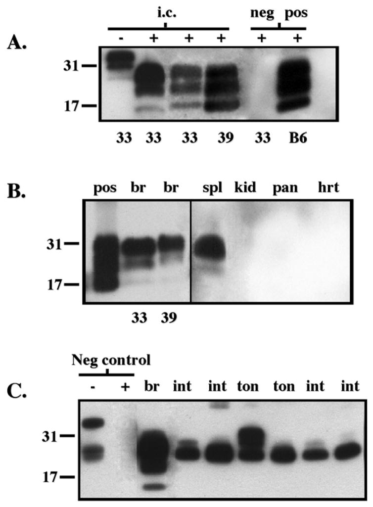
Figure 1.

Detection of PrPres deposits in CWD-infected deer PrP tg mice by Western blot. Panel A shows the presence of PrPres in brain tissue after i.c. inoculation of CWD brain. Data from clinically ill tg+/− mice from lines 33 or 39 are displayed in lanes 2–4. Similar results were obtained from over 10 additional ill CWD-infected tg mice from lines 33 and 39 at 280 to 310 dpi. Lane 1 shows Western blot in the absence of protease K treatment (−) while all the remaining lanes were blots with materials treated with proteinase K (25 μg/ml) (+). Negative (neg) control was +/− tg mouse inoculated i.c. with PBS diluent and sacrificed 400 dpi while the positive (pos) control was a C57Bl/6 mouse inoculated i.c. with murine scrapie and sacrificed at 155 dpi. PrP material was detected with monoclonal antibody D13. Panel B displays PrPres within the brain (br), spleen (spl), kidney (kid), pancreas (pan) and heart (hrt) of orally inoculated tg+/+ mice at 370 dpi using monoclonal antibody D13. Spl, kid, pan and hrt are from mouse line 39 whose br is also shown. Similar results for all these tissues were obtained from six additional CWD-infected mice, three from tg33+/+ and three from tg39+/+ strains. Brain homogenate from clinically ill CWD-infected tg+/+ mice (pos) inoculated i.c. is shown for comparison. Panel C shows PrPres deposits within the brain (br), intestine (int), and tongue (ton) of several individual tg+/+ mice infected orally with CWD. Detection was with monoclonal antibody L42. Similar to D13 antibody, L42 antibody failed to detect PrPres in kid, pan or hrt (not shown) but in contrast to D13 antibody detected PrPres in ton and int. The first two lanes on the left show the absence (−) or presence (+) of protease K (25 μg/ml) digestion of deer PrP tg mouse brain (br) inoculated with murine scrapie (neg control). The remainder of the lanes are from protease K (25 μg/ml) digestions of tissues from deer PrP tg mice infected orally with CWD.
