Covert spatial attention is the mechanism by which humans select a location for more elaborate cognitive processing without moving the eyes. Human and animal electrophysiology, studies of humans with brain damage, and other methods suggest that the movement, capture, and release of covert spatial attention involves a widely distributed network of cortical and subcortical structures. These structures include, most prominently, parts of the frontal and parietal lobes, and the superior colliculus, although their precise roles are not completely clear. The premotor theory of attention suggests that shifts of covert attention arise from plans for eye movements even when no eye movement occurs. Evidence for this theory, based on electrophysiological recording in monkey frontal eye fields (FEFs), regions implicated in generating plans for eye movements, has been controversial. Much of the evidence originates from tasks that involve eye movements, raising concerns about whether overt and covert attention could be properly dissociated. In their recent paper in The Journal of Neuroscience, Thompson et al. (2005) provide compelling evidence that covert spatial attention is dissociable from eye movement planning in the FEFs.
To direct spatial attention to a location, Thompson et al. used a visual search task. While fixating centrally, two macaque monkeys viewed displays with one item (the target) that popped out from a homogeneous set of distractor items [Thompson et al. (2005), their Fig. 1 (http://www.jneurosci.org/cgi/content/full/25/41/9479FIG1)]. In such a paradigm, attention is generally thought to be automatically and covertly directed toward the target. Unlike previous investigations requiring a saccadic response, Thompson et al. trained the monkeys to respond manually in an effort to diminish influences of saccade planning on FEF activity. During the task, they recorded from three types of FEF cells. Movement cells display above baseline activity related to the production of a saccade, whereas visual cells display above baseline activity in the presence of a visual target. Visuomovement cells display properties of both. The authors found greater activity in visual and visuomovement cells when an attended target appeared in the receptive field of the cell than when an unattended distractor was in the receptive field [Thompson et al. (2005), their Figs. 3 (http://www.jneurosci.org/cgi/content/full/25/41/9479/FIG3) and 6 (http://www.jneurosci.org/cgi/content/full/25/41/9479/FIG6)]. Because there were no eye movements during the task, this activity cannot be attributed to motor execution. To address the possibility that this activity reflected unrealized motor plans to move the eyes to the target location, the authors examined eye movements after each trial. They reasoned that if FEF activity reflected a plan to move the eyes toward the target, this plan should be executed after the end of the trial when the animals were allowed to move their eyes. Because post-trial saccades were not biased toward the target location [Thompson et al. (2005), their Fig. 2 (http://www.jneurosci.org/cgi/content/full/25/41/9479/FIG2)], Thompson et al. concluded that the target-related activity in visual neurons could not be attributed to saccade planning. Furthermore, Thompson et al. found no evidence of saccade-planning activity in movement neurons from which they recorded [Thompson et al. (2005), their Figs. 4 (http://www.jneurosci.org/cgi/content/full/25/41/9479/FIG4), 5 (http://www.jneurosci.org/cgi/content/full/25/41/9479/FIG5), and 6 (http://www.jneurosci.org/cgi/content/full/25/41/9479/FIG6)]. Previous electrophysiological studies have shown that FEF movement neurons are active when a saccade is planned toward a visual search target, regardless of whether it is actually executed. Thompson et al. likely achieved these clear results because of their saccade-free task and by testing monkeys who had never been trained to make saccades in a visual search task.
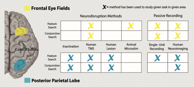
Figure 1.
The results of Thompson et al. fit nicely into ongoing work in understanding the distributed network of attention involved in visual search. The frontal eye fields (highlighted in yellow) and the posterior parietal lobe (highlighted in blue) have been studied using various methods. The table at the right side of the figure indicates which methods have been used to study the role of each area in feature search and conjunction search.
The evidence presented in this paper is consistent with evidence from human neuroimaging studies and other nonhuman electrophysiology studies. The FEFs are part of the distributed covert attention network, and activity in the FEFs does not necessarily rely on eye movement commands. But what role do the FEFs play in the attention network? A key issue is whether the FEFs generate the commands that actually cause shifts of attention or whether the target-selective FEF activity is a consequence of attentional commands generated elsewhere in the brain. Discriminating between these interpretations can be difficult (perhaps impossible) using electrophysiological recordings because there is no indication of whether the observed activity is necessary for the behavior.
Neuropsychological studies of patients, brain area inactivation, and human transcranial magnetic stimulation (TMS) can be used to establish the role of a brain area in a cognitive process (Chambers and Mattingley, 2005). Damage or temporary inactivation of a brain area will disrupt any cognitive functions in which that area plays a necessary role. A series of studies have examined various nodes in the attention network using these methods (Fig. 1). For instance, inactivation of monkey lateral intraparietal (LIP) area increases reaction time for detection of contralateral features and conjunctions of features in visual search (Wardak et al., 2004). Similar effects are observed in human patients with parietal lobe damage (Eglin et al., 1991). These findings, along with neuroimaging data, support the notion that the parietal lobe serves a necessary role in the deployment of covert spatial attention. Although researchers have used TMS to disrupt visual search by stimulating the FEFs (Muggleton et al., 2003), we are not aware of lesion or chemical inactivation studies of the FEF during visual search. Thompson et al. provide excellent groundwork for such a study. Importantly, patients with frontal damage sparing the FEFs show contralateral deficits in visual search (Eglin et al., 1991). These results emphasize the necessity of areas outside the FEF, in frontal and parietal cortices, for attentional allocation.
Another issue to consider is whether the role of the FEFs in attention may differ as a function of the type of attentional deployment. Covert attention comes in at least two forms. It can be exogenously driven by a salient target, or it can be controlled by the animal intentionally (endogenous). Thompson et al. characterize their search task as an exogenously driven movement of attention. It will be interesting to see whether the FEFs play a necessary role in both automatic and controlled attention, perhaps by comparing feature and conjunction searches [as did Wardak et al. (2004) in the LIP area].
Thompson et al. provide strong evidence for attention-related modulation of activity in subsets of FEF neurons. This solidifies the FEFs as a node in the distributed network for attention. What remains to be resolved is whether the distinct areas of the network are functionally redundant or whether more precise roles can be defined.
Editor’s Note: These short reviews of a recent paper in the Journal, written exclusively by graduate students or postdoctoral fellows, are intended to mimic the journal clubs that exist in your own departments or institutions. For more information on the format and purpose of the Journal Club, please see http://www.jneurosci.org/misc/ifa_features.shtml.
Footnotes
Review of Thompson et al. (http://www.jneurosci.org/cgi/content/full/25/41/9479)
References
- Chambers CD, Mattingley JB (2005). Neurodisruption of selective attention: insights and implications. Trends Cogn Sci 9:542–550. [DOI] [PubMed] [Google Scholar]
- Eglin M, Robertson LC, Knight RT (1991). Cortical substrates supporting visual search in humans. Cereb Cortex 1:262–272. [DOI] [PubMed] [Google Scholar]
- Muggleton NG, Juan CH, Cowey A, Walsh V (2003). Human frontal eye fields and visual search. J Neurophysiol 89:3340–3343. [DOI] [PubMed] [Google Scholar]
- Thompson KG, Biscoe KL, Sato TR (2005). Neuronal basis of covert spatial attention in the frontal eye field. J Neurosci 25:9479–9487. [DOI] [PMC free article] [PubMed] [Google Scholar]
- Wardak C, Olivier E, Duhamel JR (2004). A deficit in covert attention after parietal cortex inactivation in the monkey. Neuron 42:501–508. [DOI] [PubMed] [Google Scholar]


