Abstract
We have previously reported on the discovery of a mitochondrial specific unfolded protein response (mtUPR) in mammalian cells, in which the accumulation of unfolded protein within the mitochondrial matrix results in the transcriptional activation of nuclear genes encoding mitochondrial stress proteins such as chaperonin 60, chaperonin 10, mtDnaJ, and ClpP, but not those encoding stress proteins of the endoplasmic reticulum (ER) or the cytosol. Analysis of the chaperonin 60/10 bidirectional promoter showed that the CHOP element was required for the mtUPR and that the transcription of the chop gene is activated by mtUPR. In order to investigate the role of CHOP in the mtUPR, we carried out a deletion analysis of the chop promoter. This revealed that the transcriptional activation of the chop gene by mtUPR is through an AP-1 (activator protein-1) element. This site lies alongside an ERSE element through which chop transcription is activated in response to the ER stress response (erUPR). Thus CHOP can be induced separately in response to 2 different stress response pathways. We also discuss the potential signal pathway between mitochondria and the nucleus for the mtUPR.
Introduction
Mitochondria serve critical functions in the maintenance of cellular energy supplies, thermoregulation, synthesis of essential molecules such as phospholipids and haem, and in apoptosis. Since mitochondrial proteins are encoded by nuclear genes (at last estimate, about 1500 [1]) as well as mitochondrial genes (encoding just 13 polypeptides in mammalian species [2]), the normal functions of mitochondria require the coordination of two genomes and a system of communication between two organelles [3]–[5]. In addition, mitochondria need to respond to changes in the physiological milieu of the cell to repair damage caused by mutations in mtDNA which produces modified proteins which are unable to fold and become prone to aggregation.
Metabolic cues and other changes which occur within mitochondria can culminate in wide-ranging changes in nuclear gene expression via retrograde mitochondrial to nuclear signaling. These responses are broadly referred to as mitochondrial stress responses [6], [7] and are generally defined as a response to altered mitochondrial membrane potential or uncoupling of oxidative phosphorylation. This leads to the elevation of cytosolic Ca2+ and activation of CaMK and calcineurin responsive genes [4] which include genes involved in Ca2+ transport and storage [6] as well as a large collection of transcription factors [8]. The net effect of activation of this gene network is to facilitate recovery of the physiological functions of the mitochondrion.
A unique type of mitochondrial stress is the mitochondrial unfolded protein response (mtUPR, which we have previously called ‘the mitochondrial stress response’ [9]) where the accumulation of unfolded proteins in the mitochondrial matrix leads to an increase in nuclear encoded mitochondrial chaperones and protease, which facilitate the recovery of function by refolding or by removal of unfolded proteins [9]–[11]. Indeed, the changes in levels of these quality control proteins in the mitochondrion exactly overlap with the changes in level of protein aggregates in the organelle [9].
We have previously shown that mtUPR responsive genes are activated through a CHOP element and transcriptional activation requires the hetero-dimerisation of the C/EBP homology protein CHOP and C/EBPβ (CAAT enhancer-binding protein) [9]. However, the gene encoding CHOP is itself activated by the mtUPR suggesting that the chop promoter contains a mtUPR response element. Similarly, the erUPR also results in the transcriptional activation of the chop gene and it has recently been shown that elevation of CHOP in erUPR culminates in the elevation of the pro-apoptotic factor BIM and apoptosis [12].
In this paper, we describe the identification of an mtUPR response element and components of a signaling pathway that leads to the transcriptional activation of the chop gene in response to the accumulation of unfolded protein in the mitochondrial matrix of mammalian cells. In an accompanying paper [13], we describe features of the promoters of mtUPR responsive genes that are activated by CHOP and C/EBPβ in response to the accumulation of unfolded proteins in mitochondria.
Results
Transcriptional activation of chop
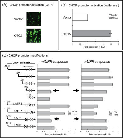
We have previously developed an experimental model for production of a mtUPR and have shown that a mutant of the mitochondrial matrix protein containing a small deletion of one of the substrate binding sites in ornithine transcarbamylase (OTCΔ) was imported into the mitochondrial matrix normally in COS-7 cells, but upon cleavage of the mitochondrial pre-sequence formed aggregates and induced genes encoding the mitochondrial chaperonins, chaperonin 60 (Cpn60) and chaperonin 10 (Cpn10) as well as the matrix protease ClpP [9]. Moreover, we showed that OTCΔ induces transcription of cpn60 and chop, but not the ER isoform of Hsp70 (Bip), in COS-7 cells. Creation of an erUPR by the addition of either tunicamycin or thapsigargin to COS-7 cells in contrast, strongly induces the ER isoform of Hsp70 and CHOP, but had only a minor effect on transcriptional activation of the cpn60 gene [9]. Thus, the accumulation of unfolded proteins leads to a specific response in each organelle, despite the fact that both UPRs induce transcription of the chop gene. Since Northern analysis measures steady-state concentration of mRNA, the experiments were repeated using chop promoter constructs. As shown in Figure 1A, expression of OTCΔ lead to an activation of a chop-gfp promoter construct of approximately 2.3 fold whereas a quantitative assay using luciferase as the reporter enzyme shows that OTCΔ activates transcription approximately 2.5 fold over the constitutive transcriptional activity obtained from cells transfected with vector without the OTCΔ insert (Figure 1B).
Figure 1. Identification of mtUPR response element in chop promoter.
(A) and (B): Chop is transcriptionally activated by mtUPR. COS-7 cells co-transfected with empty vector or OTCΔ were assayed for GFP (A) or luciferase (B) 32 h after transfection. (C): identification of an mtUPR element in the chop promoter was determined by a deletion analysis as shown. Deletions are shown as distance (bp) from the chop transcription start site. The fold activation of the promoter constructs in cells transfected with OTCΔ (slash bars) are compared with vector controls (open bars) as relative luciferase (RLU) activity. RLU activity of the promoter constructs in cells treated with or without 2 µg/ml tunicamycin (TM), to produce erUPR is shown as a control. Data represents the mean±SEM from experiments performed in triplicate.
The specificity of induction of CHOP by organelle specific UPRs suggests that the chop promoter contains separate elements for activation in response to erUPR and mtUPR.
Identification of an mtUPR element in the chop promoter
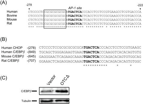
A deletion analysis of the chop promoter between bases −1015 and +17 (zero being the transcriptional start site) was carried out by assaying promoter activity using the luciferase reporter enzyme. MtUPR activity was measured by comparing the activity obtained from cells transfected with OTCΔ compared with empty vector and erUPR activity was measured by adding tunicamycin to cells transfected with the promoter-LUC construct. Deletions between −1015 and −325 had no effect on chop transcriptional activity (Fig 1C), whereas a further deletion of 103bp essentially ablated mtUPR inducible chop promoter activity. With respect to erUPR inducibility of chop, the critical element appears to lie between −105 and +1 bp (Fig 1C and Fig 2). A sequence comparison of chop promoters from human, bovine, mouse, and rat shows that this region between −278 and −222 contains an AP-1 site (Figure 2A), whereas the previously identified ERSE [14], [15] lies between −105 bp and +1 bp (data not shown). The ERSE element consists of two transcription factor binding sites, ATF-6 [15] and NF-Y [16], [17]. We deleted the AP-1, ATF-6, and NF-Y sites to determine if any of these sites were required for the regulation of CHOP expression in response to mtUPR. The deletion of the AP-1 site ablated the mtUPR responsiveness (Fig 1C). In contrast, the deletion of either ATF-6 or NF-Y elements, although substantially reducing the erUPR responsiveness (Fig 1C), did not remove the mtUPR responsiveness. Conversely the deletion of the AP-1 site had no effect on the erUPR responsiveness of the chop promoter, although the deletion of the NF-Y site did reduce the overall activity of the chop promoter.
Figure 2. Chop and c/ebpβ promoters contain AP-1 sites and are inducible by mtUPR.
(A) Nucleotide sequence alignment of the mammalian chop promoters (−278 to −222) from human, bovine, mouse and rat. Bold letters show the highly conserved bases of the AP-1 site and the asterisks show the highly conserved sequence surrounding the AP-1 site in chop promoters. The position of the putative novel element of N 30 [18] is shown in the box. (B): Nucleotide sequences of mammalian c/ebpβ promoter region around AP-1 site (Human, Mouse, and Rat) is compared with the chop promoter sequence (−278 to −233). The numbers refer to the distance from transcription initiation site of human chop or human, mouse, and Rat c/ebpβ. The asterisks indicate the conserved nucleotides around the AP-1 site. (C): C/EBPβ expression in response to mtUPR. Extracts from cells transfected with vector or OTCΔ were subjected to western blotting and probed with antibodies against C/EBPβ and tubulin as control and show that C/EBPβ, like CHOP is induced by expression of OTCΔ.
As shown in Figure 2A, the promoter region flanking the AP-1 site is highly conserved in other mammalian chop promoters. These flanking regions may contain additional information for the activation of the chop gene by mtUPR. One of these regions contains a sequence homologous to a putative element, N30, previously identified in a homology search of promoter regions in a range of animal species [18] (Fig 2A, boxed sequence). Deletion of this element had a partial effect on the mtUPR responsiveness of the chop promoter (Fig 1C).
Since we previously showed that CHOP induces transcription of mtUPR responsive genes in combination with C/EBPβ [9], it was of interest to note that the promoter of c/ebpβ gene also contains an AP-1 site with highly conserved nucleotides (CCCA) in the region flanking the AP-1 site (Fig 2B). This site in the c/ebpβ gene is also highly conserved between human, mouse, and rat promoter (Fig 2B) and therefore, we should expect both chop and c/ebpβ transcription to be elevated by mtUPR. This was confirmed by Western blot analysis (Fig 2C). It has recently been shown that CHOP combines with C/EBPα or β to activate BIM transcription and apoptosis in response to erUPR [12]. However, the c/ebpα promoter does not contain an AP-1 site (data not shown). This raises the question whether mtUPR also induces apoptosis.
Involvement of JNK2 in mtUPR signaling
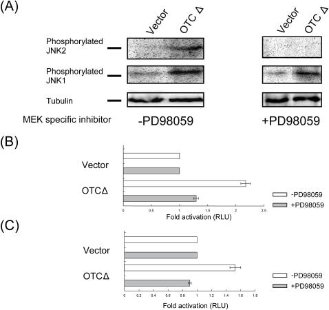
Since it is well-known that c-Jun, which is activated by JNK (c-Jun N-terminal kinase), binds to the AP-1 site [19] and it has been reported that the activation of JNK-dependent ATF2 (activated transcription factor 2) is important for the signaling from mitochondria to nucleus during the both genetic and metabolic stresses of mitochondria [3], [20], we therefore investigated the effect of mtUPR on the phosphorylation of JNK1 and JNK2 (Fig 3A). The expression of OTCΔ in COS-7 cells had a substantial effect on the phosphorylation of JNK1 and 2 (Fig 3A). To further test the potential role of JNK1 and JNK2 in mtUPR signaling, we determined the effect of the MEK inhibitor PD98059 [21], [22] on JNK phosphorylation in response to expression of OTCΔ. As shown in Figure 3A, the inhibitor completely blocked mtUPR dependent phosphorylation of JNK2, but had only a small effect on JNK1 phosphorylation. These experiments were followed up by measuring the effects of PD98059 on OTCΔ dependent activation of the mtUPR responsive promoters yme1l1 [13] (Figure 3B) and mppβ [13](Fig 3C). As shown in Figure 3B and C, 10 µM MEK inhibitor inhibited the OTCΔ inducible activation of the promoter-luciferase reporter constructs in transfected COS-7 cells. This suggests that mtUPR signaling utilizes the MEK/JNK2 pathway.
Figure 3. MtUPR increases phosphorylation of JNK and a MEK specific inhibitor blocks mtUPR.
(A): mtUPR increases phosphorylation of JNK 1&2. Extracts from cells transfected with vector or OTCΔ, and treated with or without 10 µM of MEK specific inhibitor PD98059, were subjected to Western transfers probed with antibody against p-JNK. (B) and (C): mtUPR induction of the yme1l1(B) and mppβ(C) promoter is inhibited by the MEK specific inhibitor, PD98059. COS-7 cells co-transfected with vector or OTCΔ and yme1l1and mppβ promoter-reporter constructs, with or without 10 µM of PD98059 were used for luciferase assay 32 h after transfection. The fold activation of the promoter constructs in cells expressing OTCΔ compared with those expressing vector alone, with or without of PD98059, is shown as relative luciferase (RLU) activity. Data represent the mean±SEM from experiments performed in triplicate.
Discussion
The evolution of the eukaryotic cell facilitated the development of increased metabolic and functional complexity by dividing cells into distinct, membrane enclosed compartments. However, these organelles/compartments are extremely crowded, both in terms of small solutes and macromolecules. Thus, it has been estimated that the cytosol has a protein concentration of around 350 mg/ml [23] and the concentration inside the mitochondrial matrix may approach 500 mg/ml [24]. Not surprisingly then, the cell has evolved stress response mechanisms which come into play under conditions where unfolded proteins accumulate, such as the heat shock response [25]. Equally, the cell has evolved mechanisms to respond to the accumulation of unfolded proteins in organelle compartments such as the ER, which has become known as the UPR [14], [15], [26], [27]. This response, which was initially discovered in baker's yeast [28] has been extensively investigated and is characterized by the transcriptional regulation of a large group of genes and post transcriptional regulation of proteins involved in quality control of the secretory pathway [14], [15], [26], [27].
We discovered an equivalent stress response pathway in mitochondria of mammalian cells [9], [10] and originally called it the Mitochondrial Stress Response. More recently, Ron and colleagues discovered the response in c.elegans [11] and more appropriately called the response the mtUPR, distinguishing it from the erUPR, as we have done in this paper. Surprisingly, the mtUPR has not been found in fungi and appears to be an organelle specific stress response found only in multi-cellular organisms.
We originally found that in the mammalian mtUPR responsive gene cpn60/10, the CHOP and C/EBPβ transcription factors were involved in transcription regulation [9]. However, since mtUPR also led to the transcriptional regulation of chop, this suggested that the induction of the chop gene is an early event in mtUPR. We were also intrigued by the finding that although there appears to be little overlap in the mtUPR and erUPR, both responses led to the induction of chop transcription. In this paper we describe the identification of an mtUPR response element in the promoters of both chop and c/ebpβ genes. This element is an AP-1 site, suggesting that mitochondrial to nuclear signaling of the accumulation of unfolded proteins in the mitochondrial matrix is through a JNK pathway. We show, using a specific MEK inhibitor, that this signaling is through JNK2 and that an inhibition in the phosphorylation of JNK2 also inhibits mtUPR. We suggest that the cell can discriminate between organelle specific unfolded protein responses through different pathways to activate genes that harbor different stress elements within their promoters. Recently, it has been reported that JNK2 is a positive regulator of the cJun transcription factor [29], and can regulate both mitochondrial and lysosomal death pathways in mouse embryonic fibroblasts [30]. This, taken together with the data presented here, suggests that the JNK2 pathway may play a significant role for the communication from mitochondria to the nucleus in response to mtUPR. Since both mtUPR and erUPR activate transcription of a distinct set of genes, yet both induce CHOP, it is apparent that additional factors besides CHOP and C/EBPβ account for the specificity of the mtUPR. This specificity is provided for the erUPR by the transcription factors ATF6 and NFY [14.15]. The question of the specificity of mtUPR is further explored in the accompanying paper [13].
Recently, Benedetti et al. [31] have carried out a search for genes involved in signaling of mtUPR in c.elegans and discovered the involvement of the ubl-5 gene, encoding the ubiquitin-like protein 5. Whether this pathway exists in mammalian cells, or whether this pathway in c.elegans intersects with the pathway we describe here is currently unknown, as is the question whether the CHOP based response described in this paper operates in c.elegans.
Materials and Methods
Materials
Tunicamycin was purchased from Sigma Chemical (St Louis, USA). MEK inhibitor, PD98059, anti-C/EBPβ, and anti-pJNK were purchased from Santa Cruz Biotechnology (Santa Cruz, USA). All reagents were of reagent grade quality.
Plasmid construction, transfection and promoter analysis
Mammalian expression vectors of wild-type OTC and deletion mutant OTCΔ were constructed as described previously [9]. Transfection efficiencies were between 72 and 85% as determined by transfections with a GFP construct. Based on the human genome sequence information of NCBI, the promoter region of CHOP (from −1015 to +17) was amplified by PCR [32] from human genomic DNA (Promega, Madison, USA) using 5′-CTTTTGGGAGATCTACGGGGCTAGAACAGGAGACCACCC-3′ and 5′-GATACGCTCAGAAGCTTAGACTTAAGTCTCTGACCTCGG-3′ as the upper and lower primers, respectively (mutated nucleotides to introduce BglII and Hind III are underlined), and cloned into BglII-Hind III sites of the pGL3-Basic vector (Promega, Madison, USA), which contains the firefly luciferase coding sequence but lacks eukaryotic promoter or enhancer elements. For the GFP assay of promoter constructs, luciferase was replaced by GFP cDNA using Nco I – Xba I sites in the pGL3-Basic vector. Deletion mutants of the CHOP promoter were constructed by PCR using 5′-GGGGCCAAGAGATCTGGGAGTCCCTTATAG-3′(−555), 5′-GACACCGGTTGCCAGATCTTGCATCATCCCCGCC-3′(−325), 5′-CCGTGAAGCCTCGAGATCTAAAGCCACTTCCGGG-3′(−222), and 5′-GGCGGATGCGAAGATCTGGGCGGGGCCAATGCC-3′(−105) as upper primers, respectively, and 5′-GGTGGCTTTACCAACAGTACCGGAATGCC-3′ as lower primer (mutated nucleotides are underlined) for wild type CHOP promoter introduced into PGL3-Basic vector. For disruption of ATF-6, NF-Y, and AP-1 transcription factors [14], [18] or point mutation, site-directed mutagenesis was carried out by PCR [32] using 5′-GCCGGCGGGCCACTTTCTGATTGGTAGG-3′ and 5′-CCTACCAATCAGAAAGTGGCCCGCCG-3′ for ΔATF-6, 5′-GCCGGCGTGCCACTTTCTGATGGGTAGG-3′ and 5′-CCTACCCATCAGAAAGTGGCACGCCG-3′ for ΔNF-Y, 5′-GCGCGCGCATGAAACACCCACCTCCTCCGTG-3′ and 5′-GAGGCTTCACGGAGGAGGTGGGTGTTTCATGCG-3′ for ΔAP-1, and 5′- CACTCCCCTCCGCAAACGCACATGACTCACCCACCTCCTCC-3′ and 5′- GGAGGAGGTGGGTGAGTCATGTGCGTTTGCGGAGGGGAGTG-3′ for ΔN30 [33] as the upper and lower primers, respectively (mutated nucleotides are underlined).
COS-7 cells were cultured in DME/5 % fetal calf serum and transfected at 90 % confluence using Lipofectamine 2000 (Invitrogen, California, USA). Promoter analysis using luciferase assay was carried out as described previously [9]. To induce erUPR, cells were treated with 2 µg/ml tunicamycin for 10h. Western blot analysis was carried out as described previously [9].
Acknowledgments
We are grateful for critical reading of the manuscript by Dr Jonathan Aldridge and Dr Hamsa Puthalakath.
Footnotes
Competing Interests: The authors have declared that no competing interests exist.
Funding: This work was supported by grants to Nicholas Hoogenraad from the Australian Research Council and the National Health and Medical Research Council, which provided a fellowship to Tomohisa Horibe.
References
- 1.Calvo S, Jain M, Xie X, Sheth SA, Chang B, et al. Systematic identification of human mitochondrial disease genes through integrative genomics. Nat Genet. 2006;38:576–582. doi: 10.1038/ng1776. [DOI] [PubMed] [Google Scholar]
- 2.Wallace DC. A mitochondrial paradigm of metabolic and degenerative diseases, aging, and cancer: a dawn for evolutionary medicine. Annu Rev Genet. 2005;39:359–407. doi: 10.1146/annurev.genet.39.110304.095751. [DOI] [PMC free article] [PubMed] [Google Scholar]
- 3.Butow RA, Avadhani NG. Mitochondrial signaling: the retrograde response. Mol Cell. 2004;14:1–15. doi: 10.1016/s1097-2765(04)00179-0. [DOI] [PubMed] [Google Scholar]
- 4.Kelly DP, Scarpulla RC. Transcriptional regulatory circuits controlling mitochondrial biogenesis and function. Genes Dev. 2004;18:357–368. doi: 10.1101/gad.1177604. [DOI] [PubMed] [Google Scholar]
- 5.Ryan MT, Hoogenraad NJ. Mitochondrial-Nuclear Communications. Annu Rev Biochem. 2007 doi: 10.1146/annurev.biochem.76.052305.091720. (in press) [DOI] [PubMed] [Google Scholar]
- 6.Goffart S, Wiesner RJ. Regulation and co-ordination of nuclear gene expression during mitochondrial biogenesis. Exp Physiol. 2003;88:33–40. doi: 10.1113/eph8802500. [DOI] [PubMed] [Google Scholar]
- 7.Amuthan G, Biswas G, Ananadatheerthavarada HK, Vijayasarathy C, Shephard HM, et al. Mitochondrial stress-induced calcium signaling, phenotypic changes and invasive behavior in human lung carcinoma A549 cells. Oncogene. 2002;21:7839–7849. doi: 10.1038/sj.onc.1205983. [DOI] [PubMed] [Google Scholar]
- 8.Lin J, Handschin C, Spiegelman BM. Metabolic control through the PGC-1 family of transcription coactivators. Cell Metab. 2005;1:361–370. doi: 10.1016/j.cmet.2005.05.004. [DOI] [PubMed] [Google Scholar]
- 9.Zhao Q, Wang J, Levichkin IV, Stasinopoulos S, Ryan MT, et al. A mitochondrial specific stress response in mammalian cells. Embo J. 2002;21:4411–4419. doi: 10.1093/emboj/cdf445. [DOI] [PMC free article] [PubMed] [Google Scholar]
- 10.Martinus RD, Garth GP, Webster TL, Cartwright P, Naylor DJ, et al. Selective induction of mitochondrial chaperones in response to loss of the mitochondrial genome. Eur J Biochem. 1996;240:98–103. doi: 10.1111/j.1432-1033.1996.0098h.x. [DOI] [PubMed] [Google Scholar]
- 11.Yoneda T, Benedetti C, Urano F, Clark SG, Harding HP, et al. Compartment-specific perturbation of protein handling activates genes encoding mitochondrial chaperones. J Cell Sci. 2004;117:4055–4066. doi: 10.1242/jcs.01275. [DOI] [PubMed] [Google Scholar]
- 12.Puthalakath H, O'Reilly LA, Gunn P, Lee L, Kelly PN, et al. ER stress triggers apoptosis by activating BH3-only protein. Bim Cell. 2007;129:1337–49. doi: 10.1016/j.cell.2007.04.027. [DOI] [PubMed] [Google Scholar]
- 13.Aldridge J, Horibe T, Hoogenraad NJ. Discovery of genes activated by the mitochondrial unfolded protein response (mtUPR) and cognate promoter elements. 2007 doi: 10.1371/journal.pone.0000874. (submitted to PLoS ONE) [DOI] [PMC free article] [PubMed] [Google Scholar]
- 14.Yoshida H, Okada T, Haze K, Yanagi H, Yura T, et al. ATF6 activated by proteolysis binds in the presence of NF-Y (CBF) directly to the cis-acting element responsible for the mammalian unfolded protein response. Mol Cell Biol. 2000;20:6755–6767. doi: 10.1128/mcb.20.18.6755-6767.2000. [DOI] [PMC free article] [PubMed] [Google Scholar]
- 15.Yoshida H, Haze K, Yanagi H, Yura T, Mori K. Identification of the cis-acting endoplasmic reticulum stress response element responsible for transcriptional induction of mammalian glucose-regulated proteins. J Biol Chem. 1998;273:33741–33749. doi: 10.1074/jbc.273.50.33741. [DOI] [PubMed] [Google Scholar]
- 16.Roy B, Li WW, Lee AS. Calcium-sensitive transcriptional activation of the proximal CCAAT regulatory element of the grp78/BiP promoter by the human nuclear factor CBF/NF-Y. J Biol Chem. 1996;271:28995–29002. doi: 10.1074/jbc.271.46.28995. [DOI] [PubMed] [Google Scholar]
- 17.Maity SN, de Crombrugghe B. Role of the CCAAT-binding protein CBF/NF-Y in transcription. Trends Biochem Sci. 1998;23:174–178. doi: 10.1016/s0968-0004(98)01201-8. [DOI] [PubMed] [Google Scholar]
- 18.Xie X, Lu J, Kulbokas EJ, Golub TR, Mootha V, et al. Systematic discovery of regulatory motifs in human promoters and 3′ UTRs by comparison of several mammals. Nature. 2005;434:338–345. doi: 10.1038/nature03441. [DOI] [PMC free article] [PubMed] [Google Scholar]
- 19.Weiss C, Schneider S, Wagner EF, Zhang X, Seto E, et al. JNK phosphorylation relieves HDAC3-dependent suppression of the transcriptional activity of c-Jun. Embo J. 2003;22:3686–3695. doi: 10.1093/emboj/cdg364. [DOI] [PMC free article] [PubMed] [Google Scholar]
- 20.Biswas G, Adebanjo OA, Freedman BD, Anandatheerthavarada HK, Vijayasarathy C, et al. Retrograde Ca2+ signaling in C2C12 skeletal myocytes in response to mitochondrial genetic and metabolic stress: a novel mode of inter-organelle crosstalk. Embo J. 1999;18:522–533. doi: 10.1093/emboj/18.3.522. [DOI] [PMC free article] [PubMed] [Google Scholar]
- 21.Alessi DR, Cuenda A, Cohen P, Dudley DT, Saltiel AR. PD 098059 is a specific inhibitor of the activation of mitogen-activated protein kinase kinase in vitro and in vivo. J Biol Chem. 1995;270:27489–27494. doi: 10.1074/jbc.270.46.27489. [DOI] [PubMed] [Google Scholar]
- 22.Pang L, Sawada T, Decker SJ, Saltiel AR. Inhibition of MAP kinase kinase blocks the differentiation of PC-12 cells induced by nerve growth factor. J Biol Chem. 1995;270:13585–13588. doi: 10.1074/jbc.270.23.13585. [DOI] [PubMed] [Google Scholar]
- 23.Goodsell DS. Inside a living cell. Trends Biochem Sci. 1991;16:203–206. doi: 10.1016/0968-0004(91)90083-8. [DOI] [PubMed] [Google Scholar]
- 24.Srere PA. The infrastructure of the mitochondrial matrix. Trends Biochem Sci. 1980;5:120–121. [Google Scholar]
- 25.Lindquist S. The heat-shock response. Annu Rev Biochem. 1986;55:1151–1191. doi: 10.1146/annurev.bi.55.070186.005443. [DOI] [PubMed] [Google Scholar]
- 26.Yoshida H, Matsui T, Yamamoto A, Okada T, Mori K. XBP1 mRNA is induced by ATF6 and spliced by IRE1 in response to ER stress to produce a highly active transcription factor. Cell. 2001;107:881–91. doi: 10.1016/s0092-8674(01)00611-0. [DOI] [PubMed] [Google Scholar]
- 27.Schroder M. The unfolded protein response. Mol Biotechnol. 2006;34:279–90. doi: 10.1385/MB:34:2:279. [DOI] [PubMed] [Google Scholar]
- 28.Gething MJ, Sambrook J. Protein folding in the cell. Nature. 1992;355:33–45. doi: 10.1038/355033a0. [DOI] [PubMed] [Google Scholar]
- 29.Jaeschke A, Karasarides M, Ventura JJ, Ehrhardt A, Zhang C, et al. JNK2 is a positive regulator of the cJun transcription factor. Mol Cell. 2006;23:899–911. doi: 10.1016/j.molcel.2006.07.028. [DOI] [PubMed] [Google Scholar]
- 30.Dietrich N, Thastrup J, Holmberg C, Gyrd-Hansen M, Fehrenbacher N, et al. JNK2 mediates TNF-induced cell death in mouse embryonic fibroblasts via regulation of both caspase and cathepsin protease pathways. Cell Death Differ. 2004;11:301–313. doi: 10.1038/sj.cdd.4401353. [DOI] [PubMed] [Google Scholar]
- 31.Benedetti C, Haynes CM, Yang Y, Harding HP, Ron D. Ubiquitin-like protein 5 positively regulates chaperone gene expression in the mitochondrial unfolded protein response. Genetics. 2006;174:229–39. doi: 10.1534/genetics.106.061580. [DOI] [PMC free article] [PubMed] [Google Scholar]
- 32.Kunkel TA. Rapid and efficient site-specific mutagenesis without phenotypic selection. Proc Natl Acad Sci US A. 1985;82:488–492. doi: 10.1073/pnas.82.2.488. [DOI] [PMC free article] [PubMed] [Google Scholar]


