Abstract
Borna disease virus (BDV) can persistently infect the central nervous system of a broad range of mammalian species. Mice are resistant to infections with primary BDV isolates, but certain laboratory strains can be adapted to replicate in mice. We determined the molecular basis of adaptation by studying mutations acquired by a cDNA-derived BDV strain during one brain passage in rats and three passages in mice. The adapted virus propagated efficiently in mouse brains and induced neurological disease. Its genome contained seven point mutations, three of which caused amino acid changes in the L polymerase (L1116R and N1398D) and in the polymerase cofactor P (R66K). Recombinant BDV carrying these mutations either alone or in combination all showed enhanced multiplication speed in Vero cells, indicating improved intrinsic viral polymerase activity rather than adaptation to a mouse-specific factor. Mutations R66K and L1116R, but not N1398D, conferred replication competence of recombinant BDV in mice if introduced individually. Virus propagation in mouse brains was substantially enhanced if both L mutations were present simultaneously, but infection remained mostly nonsymptomatic. Only if all three amino acid substitutions were combined did BDV replicate vigorously and induce early disease in mice. Interestingly, the virulence-enhancing effect of the R66K mutation in P could be attributed to reduced negative regulation of polymerase activity by the viral X protein. Our data demonstrate that BDV replication competence in mice is mediated by the polymerase complex rather than the viral envelope and suggest that altered regulation of viral gene expression can favor adaptation to new host species.
Viruses acquiring the ability to multiply in a new host species are of great concern for public health. Viruses of animal origin that have successfully entered the human species are the causative agents of important infectious diseases, such as AIDS, severe acute respiratory syndrome, and pandemic influenza (5, 13, 30). The molecular basis of virus adaptation to new host species is poorly understood. It is clear that adaptation can result from mutations in viral envelope components which may alter the receptor specificity or uptake efficacy of the virus. For example, massive structural alterations of the envelope seem to have occurred during adaptation of severe acute respiratory syndrome coronavirus from bats to humans (18). Recently, it became clear that alternative mechanisms can also lead to successful virus adaptation to new host species. In the case of the avian influenza A virus strain SC35, two amino acid changes in the PB2 polymerase subunit which enhance the activity of the viral polymerase complex can confer viral replication competence in mice (4). Similarly, a glutamic acid-to-lysine change at position 627 of the PB2 protein rendered an avian H5N1 influenza A virus isolate highly pathogenic for mice (8, 25). In the case of Ebola virus, adaptation to mice was associated with mutations in VP24 and in the nucleoprotein, both of which antagonize the innate antiviral immune response (3). These examples illustrate that several factors can determine the species specificity of a virus.
Borna disease virus (BDV) is a neurotropic, enveloped virus with a nonsegmented negative-strand RNA genome (2). The BDV genome of approximately 8,900 nucleotides is transcribed and replicated in the nucleus (1). Alternative splicing of polycistronic mRNA is employed to regulate viral protein levels. Based on these unique properties, BDV has been classified into a separate virus family (Bornaviridae) in the order Mononegavirales. In naturally and experimentally infected animals, BDV establishes a noncytolytic, persistent infection of the central nervous system (CNS) that frequently results in a severe immune-mediated neurological disorder (28). Successful experimental infection of a broad range of warm-blooded animals has been reported (26). Despite obvious promiscuity, most tissue culture-adapted laboratory strains of BDV do not readily infect mice but can be adapted to mice by serial passage in the CNS (7, 9, 12). In a previous study, four amino acid changes were identified in the mouse-adapted virus, two of which affected the viral glycoprotein G and two the viral polymerase L (12). However, the relative contributions of these four mutations remained undetermined due to the lack of suitable experimental systems.
We used molecularly cloned BDV strain He/80FR (10, 23) for adaptation experiments with mice. Adaptation resulted in three amino acid changes affecting the polymerase L and the polymerase cofactor P but not the viral envelope protein G. Recombinant BDV carrying these three mutations either alone or in combination all showed enhanced viral multiplication speed in Vero cells. The P mutation and one of the L mutations conferred replication competence in mice. When all three mutations were combined, a drastic increase of virulence was observed. Our data demonstrate that BDV adaptation to mice can be mediated entirely by the viral polymerase complex and suggest that minute changes in the BDV genome can generate highly virulent strains which induce disease in a new host species.
MATERIALS AND METHODS
Plasmid constructions.
The R66K mutations in the P genes of pCA-P and pBRPol II-HrBDVc (10, 22) were generated by changing the codon AGG to AAG. The L1116R and N1398D mutations in pCA-L (22) and pBRPol II-HrBDVc were inserted by changing the codon CTT to AGA and AAT to GAT, respectively. Site-directed mutagenesis PCR was performed with proofreading Turbo Pfu DNA polymerase (Stratagene) and standard reaction conditions in a model 9600 GeneAmp PCR cycler (Applied Biosystems). The integrity of all PCR-derived DNA fragments was verified by sequencing. All restriction digestions of plasmids and PCR fragments were performed using commercially available enzymes (New England Biolabs and Fermentas). Ligation reactions were done using 2.5 Weiss units of bacteriophage T4 DNA ligase (Fermentas) in a total volume of 5 μl. Ligation reaction mixtures were incubated at 16°C for at least 2 h and then used to transform 50 μl of competent Top10 bacteria (Invitrogen). Primer sequences and details of the cloning strategies are available on request.
Virus rescue.
Recombinant virus was recovered from cDNA exactly as previously described (10).
Virus stocks.
Stocks of recombinant BDV strains were prepared as previously described (1) and dialyzed for 2 days against phosphate-buffered saline, followed by titration on Vero cells. To prepare virus stocks from infected rats and mice, brain material was disrupted mechanically by multiple passages through an injection needle and ultrasonic treatment. Cell debris was removed by centrifugation.
Animals and virus infections.
Mouse breeding colonies were maintained in our local animal facility. C57BL/6 mice lacking a functional type I interferon (IFN-I) system (IFNAR10/0) (11) were originally provided by U. Kalinke, Langen, Germany. MRL/MpJ mice were originally purchased from The Jackson Laboratory (Bar Harbor, ME). Mice were infected within 96 h of birth with 10 μl of infected brain homogenate or 1,000 focus-forming units (FFU) of recombinant BDV by intracerebral injection using a Hamilton syringe. Four-week-old Lewis rats (Charles River) were anesthetized and infected by intracerebral injection of 2,000 FFU of BDV. Animals were examined daily for neurological symptoms. All animal experiments were approved by the local authorities.
Fluorescence microscopy.
Cells were seeded onto coverslips, fixed for 10 min in 3% paraformaldehyde, and permeabilized by incubation for 5 min in phosphate-buffered saline containing 0.5% Triton X-100. Virus antigen was detected as described previously (10).
Western blot analysis.
Protein extracts were prepared from homogenized brains of infected animals as described previously (17). Protein content was determined by Bradford analysis (Bio-Rad). Samples were separated by sodium dodecyl sulfate-polyacrylamide gel electrophoresis on a 10% gel and blotted onto a polyvinylidene difluoride membrane (Millipore). BDV-N was detected as described previously (19). To demonstrate the correct loading of the gel, the blot was incubated with a mouse monoclonal antibody (Sigma-Aldrich) against β-tubulin and developed with a peroxidase-coupled secondary antibody (Jackson ImmunoResearch).
Mammalian two-hybrid and minireplicon assays.
Mammalian two-hybrid and minireplicon assays were performed essentially as described previously (21, 22). However, for minireplicon assays we used human 293T cells transfected with 250 ng of a pCA plasmid encoding T7 RNA polymerase (10).
RNA preparation and Northern blot analysis.
RNA was prepared from 94-mm dishes of infected Vero cell cultures or from infected brain material with the peqGOLD TriFast reagent (PeqLab Biotechnologie, Erlangen, Germany) as recommended by the manufacturer. Northern blot analysis using 5-μg samples of total RNA was performed as described previously (22). The DNA probes for the detection of RNAs derived from the N and X/P genes were amplified by PCR using primer pairs 976(+)/1749(−). The numbering refers to the position and orientation of the primers on the BDV antigenome. PCR products were radioactively labeled by using a Prime-It II random primer labeling kit (Stratagene).
Reverse transcription-PCR and sequencing.
Two micrograms of total RNA was reverse transcribed using hexamer primers and an H-minus first-strand cDNA synthesis kit (Fermentas) according to the manufacturer's protocol. The complete BDV genome was amplified using PCR primer pairs 1(+)/1267(−), 976(+)/2268(−), 1995(+)/3266(−), 3000(+)/4261(−), 4014(+)/5263(−), 5015(+)/6294(−), 6004(+)/7268(−), 6998(+)/8246(−), and 8027(+)/8912(−). Primer sequences are available on request.
RESULTS
BDV acquires distinct mutations during serial passage in rats and mice.
Newborn MRL mice were inoculated with approximately 104 FFU of cDNA-derived BDV strain He/80FR isolated from Vero cells. Analysis of brain sections performed 8 weeks later revealed only a few infected cells in some of these animals (Fig. 1, top), but passage of extracts from such brains into new MRL mice did not yield replication-competent BDV. In a second approach, the virus was first injected into the brain of a 4-week-old Lewis rat. When the rat developed neurological disease about 3 weeks later, brain extract was prepared and inoculated into newborn MRL mice. At 6 weeks postinfection, we observed a substantial number of infected neurons in the hippocampi of such mice (Fig. 1, lower middle). When extract from one of these mouse brains was inoculated into new MRL mice, one of three animals developed neurological symptoms at 42 days postinfection (Table 1). Histological analyses revealed a widespread presence of BDV-infected cells in most brain regions of the diseased animal (data not shown). Virus was also abundantly present in the remaining two animals, which stayed healthy until analysis at 7 weeks postinfection (data not shown). In virus passage 4, widespread infection of the mouse CNS, including the cerebellum, was observed (Fig. 1, lower right), and neurological disease occurred in all animals around day 48 postinfection (Table 1). Efficacy of disease induction was slightly decreased in virus passage 5.
FIG. 1.
BDV antigen distribution in the hippocampi of infected rats and mice. Animals were inoculated intracerebrally with recombinant virus originating directly from either Vero cells, rat brain (10 μl of 10% extract), or mouse brains (10 μl of 10% extract). Animals were killed at 6 to 8 weeks postinfection or when severe neurological symptoms appeared. Brain sections were stained with a BDV-N-specific antibody. The brown stain indicates BDV-infected cells.
TABLE 1.
Amino acid changes in BDV proteins during serial passage of recombinant BDV He/80 in rats and mice
| Passage no. (species) | BDV protein | Result for indicated expt
|
|||
|---|---|---|---|---|---|
| Expt 1
|
Expt 2
|
||||
| Mutation (frequency [%])b | No. of animals diseased/no. infected | Mutation (frequency [%])b | No. of animals diseased/no. infected | ||
| 1a (rat) | L | L1116R (60) | 1/1 | Not analyzed | 1/1 |
| 2 (mouse) | L | L1116R (100) | 0/10 | L1116R (100) | 0/5 |
| N1398 (20) | |||||
| 3 (mouse) | L | Not analyzed | 1/3 | L1116R (100) | 1/5 |
| N1398D (25) | |||||
| 4 (mouse) | P | R66K (80) | 4/4 | L1116R (100) | 4/5 |
| L | L1116R (100) | ||||
| N1398D (90) | |||||
| 5 (mouse) | P | R66K (25) | 4/5 | ||
| L | L1116R (100) | ||||
| N1398D (35) | |||||
The rats were inoculated intracerebrally with 2,000 FFU of plasmid-derived BDV isolated from Vero cells.
Percentage of viral genomes encoding the mutation as estimated from the signal intensities determined for the original and mutant nucleotides with a sequence electropherogram.
Sequencing of overlapping cDNA fragments amplified from viral genomes of passage 4 revealed that four silent and three nonsilent mutations which introduced amino acid changes in P and L had occurred (Fig. 2). Bulk sequencing of PCR products indicated that the mutation responsible for the L1116R change in L was present at a frequency of virtually 100%, whereas the mutations responsible for the R66K and N1398D changes occurred less frequently (Table 1, experiment 1). From the signal intensities in the electropherogram, we estimated that the R66K mutation was present in about 80% and the N1398D mutation in about 90% of the viruses at passage 4 (Table 1). The silent mutations were present at a frequency of close to 100% (data not shown). Sequencing of the complete genome of virus from passage 1 showed that the mutation leading to the L1116R change was already present in the rat brain at a frequency of approximately 60%, while the other six mutations found after passage 4 were not present at detectable frequencies in the rat brain (Table 1 and data not shown).
FIG. 2.
Mutations in the genome acquired during adaptation of BDV to the mouse. The natures and positions of nucleotide and amino acid changes after one rat and three mouse passages are indicated. Data refer to adaptation experiment 1 (Table 1).
To gain information about the importance of the observed mutations, we performed a second independent adaptation experiment with the same stock of recombinant BDV (Table 1, experiment 2). Sequencing of relevant cDNA fragments of viruses from various passages in mice showed that the L1116R change appeared again and remained stable. The A7879G mutation causing the N1398D change in L also occurred in the second adaptation experiment. However, this mutation remained incomplete and eventually disappeared. The G1469A mutation causing the R66K change in P that was found at a high frequency in passage 4 of adaptation experiment 1 did not appear in experiment 2 (Table 1). Similarly, the two mutations in the first intergenic region (positions 1222 and 1229) were not observed in the second adaptation experiment, demonstrating that these changes are not essential for adaptation of BDV to the mouse. In experiment 2, we observed an insertion of two A nucleotides in the first intergenic region at position 1208. This insertion was present in about 30% and 5% of viruses from passages 3 and 4, respectively (data not shown). Taken together, the two independent adaptation experiments suggested a key role for the L protein and in particular for residue 1116 in successful multiplication of recombinant BDV in mice.
Amino acid changes acquired during adaptation of BDV to mice promote virus multiplication speed in cultured cells.
To analyze the individual contributions of the mutations at amino acid position 66 in P and positions 1116 and 1398 in L, we generated full-length cDNAs carrying these mutations alone or in combination and recovered the corresponding viruses. Viruses carrying the R66K, L1116R, or N1398D exchange were designated BDV-PK, BDV-LR, or BDV-LD, respectively, and the viruses carrying combinations of the L mutations or all three mutations were designated BDV-LRD or BDV-PK/LRD, respectively.
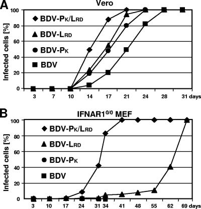
Growth analysis using Vero cells indicated that the mutations in either L or P had a substantial stimulating effect on virus multiplication, with BDV-PK/LRD being the fastest-replicating virus, followed by BDV-LRD and BDV-PK (Fig. 3A). Similarly, BDV-LR and BDV-LD showed enhanced multiplication in Vero cells compared to the original virus (data not shown).
FIG. 3.
Positive effect of adaptive mutations on BDV growth in cell culture. Kinetics of virus spread was determined in Vero cells (A) and MEF from IFNAR10/0 mice (B). Cultures were infected with the various recombinant viruses at a multiplicity of infection of 0.01 per cell. At the indicated times, the percentages of virus-infected cells were determined by analyzing samples of the cultures by indirect immunofluorescence. Growth curves of BDV-PK and wild-type BDV in IFNAR10/0 MEF were stopped on day 34 as no virus-infected cells were detected in these cultures.
A slightly different picture emerged when the growth kinetics studies with the various viruses were performed with embryo fibroblasts (MEF) of mice with defective IFN-I receptors (IFNAR10/0). This IFN-deficient cell culture system was previously reported to support the growth of mouse-adapted strains of BDV, whereas wild-type MEF do not (27). As expected, the original recombinant BDV isolated from Vero cells was unable to establish a productive infection in MEF of IFNAR10/0 mice. Similarly, the mutants BDV-PK, BDV-LR, and BDV-LD failed to grow in these cells (Fig. 3B and data not shown). In contrast, BDV-PK/LRD and to a lesser extent BDV-LRD were able to infect MEF of IFNAR10/0 mice (Fig. 3B).
Single amino acid changes in L and P independently mediate replication competence of BDV in mice.
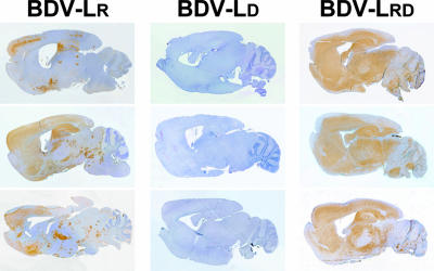
To assess the importance of the polymerase mutations at amino acid positions 1116 and 1398 for BDV adaptation to the mouse, we determined the abilities of BDV-LR, BDV-LD, and BDV-LRD to grow in mouse brains. Groups of three newborn MRL mice were infected with 1,000 FFU of each virus. None of the animals developed neurological disease by 8 weeks postinfection. Immunohistochemical analysis of brain sections showed that BDV-LR had successfully infected all three mouse brains (Fig. 4, left), whereas BDV-LD did not (Fig. 4, middle). BDV-LRD had also established a successful infection in all three animals (Fig. 4, right) and spread even more efficiently in the CNS than BDV-LR. Thus, the L1116R mutation was required for growth in mouse brains and the N1398D mutation seemed to increase viral fitness.
FIG. 4.
Growth characteristics in mouse brains of BDV variants with amino acid changes in L. Virus antigen distribution in sagittal brain sections of infected MRL mice is shown. Three animals per group were infected as newborns with 1,000 FFU of the indicated recombinant viruses and sacrificed 8 weeks later. Virus propagation was analyzed by immunohistochemistry using a rabbit antiserum specific for BDV-N. The brown stain indicates BDV-infected cells.
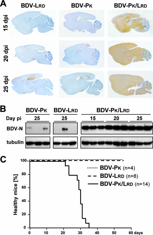
Next, we analyzed the contribution of the R66K exchange in P to BDV propagation in the CNS of mice. Since P has been reported to interfere with the induction of the IFN response (29), we performed growth analysis with C57BL/6-IFNAR10/0 mice to exclude a possible contribution of the IFN system to the outcome of the experiment. We infected groups of newborn C57BL/6-IFNAR10/0 mice with 1,000 FFU of BDV-PK, BDV-LRD, and BDV-PK/LRD. Due to the C57BL/6 background, these animals were not expected to develop neurological disease in response to BDV infection (7). Three animals of each group were sacrificed at 15, 20, and 25 days postinfection. Protein extracts were prepared from one brain hemisphere and analyzed by Western blotting for the presence of viral N protein. The other hemisphere was paraffin embedded, and sections were analyzed for virus infection by immunostaining for the N protein of BDV. Distinct foci of BDV-positive cells were present in the brains of mice infected with BDV-PK and BDV-LRD at 25 days postinfection, whereas substantially lower numbers of virus-positive cells were observed at 15 and 20 days postinfection (Fig. 5A, left and middle). By contrast, in brains of mice infected with BDV-PK/LRD large numbers of BDV-positive cells were found as early as 15 days postinfection (Fig. 5A, right). Careful analysis of the brain sections at higher-power resolution yielded no evidence for altered cell tropisms of BDV-PK/LRD (data not shown). Western blot analysis confirmed the picture of the histological analysis and showed that at all times postinfection the virus loads in brains of mice infected with BDV-PK/LRD were much higher than those in brains infected with BDV-PK or BDV-LRD (Fig. 5B). Thus, although mutations in L and P both contributed to replication competence of BDV in mice, the growth-stimulating effect was most dramatic when these mutations were combined.
FIG. 5.
P mutation R66K stimulates propagation and pathogenicity of BDV in mice. C57BL/6-IFNAR10/0 mice were infected with 1,000 FFU of the indicated recombinant viruses. At 15, 20, or 25 days postinfection, three animals of each group were sacrificed. (A) Virus propagation in one brain hemisphere was analyzed by immunohistochemistry using a rabbit antiserum specific for BDV-N. The brown stain indicates BDV-infected cells. (B) Virus propagation in the second brain hemisphere was analyzed by Western blotting using the same antiserum against BDV-N. Correct loading of the gel was verified by staining with a monoclonal antibody against β-tubulin. (C) Newborn MRL mice were infected with 1,000 FFU of the indicated recombinant viruses and observed for clinical symptoms for up to 8 weeks after infection.
BDV multiplication speed determines viral pathogenicity in mice.
To compare the pathogenic potentials of BDV-PK, BDV-LRD, and BDV-PK/LRD, we infected groups of newborn MRL mice, which are susceptible to BDV-induced neurological disorder (7). None of the 12 animals infected with either BDV-PK or BDV-LRD showed signs of neurological disorder during the entire 8-week observation period, whereas all 14 animals infected with BDV-PK/LRD developed severe neurological symptoms and had to be killed between 21 and 35 days postinfection (Fig. 5C). The good correlation between the propagation efficacies of these viruses and disease induction in mice suggested that the speed of BDV multiplication in the CNS determines viral pathogenicity.
The R66K mutation affects the interaction of P with X, resulting in reduced sensitivity of the viral polymerase complex to X-mediated inhibition.
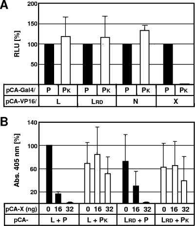
The enhanced virulence of viruses with adaptive mutations in L and P could be due to altered interactions of these proteins with other components of the BDV polymerase complex. Since L is known to interact with P (15), we used the mammalian two-hybrid system to determine whether the interactions of L and LRD with P and PK were altered. This was clearly not the case (Fig. 6A). We then asked whether the P-N and P-X interactions (24) might be influenced by the R66K mutation. Whereas the interaction of PK with N remained strong, we found that PK interacted only very poorly with X (Fig. 6A). This is of great interest as the X protein was shown to inhibit the viral polymerase activity at least in vitro (15). Although these data could not explain the enhanced virulence of BDV-LRD, they suggested a plausible explanation for the aggressive growth behavior of mutants carrying the R66K mutation in P, namely, reduced negative regulation by X.
FIG. 6.
P mutation R66K confers resistance to negative regulator X. (A) Mammalian two-hybrid assays for measuring interactions of P with L, N, and X. The relative light unit (RLU) levels measured with wild-type P were set to 100%. (B) BDV minireplicon assays containing wild-type and mutant L and P proteins as well as the indicated amounts of a plasmid encoding X. Values represent the averages for three independent experiments. Standard deviations are indicated. Abs., absorbance.
The activity of the BDV polymerase can be measured by reconstituting the complex in transfected cells, which is achieved by simultaneous expression of artificial genomic RNA encoding a reporter gene and the viral proteins N, P, and L (14, 22). In this assay, complexes containing mutant LRD showed activities similar to those of complexes containing wild-type L (Fig. 6B). Further, polymerase complexes containing wild-type or mutant P showed comparable activities (Fig. 6B). Similar results were obtained when BSR-T7 cells originating from a hamster were used instead of human 293T cells (data not shown).
BDV polymerase activity is inhibited by binding of X to P (15). Strong inhibition of complexes containing wild-type P was observed when 16 ng of plasmid encoding X was cotransfected in our standard assay (Fig. 6B). Since PK interacted poorly with X (Fig. 6A), we determined whether polymerase complexes containing this P mutant might resist inhibition by X. When 32 ng of plasmid encoding X was used, the activities of BDV polymerase complexes containing PK remained almost unaffected (Fig. 6B). These results thus strongly indicated that viruses with the R66K mutation in P are no longer responding properly to negative regulation by X, which could explain the enhanced virulence.
Enhanced transcriptional activity of BDV-LRD.
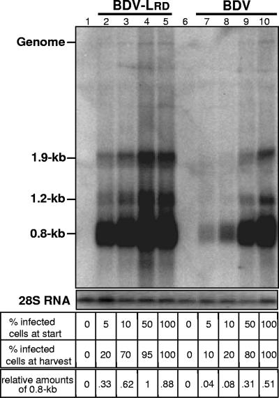
Since minireplicon assays with reconstituted viral polymerase complexes could not explain the enhanced virulence of the BDV variant carrying the LRD mutation, we turned to a less artificial assay system and performed a careful comparison of viral RNA synthesis in acutely infected Vero cells. We argued that the full transcriptional potential of the viral polymerase complex should become visible at very early times after infection of susceptible host cells. To mimic a state of acute infection, we mixed persistently infected cells with uninfected cells at various ratios and cocultured the cell mixtures for 7 days. At this stage, we determined the percentages of virus-infected cells in the cultures by immunofluorescence and measured the content of viral RNA by Northern blotting. As expected, BDV-LRD spread more efficiently to uninfected Vero cells than wild-type BDV (Fig. 7). We noted that viral transcript levels in cultures that contained about 20% acutely infected cells were substantially different. Viral mRNA was about fourfold more abundant in cultures infected to ∼20% with BDV-LRD than in cultures infected to roughly the same degree with wild-type BDV, which is shown by quantification of the 0.8-kb mRNA (Fig. 7, compare lanes 2 and 8). Approximately twofold-enhanced levels of viral transcripts were seen in cultures which contained 70 to 80% infected cells (Fig. 7, compare lanes 3 and 9). This difference was less pronounced in persistently infected cultures. These data indicate that at least during the acute phase of infection, polymerase complexes containing LRD exhibit higher activities than complexes containing wild-type L.
FIG. 7.
Viral RNA expression patterns in Vero cells infected with either wild-type or mutant BDV. Uninfected and infected Vero cells were mixed at the indicated ratios, and the mixtures were cocultured for 7 days. The percentages of infected cells were then determined by staining for the N protein of BDV. The remaining cells were lysed, and the viral RNA content in 5-μg samples was estimated by Northern blot analysis using a cDNA probe comprising nucleotides 976 to 1749 of the BDV antigenome. The identities of prominent bands are indicated. As a loading control, 28S RNA was visualized by ethidium bromide staining.
DISCUSSION
Using a reverse genetics approach, we identified viral determinants that limit BDV replication in mice. We demonstrated that the ability of recombinant BDV strain He/80FR to grow in mice was solely dependent on mutations in the viral polymerase complex and that only three amino acid changes in L and P converted a replication-incompetent virus into an effective killer of a new host species. The virulence gain can be attributed at least in part to the fact that the R66K mutation in P rendered the viral polymerase complex less sensitive to negative regulation by the viral X protein.
Virus adaptation to mice was not successful when we used molecularly cloned virus from Vero cells as the starting material but was readily achieved when the virus was amplified once in rats. A similar result was previously reported from studies with nonrecombinant BDV (9). These observations suggested that by growing BDV in the rat brain, virus mutants with increased fitness for replication in mouse brains which do not seem to accumulate in Vero cells are selected. The molecular basis for this phenomenon is unclear. As cells persistently infected with BDV are resistant to superinfection (6), BDV variants generated in persistently infected Vero cell cultures may not be able to compete with parental virus even if they carry advantageous mutations. Variants generated in the infected rat brain might spread more efficiently than wild-type virus because of the very large number of susceptible cells in this organ.
In our adaptation experiments, growth promotion of BDV in mouse brains was always accompanied by the L1116R mutation in the viral L gene. In a previous study, mouse adaptation of nonrecombinant BDV strain He/80 was associated with the mutation of two different residues in the L gene along with two mutations in the G gene (12), strongly indicating that BDV can find different ways to adapt to mice. Since the L1116R mutation appeared early in both of our two independent adaptation experiments and since this mutation was already present after the first rat brain passage in at least one experiment, it appears that the molecularly cloned virus used here is strongly biased with regard to acquiring this specific mutation in rodent cells. By contrast, the noncloned virus used in the previous study may represent a complex quasi species with substantially greater genetic flexibility.
Recombinant viruses carrying distinct amino acid changes alone or in combination unambiguously showed that the L1116R mutation in L plays a key role in the mouse adaptation process. Interestingly, the R66K mutation by itself was also able to mediate replication competence of BDV in mice, although this mutation was not detected alone at any stage of the adaptation process. This finding reinforces our conclusion that several different mutations in the viral polymerase complex may confer replication competence in mice. We assume that the biased evolutionary drift of molecularly cloned BDV toward quick acquisition of the L1116R mutation in the L polymerase subunit might suppress alternative adaptation routes.
One of the most interesting observations of this study was that by specifically screening for virus growth in mouse brains, we selected virus mutants with greatly improved virulence. We found that recombinant viruses carrying both polymerase mutations either alone (LRD) or in combination with the R66K mutation in P (PK/LRD) exhibited substantially enhanced growth kinetics in Vero cells as well as in MEF of C57BL/6-IFNAR10/0 mice (Fig. 3A and B). These results indicate that the adaptive mutations do not improve the specific interaction of the viral polymerase complex with unidentified mouse-specific factors. In agreement with this view, we recently showed that the LRD form of the polymerase can restore efficient replication in Vero cells and rats in severely attenuated BDV mutants (16, 20). Our data indicate that the mouse-adapted viruses acquired mutations which enhance polymerase performance in host cells of various species. We assume that although mouse cells provide all factors necessary for virus replication, for unknown reasons they still represent intrinsically poor host cells for BDV. We suggest that in such poor host cells standard virus may not perform well, whereas virus with improved polymerase does.
How might the adaptive mutations enhance the activity of the viral polymerase complex? Important clues came from studies aimed at measuring the strength of the interactions between the various viral polymerase subunits. We found that the R66K form of P interacted only very poorly with the viral negative regulator X (Fig. 6A). Using artificial viral genomes, we could further show that polymerase complexes containing the R66K form of P were much less sensitive to negative regulation by X (Fig. 6B). These data indicated that the enhanced performance levels of the polymerase complexes of mouse-adapted viruses carrying the R66K mutation do not result from an intrinsically enhanced activity of the complex but rather from a lack of negative regulation by X. Interestingly, two silent mutations were located in close proximity to the X initiation codon in the original mouse-adapted virus (Fig. 2). These mutations might affect X-protein expression and thereby influence virulence. They may explain why BDV-PK/LRD was more virulent in mice than the virus which appeared in the course of our adaptation experiment. In any case, our results reinforce previous conclusions from experiments with viral minireplicon systems that X negatively regulates viral polymerase activity (14, 22) and further suggest that X serves as a repressor of virulence. It is unclear at present why it is advantageous for BDV to have a negative regulator that restricts its host range. We speculate that negative regulation may favor long-term virus persistence in immune-competent hosts.
Although our biochemical analyses provided an elegant explanation for the enhanced virulence of viruses carrying the R66K mutation in P, they did not explain the virulence of viruses carrying the L1116R mutation in L. Wild-type and mutant L interacted similarly well with other polymerase subunits in mammalian two-hybrid assays (Fig. 6A). In viral minireplicon assays, mutant L further showed approximately the same activity as wild-type L (Fig. 6B). These results indicated that either these assays are not suitable for detecting minor variations in polymerase activity or the performance-enhancing effect of the L mutations is not due to increased polymerase activity. It is tempting to speculate that the virulence-enhancing effect of the mutations in L is similar in nature to that of the R66K mutation in P. Viral polymerase complexes containing mutant L might be less susceptible to negative regulation by undefined factors of viral or cellular origin than complexes containing wild-type L. Supporting evidence for this view comes from experiments in which we mixed infected and uninfected cells (Fig. 7). These experiments indicated that polymerase complexes containing mutant L are hyperactive during the acute phase but not the persistent phase of infection.
Altogether, our data demonstrate that replication competence of recombinant BDV strain He/80FR in a new species is mediated by changes in components of the polymerase complex rather than the viral envelope, and they suggest a role for virus-encoded negative regulator X in this process.
Acknowledgments
We thank Rosita Frank for excellent technical assistance and Georg Kochs, Friedemann Weber, Martin Schwemmle, and Otto Haller for comments on the manuscript.
This work was supported by grants STA 338/8-1 and SCHN 765/1-5 from the Deutsche Forschungsgemeinschaft.
Footnotes
Published ahead of print on 23 May 2007.
REFERENCES
- 1.Briese, T., J. C. de la Torre, A. Lewis, H. Ludwig, and W. I. Lipkin. 1992. Borna disease virus, a negative-strand RNA virus, transcribes in the nucleus of infected cells. Proc. Natl. Acad. Sci. USA 89:11486-11489. [DOI] [PMC free article] [PubMed] [Google Scholar]
- 2.Briese, T., A. Schneemann, A. J. Lewis, Y. S. Park, S. Kim, H. Ludwig, and W. I. Lipkin. 1994. Genomic organization of Borna disease virus. Proc. Natl. Acad. Sci. USA 91:4362-4366. [DOI] [PMC free article] [PubMed] [Google Scholar]
- 3.Ebihara, H., A. Takada, D. Kobasa, S. Jones, G. Neumann, S. Theriault, M. Bray, H. Feldmann, and Y. Kawaoka. 2006. Molecular determinants of Ebola virus virulence in mice. PLoS Pathogens 2:e73. [DOI] [PMC free article] [PubMed] [Google Scholar]
- 4.Gabriel, G., B. Dauber, T. Wolff, O. Planz, H. D. Klenk, and J. Stech. 2005. The viral polymerase mediates adaptation of an avian influenza virus to a mammalian host. Proc. Natl. Acad. Sci. USA 102:18590-18595. [DOI] [PMC free article] [PubMed] [Google Scholar]
- 5.Galvani, A. P. 2004. Emerging infections: what have we learned from SARS? Emerg. Infect. Dis. 10:1351-1352. [DOI] [PMC free article] [PubMed] [Google Scholar]
- 6.Geib, T., C. Sauder, S. Venturelli, C. Hassler, P. Staeheli, and M. Schwemmle. 2003. Selective virus resistance conferred by expression of Borna disease virus nucleocapsid components. J. Virol. 77:4283-4290. [DOI] [PMC free article] [PubMed] [Google Scholar]
- 7.Hallensleben, W., M. Schwemmle, J. Hausmann, L. Stitz, B. Volk, A. Pagenstecher, and P. Staeheli. 1998. Borna disease virus-induced neurological disorder in mice: infection of neonates results in immunopathology. J. Virol. 72:4379-4386. [DOI] [PMC free article] [PubMed] [Google Scholar]
- 8.Hatta, M., P. Gao, P. Halfmann, and Y. Kawaoka. 2001. Molecular basis for high virulence of Hong Kong H5N1 influenza A viruses. Science 293:1840-1842. [DOI] [PubMed] [Google Scholar]
- 9.Kao, M., H. Ludwig, and G. Gosztonyi. 1984. Adaptation of Borna disease virus to the mouse. J. Gen. Virol. 65:1845-1849. [DOI] [PubMed] [Google Scholar]
- 10.Martin, A., P. Staeheli, and U. Schneider. 2006. RNA polymerase II-controlled expression of antigenomic RNA enhances the rescue efficacies of two different members of the Mononegavirales independently of the site of viral genome replication. J. Virol. 80:5708-5715. [DOI] [PMC free article] [PubMed] [Google Scholar]
- 11.Muller, U., U. Steinhoff, L. F. Reis, S. Hemmi, J. Pavlovic, R. M. Zinkernagel, and M. Aguet. 1994. Functional role of type I and type II interferons in antiviral defense. Science 264:1918-1921. [DOI] [PubMed] [Google Scholar]
- 12.Nishino, Y., D. Kobasa, S. A. Rubin, M. V. Pletnikov, and K. M. Carbone. 2002. Enhanced neurovirulence of Borna disease virus variants associated with nucleotide changes in the glycoprotein and L polymerase genes. J. Virol. 76:8650-8658. [DOI] [PMC free article] [PubMed] [Google Scholar]
- 13.Paul, S. M., S. J. Costa, H. Cross, M. Pierce, W. Pizutti, D. H. Morgan, and L. Z. Ziskin. 1997. Emerging infectious diseases: new and resistant strains of HIV. N. J. Med. 94:43-47. [PubMed] [Google Scholar]
- 14.Perez, M., A. Sanchez, B. Cubitt, D. Rosario, and J. C. de la Torre. 2003. A reverse genetics system for Borna disease virus. J. Gen. Virol. 84:3099-3104. [DOI] [PubMed] [Google Scholar]
- 15.Poenisch, M., G. Unterstab, T. Wolff, P. Staeheli, and U. Schneider. 2004. The X protein of Borna disease virus regulates viral polymerase activity through interaction with the P protein. J. Gen. Virol. 85:1895-1898. [DOI] [PubMed] [Google Scholar]
- 16.Poenisch, M., S. Wille, A. Ackermann, P. Staeheli, and U. Schneider. 2007. The X protein of Borna disease virus serves essential functions in the viral multiplication cycle. J. Virol. 81:7297-7299. [DOI] [PMC free article] [PubMed] [Google Scholar]
- 17.Rauer, M., J. Gotz, D. Schuppli, P. Staeheli, and J. Hausmann. 2004. Transgenic mice expressing the nucleoprotein of Borna disease virus in either neurons or astrocytes: decreased susceptibility to homotypic infection and disease. J. Virol. 78:3621-3632. [DOI] [PMC free article] [PubMed] [Google Scholar]
- 18.Ren, W., W. Li, M. Yu, P. Hao, Y. Zhang, P. Zhou, S. Zhang, G. Zhao, Y. Zhong, S. Wang, L. F. Wang, and Z. Shi. 2006. Full-length genome sequences of two SARS-like coronaviruses in horseshoe bats and genetic variation analysis. J. Gen. Virol. 87:3355-3359. [DOI] [PubMed] [Google Scholar]
- 19.Schneider, U. 2005. Novel insights into the regulation of the viral polymerase complex of neurotropic Borna disease virus. Virus Res. 111:148-160. [DOI] [PubMed] [Google Scholar]
- 20.Schneider, U., A. Ackermann, and P. Staeheli. 2007. A Borna disease virus vector for expression of foreign genes in neurons of rodents. J. Virol. 7293-7296. [DOI] [PMC free article] [PubMed]
- 21.Schneider, U., M. Naegele, and P. Staeheli. 2004. Regulation of the Borna disease virus polymerase complex by the viral nucleoprotein p38 isoform. Arch. Virol. 149:1409-1414. [DOI] [PubMed] [Google Scholar]
- 22.Schneider, U., M. Naegele, P. Staeheli, and M. Schwemmle. 2003. Active Borna disease virus polymerase complex requires a distinct nucleoprotein-to-phosphoprotein ratio but no viral X protein. J. Virol. 77:11781-11789. [DOI] [PMC free article] [PubMed] [Google Scholar]
- 23.Schneider, U., M. Schwemmle, and P. Staeheli. 2005. Genome trimming: a unique strategy for replication control employed by Borna disease virus. Proc. Natl. Acad. Sci. USA 102:3441-3446. [DOI] [PMC free article] [PubMed] [Google Scholar]
- 24.Schwemmle, M., M. Salvatore, L. Shi, J. Richt, C. H. Lee, and W. I. Lipkin. 1998. Interactions of the borna disease virus P, N, and X proteins and their functional implications. J. Biol. Chem. 273:9007-9012. [DOI] [PubMed] [Google Scholar]
- 25.Shinya, K., S. Hamm, M. Hatta, H. Ito, T. Ito, and Y. Kawaoka. 2004. PB2 amino acid at position 627 affects replicative efficiency, but not cell tropism, of Hong Kong H5N1 influenza A viruses in mice. Virology 320:258-266. [DOI] [PubMed] [Google Scholar]
- 26.Staeheli, P., C. Sauder, J. Hausmann, F. Ehrensperger, and M. Schwemmle. 2000. Epidemiology of Borna disease virus. J. Gen. Virol. 81:2123-2135. [DOI] [PubMed] [Google Scholar]
- 27.Staeheli, P., M. Sentandreu, A. Pagenstecher, and J. Hausmann. 2001. Alpha/beta interferon promotes transcription and inhibits replication of Borna disease virus in persistently infected cells. J. Virol. 75:8216-8223. [DOI] [PMC free article] [PubMed] [Google Scholar]
- 28.Stitz, L., T. Bilzer, and O. Planz. 2002. The immunopathogenesis of Borna disease virus infection. Front. Biosci. 7:d541-d555. [DOI] [PubMed] [Google Scholar]
- 29.Unterstab, G., S. Ludwig, A. Anton, O. Planz, B. Dauber, D. Krappmann, G. Heins, C. Ehrhardt, and T. Wolff. 2005. Viral targeting of the interferon-{beta}-inducing Traf family member-associated NF-{kappa}B activator (TANK)-binding kinase-1. Proc. Natl. Acad. Sci. USA 102:13640-13645. [DOI] [PMC free article] [PubMed] [Google Scholar]
- 30.Webster, R. G., S. M. Wright, M. R. Castrucci, W. J. Bean, and Y. Kawaoka. 1993. Influenza—a model of an emerging virus disease. Intervirology 35:16-25. [DOI] [PubMed] [Google Scholar]


