Abstract
Objective
To evaluate the validity of using the conventional anteroposterior (AP) radiograph of the knee in order to identify joint space narrowing (JSN) at an early stage of osteoarthritis (OA).
Methods
Grading of JSN using a 0–5 score and quantitative measurement of joint space width (JSW) of the medial and lateral compartments of the tibiofemoral joint in AP and fluoroscopically assisted posteroanterior Lyon schuss (LS) radiographs of 202 patients with knee OA.
Results
Knees without definite JSN (score <2) were twice as common in AP than in LS radiographs (36.1% vs 18.8%). The number of knees showing definite medial JSN was identical in both views but four knees showing a medial OA in AP view were classified differently in the LS radiographs (three bicompartmental OA and one lateral OA). The frequency of lateral JSN was approximately twice as great in the LS view as in the AP view. JSN score was significantly higher (p<0.001) and JSW was significantly smaller (p<0.01) in the LS view than in the AP view. In knees with definite JSN, JSW of the compartment with no narrowing was significantly (p<0.04) larger than in knees that did not exhibit definite JSN. Medial JSW and lateral JSW were inversely correlated (p<0.001).
Conclusions
The standing AP radiograph performed poorly in identifying both the location of JSN in patients with early tibiofemoral OA (especially, lateral OA) and the severity of JSN. The LS radiographs are preferable to standing AP views for the selection of patients for therapeutic trials of structure‐modifying OA drugs.
Most studies of knee osteoarthritis (OA) use the American College of Rheumatology (ACR) symptomatic and radiographic definition of the disease.1 In clinical trials of structure‐modifying OA drugs (SMOADs), the definition has been used to select patients with medial tibiofemoral OA in whom joint space narrowing (JSN) was measured only in the medial compartment.2,3 However, the ACR definition does not indicate which compartment of the knee is affected by OA.
Among patients with tibiofemoral OA, JSN is usually localised to one or the other femoral tibiofemoral compartment—most commonly, the medial.4,5,6,7 In general, the terms medial OA and lateral OA are used to indicate the location of radiographic tibiofemoral JSN. At an advanced stage of the disease, identification of medial or lateral OA is easy, but validated definitions of early medial and lateral OA do not exist.
Radiographic progression of tibiofemoral JSN in OA of the knees has been noted in several studies that have used the standing anteroposterior (AP) view.5,6,8,9,10,11,12,13,14 However, neither the validity of using JSN to define medial or lateral knee OA at baseline, nor quantisation of the changes in joint space width (JSW) in the tibiofemoral compartment adjacent to the narrowed compartment have often been considered. Therefore, although it is expected that JSN will preferentially affect one tibiofemoral compartment, quantitative changes in JSW that occur simultaneously in both compartments during the progression of OA are not well documented.
For prospective studies evaluating the progression of OA, it is essential to accurately identify, at an early stage of the disease, the tibiofemoral compartment in which JSN is anticipated to progress. This is especially true for therapeutic trials of SMOADs, insofar as it facilitates recruitment of patients at a stage of OA that makes them most suitable for enrolment in a randomised clinical trial. Felson et al7 defined medial and lateral tibiofemoral OA as the combination of a Kellgren and Lawrence grade >2 and a JSN score >1. However, whether the location of an osteophyte is a valid predictor, which tibiofemoral compartment in the OA knee will be affected by JSN, is unclear.15,16 Finally, the definition of medial or lateral OA relies mainly on the location of JSN, which, at an early stage of the disease, is difficult to ascertain.17,18,19,20,21 This difficulty is especially pertinent to the standing AP radiograph, which is known to lack accuracy and sensitivity for the assessment of minimum JSW, the parameter on which quantitative measurements of tibiofemoral JSN are usually based. Radiographs of the knee in flexion have been shown to improve detection of tibiofemoral JSN.22,23,24,25,26 However, the ability of such views to identify early medial or early lateral OA in the presence and absence of the other has not been investigated before.
In the present cross‐sectional study, JSW was assessed in both tibiofemoral compartments of patients with a wide range of severity of medial and/or lateral tibiofemoral OA who underwent a conventional standing AP radiograph with the knee in extension and a concurrent posteroanterior (PA) radiograph of the knee in flexion. The aim was to define changes in JSW in the medial and lateral compartments and, in particular, to evaluate the validity of using the AP radiograph to identify the location of JSN at an early stage of OA, as is commonly required for a clinical trial of an SMOAD.
Patients and methods
The radiographs examined in the present cross‐sectional study were obtained from a series of 1236 patients with knee OA symptoms who were screened for entry into a therapeutic trial with a standing AP radiograph of both knees and a concomitant fluoroscopically assisted PA view of each knee in 20–30° of flexion (the Lyon schuss (LS) radiograph).22,27,28
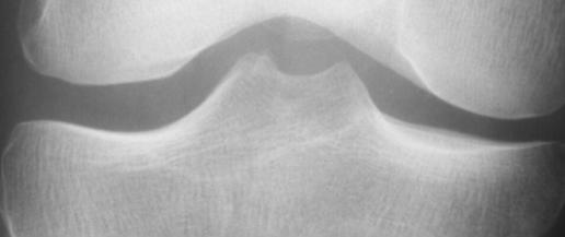
As the AP view is the one generally used for the assessment of OA in subjects who are being considered for enrolment in a clinical trial, as a first step, we selected all knees of subjects who fulfilled ACR clinical and radiographic criteria for knee OA1 (ie, who had clinical symptoms and whose AP knee radiograph showed a definite osteophyte). Quality control criteria included centring of the tibial spines within the femoral notch and acceptable alignment (<1.5 mm) between the anterior and posterior margins of the medial and lateral tibial plateaus at the point of the minimum interbone distance (fig 1).
Figure 1 Illustration of the quality of knee radiographs for film selection. Quality control criteria included centring of the tibial spines within the femoral notch and acceptable alignment of both the medial and lateral tibial pateaus at the point of minimum interbone distance.
Semiquantitative grading of JSN
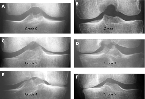
In the standing AP view of both knees, JSN was graded separately in each tibiofemoral compartment by an observer (FMV) using a validated 0–5 scale22: 0, none; 1, doubtful; 2, mild but definite; 3, large; 4, contact between bone edges of femur and tibia; and 5, bone erosion. The JSN grade was determined with the help of the examples illustrated in fig 2. It should be emphasised that JSN, as used herein, refers to a semiquantitative grading of reduction of JSW in a single film, and not to a dynamic process.
Figure 2 Atlas of radiographs used by the observer for scoring of joint space narrowing.
One knee in each AP radiograph was selected, so as to provide subgroups of at least 20 knees representing all possible combinations between a 0–4 JSN grade in the medial compartment and a 0–4 JSN grade in the lateral compartment (table 1). Only one knee of each patient was selected for examination. In some subgroups, the desired number of knees could not be achieved (table 1). A total of 202 knees (from 131 women and 71 men; mean (SD) age, 62.2 (8) years) were finally selected.
Table 1 Subgroups of tibiofemoral osteoarthritis formed by the medial and/or lateral location of joint space narrowing (JSN) and the various possible JSN scores in medial and lateral compartments.
| Compartment | M/L | M/L | M/L | M/L | M/L | M/L | M/L | M/L | M/L | M/L | M/L |
|---|---|---|---|---|---|---|---|---|---|---|---|
| JSN score | 0/0 | 1/0 | 2/0 | 3/0 | 4/0 | 0/1 | 0/2 | 0/3 | 0/4 | 1/1 | >1/>1 |
| Expected (n) | >20 | >20 | >20 | >20 | >20 | >20 | >20 | >20 | >20 | >20 | >20 |
| Observed (n) | 11 | 40 | 33 | 42 | 17 | 14 | 16 | 10 | 0 | 8 | 11 |
JSN, joint space narrowing; L, lateral; M, medial.
Expected and observed number of knees in each subgroups.
JSN in the LS radiograph of the same knee was then graded by the same observer, who was blinded to the grade that has been assigned to the corresponding AP view.
Quantitative measurement of JSW
JSW was quantified in mm by an experienced observer (EV) using a digitised image analysis system (Holy's software, Actibase, Lyon, France).27,28 The computer provided automated detection of the bone edges in both the medial and lateral tibiofemoral compartment. Location of the minimum JSW of both the compartments was illustrated on the screen and its measurement was automatically made.
The reproducibility of the JSW measurements in both the areas was determined by blinded remeasurement of 30 randomly selected LS radiographs by the same observer, who was unaware of the previous reading. The coefficients of variation for the medial and lateral compartment were 1.15% and 1.50%, respectively. The intraclass correlation coefficients for the medial and lateral compartment were 0.99 and 0.93, respectively.
Statistical analysis
Comparison of the location (medial/lateral) of JSN in AP and LS radiographs was made by using the χ2 test. Comparison of medial and lateral JSN scores and JSW in AP and LS radiographs of the same knees was made with a paired Student's t test. Comparison of medial JSW and lateral JSW in the various OA groups (medial OA, lateral OA and no JSN) was made by analysis of variance. The correlation between medial JSW and lateral JSW was made with a simple regression analysis.
Results
Location of JSN and semiquantitative grade of JSN in the AP and LS radiographs
The 202 knees were classified into four groups, according to the location of definite JSN (ie, JSN grade >1):
Medial compartment JSN, medial JSN score 2–4 and lateral JSN 0–1.
Lateral compartment JSN, lateral JSN score 2–4 and medial JSN 0–1.
No JSN, JSN score <2 in both the medial and lateral compartment.
Bicompartmental JSN, JSN score >1 in both the medial and lateral compartment.
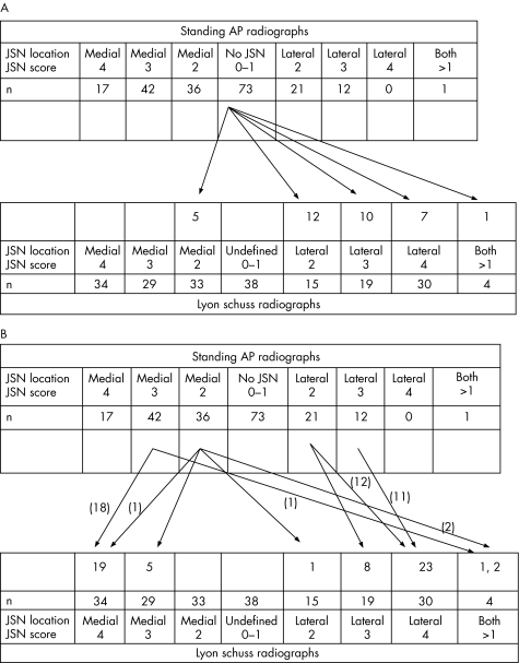
In the AP radiographs, 95 knees showed definite medial, 33 definite lateral and 1 bicompartmental JSN. The corresponding numbers for the LS radiographs were 96, 64 and 4, respectively. Thus, although LS view and AP view were comparable with respect to their ability to detect medial JSN, LS view was nearly twice as likely to reveal lateral tibiofemoral compartment JSN and also more likely to detect bicompartmental JSN than the AP view, which detected only medial compartment narrowing (fig 3A,B). In addition, among the knees examined, 73 (36.1%) of the AP views, but only half as many of the LS views (38, 18. 8%) were reported to not have JSN in either tibiofemoral compartment.
Figure 3 Location of joint space narrowing (JSN) and the JSN score in standing anteroposterior (AP) and Lyon schuss (LS) radiographs of 202 osteoarthritis knees. (A) Identification of definite JSN (score >1 in either tibiofemoral compartment) in the LS radiograph among the 73 knees classified as not having definite JSN (score <2 in both tibiofemoral compartments) in the standing AP radiographs. Most often, lateral JSN was identified in the LS radiograph. Among the 38 knees without definite JSN in the LS radiograph, none was found to have definite JSN in the concurrent standing AP radiograph. (B) Differences between the standing AP and the LS radiographs with respect to the location of JSN and to JSN in knees with definite (score >1) JSN in the standing AP radiograph.
Among the LS radiographs, the number of knees with definite medial JSN was nearly identical to that in the AP view (47.5%). However, among four knees that exhibited definite JSN of the medial compartment in the AP view, three were classified as having bicompartmental JSN in the LS view and one as having lateral compartment JSN (fig 3B).
Among the 73 knees that did not exhibit JSN in the AP radiograph, the concurrent LS view showed definite JSN medially in 5 (6.8%), laterally in 29 (39.7%) and in both tibiofemoral compartments in 1 (1.4%; fig 3A).
These differences between the LS and AP views with respect to the localisation of tibiofemoral compartment OA were highly significant (p<0.001).
The grade of JSN was higher in the LS than in the AP view in 98 (48.5%) knees (fig 3). Among knees with medial tibiofemoral OA in both images (n = 91), the mean (SD) JSN grade was 2.8 (0.7) in the AP view and 3.0 (0.8) in the LS view (p<0.001). Among knees with lateral tibiofemoral OA in both views (n = 33), the mean (SD) JSN grade was 2.3 (0.4) and 3.7 (0.6) in the AP and LS views, respectively (p<0.001).
Quantitative measurement of JSW in knees without definite JSN
Figure 2 shows the measurements of JSW in both compartments of knees that did not show definite JSN. In those knees, medial JSW was smaller (p<0.05) and lateral JSW was larger (p = NS) in the LS than in the AP radiographs. The difference between medial and lateral JSW was considerably larger (0.66 vs 1.38 mm, p<0.01) in the LS than in the AP view.
Quantitative measurement of JSW in the narrowed tibiofemoral compartment
Table 2 shows the measurements of JSW in the narrowed tibiofemoral compartment of knees with medial or lateral OA and in the medial and lateral compartments of knees without definite JSN (ie, semiquantitative grade <2 in both compartments). Knees with bicompartmental JSN were excluded from this analysis.
Table 2 Minimum (SD) joint space width (mm) in knees with no definite joint space narrowing in either the medial or lateral tibiofemoral compartment and in the narrowed compartment of knees with medial or lateral osteoarthritis.
| JSN location | None | Medial | None | Lateral |
|---|---|---|---|---|
| Compartment JSW | Medial | Medial | Lateral | Lateral |
| Standing AP radiograph | 4.33 (1.00), n = 73 | 2.40 (1.10), n = 95 | 4.99 (1.27), n = 73 | 3.36 (1.19), n = 33 |
| Lyon schuss radiograph | 3.94 (0.88), n = 38 | 1.61 (1.2), n = 96 | 5.32 (1.16), n = 38 | 1.44 (1.30), n = 64 |
| p Value | 0.045 | <0.001 | NS | <0.001 |
AP, anteroposterior; JSN, joint space narrowing; JSW, joint space width; NS, not significant.
Among knees without definite JSN, medial compartment JSW was smaller by 9% (p<0.05) and lateral compartment JSW was, on an average, larger by 6% (p = NS) in the LS view than in the AP view. Among knees with definite medial tibiofemoral OA, the mean JSW of the narrowed compartment was 33% smaller in the LS view than in the AP view (p<0.001). Among knees with definite lateral compartment OA, it was 57% smaller (p<0.001).
Quantitative measurement of JSW in the tibiofemoral compartment adjacent to the narrowed compartment
Table 3 shows the measurements of JSW in the non‐narrowed tibiofemoral compartment of knees with medial or lateral OA and in the medial and lateral compartments of knees without JSN (ie, semiquantitative grade <2 in both compartments). Knees with bicompartmental JSN were excluded from this analysis.
Table 3 Minimum (SD) joint space width (mm) of the compartment adjacent to the narrowed compartment in knees with medial or lateral osteoarthritis.
| JSN location | No JSN | Medial | No JSN | Lateral |
|---|---|---|---|---|
| Compartment JSW | Lateral | Lateral | Medial | Medial |
| Standing AP Radiograph | 4.99 (1.27), n = 73 | 5.64 (1.47), n = 95 | 4.33 (1.00), n = 73 | 5.11 (1.14), n = 33 |
| p Value | <0.001 | <0.006 | ||
| Lyon schuss radiograph | 5.32 (1.16) n = 38 | 5.78 (1.13), n = 96 | 3.94 (0.88) n = 38 | 5.28 (1.10) n = 64 |
| p Value | <0.038 | <0.001 | ||
AP, anteroposterior; JSN, joint space narrowing; JSW, joint space width.
Comparison with the minimum JSW of the same compartment in knees with no JSN.
Among patients who exhibited either had medial or lateral tibiofemoral compartment JSN, JSW in the adjacent non‐narrowed compartment was increased in comparison with that in the corresponding compartment of knees that did not exhibit radiographic JSN. In knees with either medial or lateral OA, the increase was significant, both in the AP view (p<0.001 and p<0.006, respectively) and in the LS view (p<0.038 and p<0.001, respectively). The most marked widening was found in the LS view of the medial compartment of knees with lateral tibiofemoral OA.
Association between medial and lateral tibiofemoral compartment JSW
The association between quantitative JSW in the two tibiofemoral compartments of the 202 knees was examined initially by regression analysis. In both the AP and LS views, a highly significant (p<0.001) inverse correlation between JSW in the two compartments existed, with the relationship being much stronger in the LS view (r = 0.65 and r = 0.31, respectively).
The association between JSW in the two compartments was further analysed separately in knees with medial OA and with lateral OA, as shown below.
In AP radiographs of knees with medial OA and in those without JSN in either compartment, JSW in the medial or lateral compartment were inversely related but the correlation was weak and not significant (n = 168, r = 0.13, p = 0.09). Similarly, in AP radiographs of knees with lateral OA and those that did not exhibit JSN, no correlation between medial and lateral JSW was noted.
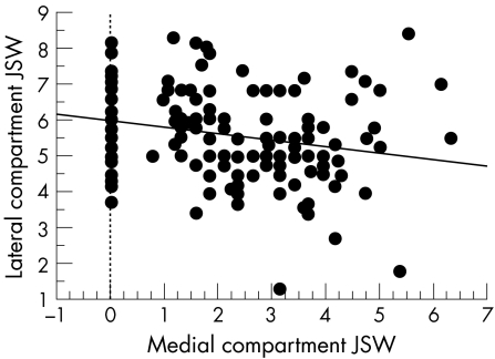
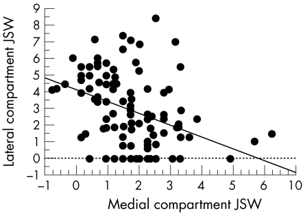
In marked contrast to the findings in the AP view, in the LS view of knees with medial OA and knees that showed no evidence of JSN, the inverse correlation between medial and lateral JSW was highly significant (n = 134, r = 0.22, p = 0.008; fig 4). Among knees with definite lateral OA and those with no JSN, the inverse correlation between the two compartments was even stronger (n = 102, r = 0.39, p<0.001; fig 5). Based on regression analysis, a 1 mm decrease in medial compartment JSW in the LS view was associated with a 0.19 mm increase in lateral compartment JSW and a 1 mm decrease in lateral compartment JSW was accompanied by a 0.70 mm increase in medial compartment JSW.
Figure 4 Correlation between medial compartment joint space width (JSW) and lateral compartment JSW (mm) in knees with medial tibiofemoral osteoarthritis. Lyon schuss radiographs.
Figure 5 Correlation between medial compartment joint space width (JSW) and lateral compartment JSW (mm) in knees with lateral tibiofemoral osteoarthritis and knees with no definite joint space narrowing. Lyon schuss radiographs.
Discussion
Radiographic JSN in patients with tibiofemoral OA is most commonly located in one or the other tibiofemoral compartment,4,5,6,7 and is generally expected to progress in that compartment and to be associated with a widening of the adjacent compartment. However, concurrent progression of JSN in the two tibiofemoral compartments has rarely been evaluated quantitatively.
Ledingham et al5 reported that progression of JSN was less common in the lateral than in the medial compartment (26% vs 46%, respectively). Although Dieppe et al10 found that JSW decreased, on an average, about 0.2 mm over 3 years in both the medial and lateral compartments, they reported increases in tibiofemoral JSW as high as 2 mm during this period. However, whether enlargement of one compartment was accompanied by narrowing of the other was not noted. Boegard et al14 reported a decrease in medial tibiofemoral JSW and an increase in lateral tibiofemoral JSW over 2 years, but neither change was statistically significant. Notably, Boegard et al14 used a non‐fluoroscopically assisted PA view of the knee in flexion, whereas the other studies cited used a conventional standing AP view.
In the present study, we measured JSW with a reliable method that permitted comparison of the two tibiofemoral compartments in subjects exhibiting a broad range of JSW. A significant inverse correlation between the medial compartment and the lateral compartment was demonstrated in both the AP and LS views. The result did not firmly demonstrate an enlargement of JSW of the compartment adjacent to the narrowed one; it could mainly reflect the fact that JSN was only occuring in one compartment. However, in the LS view but not in the PA view, an increased JSW in the compartment adjacent to the narrowed one was demonstrated in knees with either isolated medial or isolated lateral OA.
Thus, the results of this cross‐sectional study suggest that in subjects with knee OA, JSN generally progresses in only one or the other tibiofemoral compartment. However, as longitudinal MRI studies have shown clearly that articular cartilage lesions in OA usually progress in both compartments,29,30,31,32 the increase in JSW seen radiographically in the compartment adjacent to the narrowed compartment is probably artefactual and associated with change in load distribution over the two compartments. Widening of the adjacent non‐narrowed compartment is probably more accentuated in patients with valgus or varus deformity, but it was not possible to evaluate the latter in the present study.
This finding indicates that for prospective studies on the progression of OA, it is important to accurately identify, at an early stage of the disease, the tibiofemoral compartment in which JSN will progress. To adequately evaluate JSN progression, therapeutic trials of SMOADs have preferentially used knees with early OA as seen in the conventional AP radiographs.2,3 The present findings strongly suggest that a number of patients in such studies were erroneously selected for the presence of medial OA. This could explain a proportion of cases in which an unexpected enlargement of medial JSW was found.Also, the erroneous identification of medial OA at baseline probably made the accurate JSN of tibiofemoral OA to be underestimated. Obviously, the point requires a validated definition of early medial and lateral OA.
Although Felson et al7 defined medial and lateral OA by the combination of a Kellgren and Lawrence grade >2 and JSN score >1, the validity of that definition is debatable. Osteophytosis is considered to be the most sensitive feature defining the presence of radiographic OA and can be assessed reliably.17,18,19,20,21 The standing AP view is more sensitive than the tunnel view for the detection of osteophytes and comparable to the semiflexed and LS views in this respect.2,33 However, whether the location of an osteophyte is a valid predictor of which tibiofemoral compartment in the OA knee will be affected by JSN is unclear.15,16 Although the specificity of medial osteophytosis for medial compartment cartilage defects, as identified by MRI, was reported to be 97%,14 sensitivity was only 44% and the association between lateral tibiofemoral osteophytosis and lateral compartment cartilage defects was much weaker.16
Defining OA on the basis of semiquantitative grading of JSN is also difficult. Several scoring systems have been described,20 and JSN grades have only marginal reliability.17,18,19,20,21 Indeed, the validity of a JSN grade has been questioned by Brandt et al,21 who noted that a JSN grade of 1 or 2 (out of 4) was common in the standing AP radiographs of knees in which the articular cartilage was normal at arthroscopy. It has been proposed that cut‐off values for minimum JSW in both compartments be used to define knee OA,17,34 but a quantitative definition of medial/lateral OA such as this suffers from the large variability among normal individuals.
In the present study, we confirmed that radiographs of the knee in the flexion perform much better than the conventional standing AP view for the identification of tibiofemoral compartment narrowing. The number of knees without definite JSN (JSN score <2) in the AP radiographs was nearly halved in the LS views. Among the 73 knees that did not exhibit definite JSN in the AP radiograph, examination of the concurrent LS view identified JSN medially in 6.8% and laterally in 39.7%. Some knees with definite (JSN grade >2) medial OA in the AP radiograph were also found to exhibit lateral JSN in the LS view or showed bicomparmental narrowing in that view.
The present work also showed that JSN of the narrowed compartment was significantly greater in the flexed LS view than in the standing AP view, in agreement with a number of previous reports.22,23,24,25,26 The increase in JSN score was clearly related to a JSW that was significantly smaller in the flexed LS view than in the standing AP view. Thus, the LS view, by illustrating the smallest JSW within the tibiofemoral compartment—that is, by more accurately imaging the site of maximum cartilage damage in the OA knee22,23,24,25,26—optimises the sensitivity of grading of JSN and, consequently, identification of early OA.
In knees without definite JSN, lateral compartment JSW was larger than medial compartment JSW in both the AP and LS views, but the difference between the two compartments was much greater in the LS than in the AP view. Thus, existence of radiographic lateral tibiofemoral OA should be suspected in the LS view whenever lateral compartment JSW is less than the JSW of the medial compartment of the same knee.
As the conventional AP radiograph of the knee in extension performs poorly in identifying the location of JSN in early tibiofemoral OA and, especially, in early lateral compartment disease, we suggest that radiographs obtained with the degree of knee flexion afforded by the LS view are preferable to the standing AP view in extension for the selection of patients for therapeutic trials of SMOADs.
Abbreviations
ACR - American College of Rheumatology
AP - anteroposterior
JSN - joint space narrowing
JSW - joint space width
LS - Lyon schuss
OA - osteoarthritis
PA - posteroanterior
SMOAD - structure‐modifying OA drug
Footnotes
Funding: This work was supported in part by NIH grants AR20582, AR43348 and AR39250.
Competing interests: None declared.
References
- 1.Altman R, Asch E, Bloch D, Bole G, Borenstien D, Brandt K D.et al Development of criteria for the classification and reporting of osteoarthritis. Arthritis Rheum 1986291039–1049. [DOI] [PubMed] [Google Scholar]
- 2.Reginster J Y, Deroisy R, Rovati L C, Lee R L, Lejeune E, Bruyere O.et al Long term effects of glucosamine sulfate on osteoarthritis progression: a randomized, placebo‐controlled clinical trial. Lancet 2001357251–256. [DOI] [PubMed] [Google Scholar]
- 3.Pavelka K, Gatterova J, Olejarova M, Machacek S, Giacovelli G, Rovati LC. Glucosamine sulfate use and delay of progression of knee osteoarthritis: a 3 year, double‐blind study. Arch Intern Med 20021622113–2123. [DOI] [PubMed] [Google Scholar]
- 4.McAlindon T E, Snow S, Cooper C, Dieppe P A. Radiographic patterns of osteoarthritis of the knee joint in the community: the importance of the patellofemoral joint. Ann Rheum Dis 199251844–849. [DOI] [PMC free article] [PubMed] [Google Scholar]
- 5.Ledingham J, Regan M, Jones A, Doherty M. Factors affecting radiographic progression of knee osteoarthritis. Ann Rheum Dis 19955453–58. [DOI] [PMC free article] [PubMed] [Google Scholar]
- 6.Dieppe P, Cushnaghan J, Young P, Kirwan J. Prediction of the progression of joint space narrowing in osteoarthritis of the knee by bone scintigraphy. Ann Rheum Dis 199352557–563. [DOI] [PMC free article] [PubMed] [Google Scholar]
- 7.Felson D T, Nevitt M C, Zhang Y, Aliabadi P, Baumer B, Gale D.et al High prevalence of lateral knee osteoarthritis in Bejing Chinese compared with Framingham Caucasian subjects. Arthritis Rheum 2002461217–1222. [DOI] [PubMed] [Google Scholar]
- 8.Schouten J S A G, Van den Ouweland F, Valkenburg H A. A 12 year follow up study in the general population on prognostic factors of cartilage loss in osteoarthritis of the knee. Ann Rheum Dis 199251932–937. [DOI] [PMC free article] [PubMed] [Google Scholar]
- 9.Spector T D, Dacre J E, Harris P A, Huskinsson E C. Radiological progression of osteoarthritis: an 11 year follow up study of the knee. Ann Rheum Dis 1992511107–1110. [DOI] [PMC free article] [PubMed] [Google Scholar]
- 10.Dieppe P A, Cushnaghan J, Shepstone L. The Bristol ‘OA500' study: progression of osteoarthritis (OA) over 3 years and the relationship between clinical and radiographic changes at the knee joint. Osteoarthritis Cartilage 1997587–97. [DOI] [PubMed] [Google Scholar]
- 11.Dieppe P, Cushnaghan J, Tucker M, Browning S, Shepstone L. The Bristol ‘OA500 study': progression and impact of the disease after 8 years. Osteoarthritis Cartilage 2000863–68. [DOI] [PubMed] [Google Scholar]
- 12.Cooper C, Snow S, McAlindon T E, Kellingray S, Stuart B, Coggon D.et al Risk factors for the incidence and progression of radiographic knee osteoarthritis. Arthritis Rheum 200043995–1000. [DOI] [PubMed] [Google Scholar]
- 13.Wolfe F, Lane N E. The longterm outcome of osteoarthritis: rates and predictors of joint space narrowing in symptomatic patients with knee osteoarthritis. J Rheumatol 200229139–146. [PubMed] [Google Scholar]
- 14.Boegard T L, Rudling O, Petersson I F, Jonsson K. Joint space width of the tibiofemoral and the patellofemoral joint in chronic knee pain with or without radiographic osteoarthritis: a 2 year follow‐up. Osteoarthritis Cartilage 200311370–376. [DOI] [PubMed] [Google Scholar]
- 15.Nagaosa Y, Lanyon P, Doherty M. Characterisation of size and direction of osteophyte in knee osteoarthritis: a radiographic study. Ann Rheum Dis 200261319–324. [DOI] [PMC free article] [PubMed] [Google Scholar]
- 16.Boegard T, Rudling O, Petersson I F, Jonsson K. Correlation between radiographically diagnosed osteophytes and magnetic resonance detected cartilage defects in the tibiofemoral joint. Ann Rheum Dis 199857401–407. [DOI] [PMC free article] [PubMed] [Google Scholar]
- 17.Spector T D, Hart D J, Byrne J, Harris P A, Dacre J E, Doyle D V. Definition of osteoarthritis of the knee for epidemiological studies. Ann Rheum Dis 199352790–794. [DOI] [PMC free article] [PubMed] [Google Scholar]
- 18.Sun Y, Gunther K P, Brenner H. Reliability of radiographic grading of osteoarthritis of the hip and knee. Scand J Rheumatol 199726155–165. [DOI] [PubMed] [Google Scholar]
- 19.Lanyon P, O'Reilly S, Jones A, Doherty M. Radiographic assessment of the symptomatic knee osteoarthritis in the community: definition and normal joint space. Ann Rheum Dis 199857595–601. [DOI] [PMC free article] [PubMed] [Google Scholar]
- 20.Gunther K P, Sun Y. Reliability of radiographic assessment in hip and knee osteoarthritis. Osteoarthritis Cartilage 19997239–246. [DOI] [PubMed] [Google Scholar]
- 21.Brandt K D, Fife R S, Braunstein E M, Katz B. Radiographic grading of the severity of knee osteoarthritis : relation of the Kellgren and Lawrence grade to a grade based on joint space narrowing, and correlation with arthroscopic evidence of articular cartilage degeneration. Arthritis Rheum 1991341381–1386. [DOI] [PubMed] [Google Scholar]
- 22.Piperno M, Hellio M P, Conrozier T, Bochu M, Mathieu P, Vignon E. Quantitative evaluation of joint space width in tibiofemoral osteoarthritis: comparison of three radiographic views. Osteoarthritis Cartilage 19986252–259. [DOI] [PubMed] [Google Scholar]
- 23.Rosenberg T D, Lonnie E P, Richard D P, Coward D B, Scott S M. The forty‐five‐degree posteroanterior flexion weight‐bearing radiograph of the knee. J Bone Joint Surg 198870(A)1479–1483. [PubMed] [Google Scholar]
- 24.Dervin G F, Feibel R J, Rody K, Grabowski J. 3‐Foot standing AP versus 45 degrees PA radiograph for osteoarthritis of the knee. Clin J Sport Med 20011110–16. [DOI] [PubMed] [Google Scholar]
- 25.Inoue S, Nagamine R, Miura H, Urabe K, Matsuda S, Sakaki K.et al Anteroposterior weight‐bearing radiography of the knee with both knees in semiflexion, using new equipment. J Orthop Sci 20016475–480. [DOI] [PubMed] [Google Scholar]
- 26.Ritchie J F, Al‐Sarawan M, Worth R, Conry B, Gibb P A. A parallel approach: the impact of schuss radiography of the degenerate knee on clinical management. Knee 200411283–287. [DOI] [PubMed] [Google Scholar]
- 27.Vignon E, Piperno M, Le Graverand M P, Mazzuca S A, Brandt K D, Mathieu P.et al Measurement of radiographic joint space width in the tibiofemoral compartment of the osteoarthritic knee: comparison of standing anteroposterior and Lyon schuss views. Arthritis Rheum 200348378–384. [DOI] [PubMed] [Google Scholar]
- 28.Conrozier T, Favret H, Mathieu P, Piperno M, Provvedini D, Taccoen A.et al Influence of the quality of tibial plateau alignment in the reproducibility of computer joint space measurement from Lyon‐schuss radiographic views of the knee in patients with knee osteoarthritis. Osteoarthritis Cartilage 200412765–770. [DOI] [PubMed] [Google Scholar]
- 29.Raynauld J P, Martel‐Pelletier J, Berthiaume M J, Labonte F, Beaudoin G, de Guise J A.et al Quantitative magnetic resonance imaging evaluation of knee osteoarthritis progression over two years and correlation with clinical symptoms and radiologic changes. Arthritis Rheum 200450476–487. [DOI] [PubMed] [Google Scholar]
- 30.Wluka A E, Stuckey S, Brand C, Cicuttini F M. Supplementary vitamin E does not affect the loss of cartilage volume in knee osteoarthritis: a 2 year double blind randomized placebo controlled study. J Rheumatol 2002292585–2591. [PubMed] [Google Scholar]
- 31.Wluka A E, Stuckey S, Snaddon J, Cicuttini F M. The determinants of change in tibial cartilage volume in osteoarthritic knees. Arthritis Rheum 2002462065–2072. [DOI] [PubMed] [Google Scholar]
- 32.Cicuttini F M, Wluka A E, Stuckey S L. Tibial and femoral cartilage changes in knee osteoarthritis. Ann Rheum Dis 200160977–980. [DOI] [PMC free article] [PubMed] [Google Scholar]
- 33.Buckland‐Wright J C, Wolfe F, Ward R J, Flowers N, Hayne C. Substantial superiority of semiflexed (MTP) views in knee osteoarthritis : a comparative radiographic study, without fluoroscopy, of standing extended, semiflexed AP, and schuss views. J Rheumatol 1999262664–2674. [PubMed] [Google Scholar]
- 34.Dacre J E, Scott D L, Da Silva J A P, Welsh G, Huskinsson E C. Joint space in radiologically normal knees. Br J Rheumatol 199130426–428. [DOI] [PubMed] [Google Scholar]


